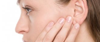
Otitis is one of the most common diagnoses in the daily practice of an otolaryngologist. In acute otitis media, we observe an inflammatory process affecting one of the parts of the human hearing organ. The appearance of acute pain in the ear is the main symptom signaling the onset of inflammation.
The disease is common among both children and adults. Although children are at increased risk of developing acute inflammation. This is due to the structural features of the child’s ear and weak, fragile immunity.
Diseases of the hearing organ, like any other disease concentrated in the head area, must be treated carefully and responsibly, since an infection through the bloodstream can easily reach the brain and cause irreversible consequences. Therefore, it is necessary to treat an acute inflammatory process as soon as the first prerequisites for the disease appear. Treatment of the disease should be carried out in a hospital, under the supervision of a competent doctor.
In this article we will look at how the disease develops, what treatment methods are available today, how complications of otitis manifest themselves and how to avoid them.
Types of disease
Inflammation that occurs in the organ of hearing can be chronic or acute. In acute cases of otitis, the disease lasts for up to three weeks, in chronic cases - more than three months. The chronic process starts when treatment of the acute form of otitis was not carried out or was not carried out at the proper level. There is also an intermediate form - subacute, when the duration of the disease ranges from three weeks to three months.
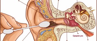
The human hearing organ is divided into three parts: the outer, middle and inner ear. Otitis may appear in each of these areas. Based on the location of the inflammation, acute otitis media is distinguished, and inflammation of the inner ear, otherwise known as labyrinthitis.
External manifestations of inflammation, in turn, are divided into limited, manifesting mainly in the form of a boil of the auricle, and diffuse otitis media. With diffuse otitis, a significant area of the outer ear is affected.
Acute inflammation of the middle ear involves the tympanic cavity of the ear, the auditory (Eustachian) tube and the mastoid process. This type of hearing disease is the most common.
Make an appointment right now!
Call us by phone or use the feedback form
Sign up
The disease of the internal part is called labyrinthitis (this part of the ear is called the labyrinth because of the similarity of its shape to the cochlea). As a rule, inflammation covers the internal part if the treatment of inflammatory disease of the middle ear was carried out late or the treatment for otitis media was chosen incorrectly.
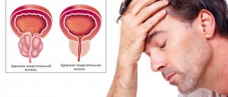
Based on the causes of occurrence, infectious otitis media is distinguished, caused by various pathogens, and non-infectious (for example, arising due to exposure to allergens or ear injuries).
Otitis in acute form can occur in catarrhal (without the formation of secretion in the ear cavity), exudative (with the formation of fluid in the tympanic cavity) and purulent (with the presence of purulent masses) forms.
ACUTE OTITIS MEDIUM
What pathogenetic factor is leading in the development of otitis media?
What are the stages of acute otitis?
Is there a drug of choice in the treatment of acute otitis media?
What can be considered a criterion for recovery in acute otitis media?
Inflammatory diseases of the middle ear occur in all age groups. Among the total number of patients with various diseases of the ENT organs, acute otitis media is diagnosed in 20-30% of cases. This disease develops especially often in children, with the peak incidence occurring between 6 and 18 months; Before the age of three, 90% of children experience acute inflammation of the middle ear at least once [1]. Timely diagnosis and adequate treatment of otitis are often carried out with the participation of a general practitioner, so knowledge of the features of diagnosis and treatment of these diseases is extremely important to prevent possible adverse consequences of otitis.
Based on the nature of inflammation, catarrhal, serous and purulent otitis media are distinguished. Fibrinous, hemorrhagic inflammation and its mixed forms are also possible [4].
More often than other types of inflammation of the middle ear, catarrhal otitis media is observed, also called eustachitis, tubootitis, salpingootitis, etc. and developing as a result of dysfunction of the auditory tube. The cause of catarrhal otitis media is a more or less pronounced dysfunction of the auditory tube, leading to impaired ventilation of the tympanic cavity. This is possible for acute respiratory, acute infectious diseases, as well as influenza. The spread of infection from the upper respiratory tract to the mucous membrane of the auditory tube can lead to disruption of its patency, primarily in the area of the pharyngeal mouth. Tubootitis can also be caused by sudden changes in atmospheric pressure during the ascent and descent of an aircraft (aerootitis), or during the diving and ascent of divers and submariners (mareotitis).
Impaired ventilation of the tympanic cavity leads to the fact that the air contained in it is absorbed by the mucous membrane, and its replenishment is difficult due to the obstruction of the patency of the auditory tube. As a result, the pressure in the tympanic cavity decreases.
The main complaints with tubo-otitis are ear congestion, decreased hearing, sometimes tinnitus, autophony (the resonance of one’s own voice in the affected ear). Ear pain is usually absent or mild, the general condition remains satisfactory. Otoscopy data are very important for making a diagnosis. In this case, a retraction of the tympanic membrane is noted, accompanied by the following characteristic signs: apparent shortening of the handle of the malleus, a sharp protrusion of the short process towards the ear canal; disappearance or deformation of the light cone. Sometimes a radial injection of the vessels of the tympanic membrane along the handle of the malleus or a circular injection in the area of the annulus tympanicus is determined.
Hearing in acute tubo-otitis is slightly reduced, due to the type of sound conduction disturbance, mainly at low frequencies. Sometimes patients note improved hearing after yawning or swallowing saliva, accompanied by the opening of the lumen of the auditory tube.
Acute inflammatory processes of the upper respiratory tract are the causes of the development of temporary dysfunctions of the auditory tube. These disorders are more persistent in adenoid vegetations, various chronic diseases of the nasal cavity and paranasal sinuses (chronic purulent or polypous rhinosinusitis, especially with choanal polyps, deviated nasal septum, hypertrophy of the posterior ends of the inferior turbinates, etc.), and tumors of the nasopharynx.
Against the background of dysfunction of the auditory tube, exudative otitis media can develop, characterized by the presence of serous-mucosal effusion in the tympanic cavity. Today, various designations for the disease are accepted: “secretory otitis media,” “serous otitis media,” “mucous,” etc. The leading pathogenetic factor of exudative otitis media is also a persistent violation of the ventilation and drainage functions of the auditory tube. The very name of this form of otitis indicates increased secretion of mucus and a protracted course of the disease. Its characteristic signs are the appearance of a thick viscous secretion in the tympanic cavity, slowly increasing hearing loss and the absence of perforation of the eardrum. In the development of the disease, along with persistent tuberculous dysfunction, a change in the immunobiological properties of the body, a decrease in general and local resistance, also plays an important role.
Against the background of rarefaction in the non-ventilated tympanic cavity, transudation occurs, the migration of a small number of neutrophilic leukocytes and lymphocytes; transudate leaks into the tympanic cavity.
During this period, the patient notes ear congestion; sometimes mild autophony and hearing loss are observed. During otoscopy, the eardrum is retracted, gray in color, with injected vessels along the handle of the malleus; sometimes air bubbles are visible in the tympanic cavity.
The appearance of fluid in the tympanic cavity is subjectively manifested by a feeling of fullness and pressure in the ear, sometimes noise in the ear and more severe conductive hearing loss. There is often a sensation of fluid transfusion (splashing) when the head position changes, accompanied by improved hearing. This can be explained by the fact that when the head is tilted, the fluid in the tympanic cavity moves, thereby opening up the niches of the windows of the labyrinth, which leads to improved hearing. During otoscopy during this period, fluid is often visible through the eardrum, the level of which is determined in the form of an arcuate line that moves when the position of the head changes.
Restoring ventilation of the tympanic cavity can lead to recovery. However, with continued disruption of the tubular function, secretory otitis media takes a chronic course, turning into fibrosing otitis media, characterized by the appearance of a scar process in the tympanic cavity - the so-called adhesive otitis media develops, leading to severe persistent hearing loss.
Diagnosis of exudative otitis media is complex and not always timely. This is due to the asymptomatic course of the disease, which does not cause any significant pain and does not lead to disruption of the general condition of the patient. The patient gets used to a moderate decrease in hearing in one ear, gradually increasing, and stops paying attention to it, especially if the other ear hears normally. The doctor should take into account that the asymptomatic course of exudative otitis media is now becoming more common. Otoscopy, preferably with magnification, is of great importance in diagnosis. To clarify the diagnosis, a study of the function of the auditory tube is performed using publicly available tests; Impedance measurement is also performed, which reveals a flattened curve. Hearing is examined using tuning forks and audiometry.
Acute purulent otitis media is an acute inflammation of the mucous membrane of the tympanic cavity, and all parts of the middle ear are involved to one degree or another in the process.
This is a widespread disease of the middle ear, which can be either mild or, rapidly developing, cause a severe general inflammatory reaction of the body. However, in both cases, it often leaves behind an adhesive process, accompanied by difficult-to-treat hearing loss, or becomes chronic, often progressive, which also leads to hearing loss and often to severe complications. A distinctive feature of this disease at present is a less acute onset and sluggish course, and in childhood - a tendency to relapse.
The cause of the disease is infection in the tympanic cavity with reduced local and general resistance. Most often (up to 80%) the causative agents of acute purulent otitis media in adults and children are S. pneumoniae and H. influenzae, somewhat less often - M. catarrhalis, S. pyogenes, S. aureus or associations of microorganisms. Viral otitis is observed mainly during epidemics of viral diseases.
The most common route of infection is tubogenic - through the auditory tube. With various general infectious diseases, local inflammatory processes in the upper respiratory tract, the protective function of the epithelium of the auditory tube is disrupted, and the microflora penetrates into the tympanic cavity. Less commonly, the infection enters the middle ear through a damaged eardrum due to injury or through a wound to the mastoid process. In this case, they talk about traumatic otitis media. The third route of infection into the middle ear, hematogenous, is relatively rarely diagnosed; it is possible with infectious diseases such as influenza, scarlet fever, measles, typhoid, tuberculosis.
The inflammatory reaction in acute purulent otitis media from the very beginning affects not only the mucous membrane of the middle ear, but also the periosteum closely adjacent to it. The middle ear is filled with inflammatory exudate, which may initially be serous and then becomes purulent. The mucous membrane becomes sharply thickened, erosions and ulcerations appear on its surface. At the height of inflammation, the tympanic cavity is filled with exudate, granulations and thickened mucous membrane. If the drainage function of the auditory tube is impaired, this leads to bulging of the eardrum; as a result of strong pressure of purulent exudate and circulatory disorders, melting of some area and perforation of the eardrum often occurs, followed by otorrhea.
As the inflammatory changes subside, the amount of discharge decreases and suppuration stops completely. After the discharge from the ear stops, the perforation of the eardrum may heal, but ear congestion persists for some time. The criterion for recovery is normalization of the otoscopic picture and complete restoration of hearing.
In its course, acute purulent otitis media can be mild and quickly resolving, sluggish and protracted, acute and violent; as a rule, it ends with complete recovery; if this does not happen, it can cause chronic otitis media. In some cases, acute purulent otitis media is complicated by mastoiditis or even the development of intracranial complications or sepsis, although the latter conditions more often occur with chronic purulent inflammation of the middle ear.
The clinical picture of a typical acute purulent otitis media is characterized by a staged course. Local and general symptoms of the disease are expressed differently, depending on the stage and severity of the process. It is customary to distinguish pre-perforation, perforation and reparative stages of acute purulent otitis media. The process does not always go through all three stages. Thanks to the mobilization of the body's natural defenses, as well as during intensive therapy, the disease can already acquire an abortive course at the first stage.
The initial, pre-perforative stage of the disease is characterized by pronounced local and general symptoms. The leading complaint is pain in the ear, often very sharp, radiating to the temple and crown. Steadily growing, it sometimes becomes painful and unbearable. Pain occurs as a result of inflammatory infiltration of the mucous membrane of the tympanic cavity and the accumulation of exudate in it; in this case, irritation of the receptor endings of the branches of the trigeminal and glossopharyngeal nerves occurs. Sometimes there is pain on palpation and percussion of the mastoid process, which is caused by inflammation of its mucous membrane. At the same time, congestion and noise in the ear occur, and conductive hearing loss is detected with a slight deterioration in bone conduction of sound. With influenza, as well as measles and scarlet fever, the inner ear is sometimes involved in the process, which is manifested by a more significant impairment of sound perception. During this period, the general condition of the patient is often disturbed - signs of intoxication appear, body temperature rises to 38-39°C, changes characteristic of the inflammatory process are detected in the peripheral blood.
During otoscopy, the injection of blood vessels along the handle of the malleus and the radial vessels of the membrane is first visible, accompanied by shortening of the light cone. Then the hyperemia of the eardrum increases, becomes diffuse, its identifying points disappear, the membrane protrudes, becomes infiltrated, and sometimes becomes covered with a whitish coating. The duration of the initial stage of acute otitis media ranges from several hours to two to three days.
The perforated stage is characterized by perforation of the eardrum and the appearance of suppuration. In this case, the pain in the ear quickly subsides, the patient’s well-being improves, and the body temperature decreases. Discharge from the ear is initially profuse, mucopurulent, sometimes mixed with blood. During otoscopy, a so-called “pulsatile reflex” can be observed, when pus enters through the perforation in portions, synchronously with the pulse.
Gradually, the amount of discharge decreases, it becomes thick and acquires a purulent character. Suppuration usually lasts five to seven days. Perforation in acute otitis media is usually small; more extensive perforations occur with scarlet fever, measles, and tuberculosis.
The reparative stage is characterized not only by the cessation of suppuration and, in most cases, spontaneous scarring of the perforation, but also by the gradual restoration of hearing. Along with the cessation of discharge, hyperemia and infiltration of the eardrum disappear, its shine appears, and identifying contours become visible. Small perforations (up to 1 mm) close quite quickly, leaving no marks. With a large perforation, the middle fibrous layer at the site of the defect usually does not regenerate; in this case, if the perforation does close, this area looks atrophic, sometimes there are deposits of lime salts. Fibrous adhesive changes after otitis media often remain in the tympanic cavity itself, limiting the mobility of the auditory ossicles.
The typical course of acute purulent otitis media can be disrupted at any stage of the process. In some cases, the disease immediately takes on a sluggish, protracted nature, accompanied by mild general symptoms. Perforation of the eardrum does not occur, but a viscous, thick secretion accumulates in the tympanic cavity, which is difficult to evacuate. Following this, an adhesive (adhesive) process often develops in the tympanic cavity. Sometimes, on the contrary, from the very beginning the course of the disease can be extremely severe, with high fever, severe headache, vomiting, dizziness and a sharp deterioration in general condition. In some cases, even before perforation, the infection can quickly spread from the middle ear into the cranial cavity and lead to severe intracranial complications.
If, despite the perforation of the eardrum, the temperature does not decrease and the patient’s condition does not improve, this is usually associated with the transition of inflammation to the mastoid process, i.e., with the development of mastoiditis. Suppuration that does not stop for a long time (three to four weeks), when after cleaning the ear pus fills the ear canal again, indicates empyema of the mastoid process, which, as a rule, causes melting of its bone bridges.
In the normal course of otitis, changes in the peripheral blood are manifested by moderate leukocytosis without a pronounced shift of the formula to the left, and a mild increase in ESR. With a severe disease, pronounced leukocytosis is observed, sometimes up to 20.0•109/l and higher, with a noticeable shift to the left.
Treatment of acute otitis media is carried out differentially, depending on the specific nosological form, the severity of clinical symptoms and the characteristics of the patient’s somatic status. In all cases, the earlier it is started, the higher the effectiveness of treatment.
Considering the important role of tubular dysfunction in the pathogenesis of various forms of acute otitis media, first of all, measures are taken to restore the aeration of the tympanic cavity through the lumen of the auditory tube. In order to reduce swelling of the mucous membrane in the area of its pharyngeal mouth, the patient is prescribed vasoconstrictor nasal drops: naphthyzin, sanorin, tizin, nazivin, a combination drug - polydex with phenylephrine, xylometazoline [3], etc. Sometimes antihistamines (diphenhydramine) help reduce swelling of the mucous membrane , Suprastin, Tavegil, Claritin, Telfast, etc.). To prevent infected mucus from entering the nasopharynx through the auditory tube into the tympanic cavity, the patient should be warned against blowing his nose too vigorously. The nose should be cleaned one nostril at a time, without straining too much. For the same purpose, in the presence of inflammatory changes in the nasopharynx, it is not recommended to blow the auditory tubes according to Politzer; preference is given to catheterization of the auditory tube, performed after thorough anemization of its pharyngeal mouth. Through a catheter, a few drops of a 0.1% solution of adrenaline or dexamethasone can be injected into the lumen of the auditory tube. In case of acute eustachitis, the complex of treatment measures includes various physiotherapeutic procedures: ultraviolet radiation, UHF on the nose, laser therapy on the area of the mouth of the auditory tube, pneumomassage of the eardrum.
With adequate treatment, eustachitis usually goes away within a few days. The effectiveness of treatment of this disease depends on the timely elimination of pathology of the nasal cavity, paranasal sinuses and nasopharynx, which provoke the occurrence and further course of tubo-otitis.
Treatment of exudative otitis media should be comprehensive. First of all, one should also strive to restore the function of the auditory tube using the above methods. To improve tubular function, the ears are blown using the Politzer method or through an ear catheter, with simultaneous massage of the eardrum using a Siegle funnel. Dexamethasone, antibiotics, and chymotrypsin are injected into the lumen of the auditory tube through a catheter. The introduction of proteolytic enzymes and lidase through endaural electrophoresis is quite effective. Vasoconstrictor drugs are used in the nose in the form of drops, but their long-term use is undesirable, since the substances they contain reduce the mucociliary activity of the ciliated epithelium of the nasal cavity and auditory tube.
The prescription of antihistamines is recommended only in cases where serous otitis media develops against the background of allergies. General strengthening agents and vitamins are also indicated. The complex of therapeutic measures has recently increasingly included the anti-inflammatory drug fenspiride (erespal) [2], as well as immunocorrectors (for example, polyoxidonium 0.006 g intramuscularly every other day - a total of six to ten injections; derinat intramuscularly 5.0 ml every other day - five injections).
In cases where the function of the auditory tube is not restored, the exudate does not resolve and hearing does not improve, surgical methods are used to evacuate secretions from the tympanic cavity. The most widely used is tympanic cavity bypass.
For acute purulent otitis media, treatment is complex, and it is prescribed depending on the stage of the disease. An outpatient regimen is recommended, and in case of a pronounced increase in temperature or general malaise, bed rest. If there is a suspicion of an incipient complication, the patient should be urgently hospitalized.
The measures discussed above are carried out in order to restore or improve the ventilation and drainage functions of the auditory tube.
An important place in the treatment of acute purulent otitis media is occupied by catheterization of the auditory tube. Blowing of the auditory tube in acute otitis media using a catheter is performed with the aim of draining the middle ear, eliminating the vacuum in the tympanic cavity that always occurs with this disease, as well as introducing medications into it. A solution of amoxicillin clavulanate and dexamethasone in a 3:1 ratio with the addition of one or two drops of a 0.1% adrenaline solution is injected through an ear catheter. Catheterization helps normalize the function of the auditory tube and eliminates inflammation. Catheterization is carried out from the very beginning of the disease, which often makes it possible to achieve an abortive course of the process; at stages II-III of acute inflammation of the middle ear, blowing with a catheter also gives a good therapeutic effect.
The basis of drug treatment for acute suppurative otitis media is antibiotic therapy.
The prescription of antibiotics is, of course, indicated already in the pre-perforation stage. The drug of choice for the treatment of uncomplicated forms of otitis in adults is amoxicillin orally, 0.25–0.5 g three times a day for 10 days. If there is no effect after three days of amoxicillin therapy, the drug should be changed to augmentin (0.375 or 0.625 g orally two to three times a day) or cefuroxime axetil (0.25 or 0.5 g orally twice a day) [5]. In case of intolerance to b-lactam antibiotics, modern macrolides are prescribed (Rulid 0.15 orally twice a day; spiramycin 1.5 million IU orally twice a day). For complicated forms of otitis, fluoroquinolone drugs of III-IV generations are prescribed: Sparflo 400 mg orally on the first day, then 200 mg per day; avelox 400 mg orally once a day, the duration of treatment depends on the severity of the disease.
Even with a sharp improvement in the patient’s general condition and mitigation of local symptoms, the course of antibiotic therapy should not be stopped prematurely; its duration should be at least 8-10 days. Premature discontinuation of drugs can lead to relapse of the disease and the formation of adhesions in the tympanic cavity, which, in turn, can lead to persistent hearing loss [6].
For pain relief in the initial (preperforative) stage of the disease, paracetamol 1 g is prescribed four times a day. A good analgesic effect at this stage is provided by an endaural microcompress according to Tsytovich with an alcohol-glycerin mixture (equal parts of a 3% alcohol solution of boric acid and glycerin). A gauze or cotton wool moistened with this mixture is inserted into the external auditory canal until it comes into contact with the eardrum; the opening of the external auditory canal is obstructed with cotton wool moistened with Vaseline or greasy cream. Such a compress can be left in the ear for 4–6 hours. Otipax ear drops, which contain lidocaine hydrochloride, phenazone, sodium thiosulfate, ethyl alcohol and glycerin, have a pronounced analgesic, anti-inflammatory and anti-exudative effect.
A warming semi-alcohol compress is also applied topically to the ear, which accelerates the resolution of the inflammatory process. However, when, after applying a compress, the patient notices increased pain in the ear, the compress should be removed immediately so as not to provoke the development of complications.
If, despite the treatment, the patient’s condition does not improve, he is still bothered by severe pain in the ear, a high temperature persists, pain is detected when pressing on the mastoid process, and a protrusion of the eardrum is observed during otoscopy, then paracentesis is performed - an incision of the eardrum. Paracentesis should be performed according to emergency indications when signs of irritation of the inner ear or meninges appear (dizziness, vomiting, severe headache, etc.).
In the presence of perforation of the eardrum, the main attention is to ensure the free outflow of pus. Turundas should be changed frequently, while clearing the ear canal of pus. If the pus thickens, it can be removed by infusing a 3% solution of hydrogen peroxide, which, when combined with pus, forms foam. Foam and pus are removed from the depths of the ear canal using a probe with cotton wool wound around it. The patient should be instructed how to independently remove purulent secretions from the depths of the external auditory canal two to three times a day.
Medications can be administered into the middle ear using transtympanic injection. The above mixture of antibiotic and dexamethasone (and subsequently enzymes that prevent the formation of scars in the tympanic cavity - trypsin, chymopsin, lidase, etc.) is poured into the external auditory canal in an amount of 1 ml and injected by gently pressing the tragus into the external opening of the auditory canal . In this case, the medicinal substance passes through the tympanic cavity, the auditory tube and can enter the nasal cavity and mouth. Catheterization and transtympanic drug injection are effective treatment methods.
For thick purulent discharge, mucolytics are prescribed internally (fluimucil, ACC, fluifort, sinupret), erespal is an anti-inflammatory drug that reduces hypersecretion and swelling of the mucous membrane and stimulates the function of the ciliated epithelium of the auditory tube. At home, physiotherapeutic procedures (UV irradiation, UHF or microwave therapy, laser therapy) and warm compresses on the ear also contribute to a faster recovery.
After removing the purulent secretion, a medicinal solution prescribed by a doctor, heated to 37°C, is poured into the ear. This can be a 0.5-1% dioxidine solution, cypromed ear drops, which contain the antibacterial drug ciprofloxacin with a wide spectrum of antibacterial action; Otofa drops containing the active substance rifamycin, etc. It is not recommended to prescribe alcohol drops in the second stage of otitis, since alcohol often causes irritation of the mucous membrane of the tympanic cavity and severe pain.
Suppuration usually stops after a few days, which marks the transition of the disease to the final reparative stage. The perforation of the eardrum most often closes, forming an inconspicuous scar. During this period, it is important to achieve as complete a hearing restoration as possible. Antibiotic therapy is canceled, ear toilet is stopped, thermal procedures are also completed. After the disappearance of perforation, the main attention should be paid to restoring the ventilation function of the auditory tube and increasing the body's resistance. The auditory tube is blown through the Politzer method or through a catheter, and it is possible to introduce enzyme preparations into the tympanic cavity that prevent the formation of adhesions. For the same purpose, pneumomassage of the eardrum is performed using a pneumatic Siegle funnel, and endaural iontophoresis with lidase. It is recommended to continue vitamin therapy; biostimulants are prescribed - apilak, actovegin, cigapan, etc.
To ensure the restoration of hearing function, control audiometry is performed. In a typical favorable course, recovery occurs with the elimination of the inflammatory process and complete restoration of hearing.
Literature.
- Bogomilsky M. R., Chistyakova V. R. Pediatric otorhinolaryngology // Textbook for universities. - M.: GEOTAR-MED, 2001. - P. 78-95.
- Levina Yu. V., Luchikhin L. A., Krasyuk A. A. The use of erespal in the treatment of exudative otitis media // Vestn. otorinolar., 2003. - 4. - pp. 35-37.
- Palchun V. T., Polyakova T. S., Luchikhin L. A. New dosage form of xylometazoline // Proceedings of the Russian Conference of Otolaryngologists November 19-20, 2002 - pp. 371-373.
- Palchun V. T., Magomedov M. M., Luchikhin L. A. Otorhinolaryngology. — M.: Medicine. - 2002. - P. 382-408.
- Strachunsky L. S., Kozlov S. N. Modern antimicrobial therapy // Guide for doctors. - M.: Borges, 2002. - 436 p.
- Butler CC, Williams RG The etiology, Pathophysiology and Management of Otitis Media with Effusion. — Curr. Infect. Dis. Rep., 2003, 3: 205-213.
L. A. Luchikhin, Doctor of Medical Sciences, Professor of Russian State Medical University, Moscow
Acute otitis media of the middle ear: what causes inflammation?
The inflammatory process is always caused by pathogenic microorganisms, which means that the prerequisites for their activation must be present in the body. The causes of otitis media are:
- hypothermia;
- diseases caused by infection (flu, ARVI, measles);
- inflammatory processes of the ENT organs (the tympanic cavity is connected to the nasopharynx via the Eustachian tube, it is not surprising that the infection from the nasopharynx easily penetrates into the middle ear);
- improper nose blowing;
- hypertrophy of adenoid vegetations;
- rhinitis, sinusitis;
- allergic reactions;
- deviated nasal septum;
- foreign object in the ear;
- damage to the hearing organ.
Diagnostics
An otolaryngologist diagnoses the disease. First of all, when a patient contacts, the doctor collects an anamnesis of the disease and. After listening to all the patient’s complaints, he examines the ear (otoscopy). During the examination, changes in the structure of the eardrum and its deformation are determined, and if the membrane is thinned, the volume of accumulated exudate is determined. The main examination methods used for suspected exudative otitis media are:
- audiometry – it is used to determine the patient’s hearing level, as well as his sensitivity to sounds of different frequencies;
- determination of the ventilation functions of the Eustachian tube;
- determination of the mobility of the eardrum, for which the Valsalva maneuver is used;
- endoscopic examination of the ear cavity and Eustachian tube - the same examination of the nasal cavity may be required;
- X-ray – prescribed if there are suspicions of cellular disorders;
- computed tomography is necessary in a situation where, after all the examinations, difficulties arise in making an accurate diagnosis due to the receipt of somewhat contradictory data.
In some exceptional cases, the patient may be referred for a general blood and urine test, as well as an ear smear. Usually it is quite easy for a doctor to make a correct diagnosis. Difficulties can arise only when pathology appears in a small child who cannot explain what is happening to them. Therefore, in children under three years of age, the disease most often becomes chronic, since there is no treatment, due to the fact that the disorder goes unnoticed.
Outer and inner ear: causes of inflammation
Otitis externa can develop due to improper ear hygiene. If you don't take care of your ears, dirt will accumulate in them, and this is a favorable environment for the growth of bacteria. Excessive hygiene is also harmful: earwax is a natural barrier against the penetration of bacteria into the ear. If you diligently clean the ear canals every day, a person loses this barrier and opens the way for pathogens. Another mistake that leads to acute ear inflammation is cleaning the ears with sharp objects that are not intended for this (toothpicks, matches, hairpins). Such actions can lead to damage to the auricle, which in turn leads to infection entering the wounds. Another factor is dirty water that gets into the ear, which contains pathogens. “Swimmer’s ear” is another name for this type of disease.
As we have already said, inflammation of the internal region occurs due to undertreated otitis media, if due attention has not been paid to the treatment of otitis media. Bacteria can also get here from the meninges, for example, with meningitis. This type of inflammation can be caused by injuries and fractures of the skull or temporal bone.
In order to recognize the disease in time and choose the right treatment, you need to be able to identify its signs.
Forms
Doctors divide exudative otitis media by whether it affects one or two ears, and by the duration of the course. The first classification distinguishes unilateral (left-sided or right-sided) otitis and bilateral. The second divides the disease into the following categories:
- acute exudative otitis - the acute form includes a disease that is completely cured within a period of up to three weeks;
- subacute exudative otitis - its duration is more than three weeks, but not more than 8 weeks;
- chronic exudative otitis media - the disease of this form lasts more than 8 weeks. Its treatment is more complex and lengthy than acute and subacute forms. It also significantly increases the likelihood of complications.
Not only the doctor, but also the patient himself can determine which otitis media occurs in a particular case, if he remembers exactly when the first signs of pathology appeared. In some cases, symptoms of exudative otitis media may be absent, and then it is discovered by chance during a routine medical examination or when contacting a specialist for another reason. As a rule, this happens if the disease is chronic.
Symptoms
The acute course of the disease is characterized by a rapid onset and pronounced symptoms.
With a disease of the outer ear, a person experiences pain inside, which intensifies when pressing on it from the outside. Acute pain occurs when swallowing and chewing food. The ear itself swells and turns red. The skin of the auricle is itchy, the patient's complaints are reduced to a state of stuffiness and ringing in the ear.
In acute otitis media, the main sign of inflammation is the sudden appearance of sharp shooting pains, which become stronger by night. The pain can radiate to the temples, left or right frontal parts, to the jaw - it is very difficult to endure even for an adult, not to mention children. The following symptoms are also characteristic of acute otitis media:
Friends! Timely and correct treatment will ensure you a speedy recovery!
- fever (up to 39°C);
- tinnitus;
- hearing loss;
- lethargy, malaise, loss of appetite;
- in the exudative form, discharge comes from the ear (usually this discharge is transparent or white);
- Acute purulent otitis media is characterized by suppuration from the ear.
The main symptom of labyrinthitis is dizziness. They can last a few seconds, or they can last for several days.
If you notice one or more of the symptoms described above, you should immediately consult a doctor for treatment.
What is prohibited for otitis media
- Under no circumstances should foreign bodies be introduced into the ear (geranium leaves, ear phyto-candles). This will make diagnosis difficult and may lead to a worsening of the condition (for example, leaves that have not been removed begin to rot and become a source of infection).
- If the pain is severe, do not apply a heating pad to your ear or apply warm compresses. This is dangerous if purulent inflammation has begun in the ear. Compresses can only help at stages 1-2 of the disease.
- You should not put melted oil in your ear: if there is a perforation, the oil will end up in the tympanic cavity.
- You should not put camphor oil or camphor alcohol into your ear - it can burn the walls of the ear canal and irritate the eardrum, which will increase ear pain.
At MedicCity you will be denied professional help for otitis media and other ENT diseases. Our otolaryngologists will conduct a comprehensive examination of the patient and prescribe a treatment regimen, depending on the cause and stage of the disease. However, the success of treatment depends no less on the patient himself: the sooner he consults a doctor, the more effective the result will be and the lower the likelihood of complications. It is also important to follow preventive measures. So, in the cold season, to prevent otitis media, it is important to wear a hat, protect your ears from drafts, and of course, boost your immunity!
Stages of disease development
Treatment of acute otitis lasts from one to three weeks. There are several stages in the development of the disease. But it is not at all necessary that the patient will go through all of them. If treatment for infectious otitis is started on time and the acute disease is treated by a competent ENT doctor, recovery will not take long.
So, the course of the disease is conventionally divided into several stages:
- Catarrhal. Pathogenic microorganisms begin to actively multiply, triggering an inflammatory process in the ear. At this time, catarrhal edema and inflammation are observed.
- Exudative. Inflammation leads to active formation of fluid (secret). It accumulates and pathogenic microorganisms continue to multiply here. Timely treatment at this stage will allow you to cure otitis media, avoiding complications.
- Purulent. Acute purulent inflammation is characterized by increased formation of purulent masses in the middle ear cavity. They accumulate, the patient experiences pressure from the inside. The state of congestion does not go away. This phase usually lasts from several days to several hours.
- Perforated. At this stage, accumulated pus causes a rupture of the eardrum, and purulent masses emerge from the tympanic cavity to the outside. At this moment, the patient begins to feel noticeable relief, the high temperature decreases, and the pain gradually disappears. It happens that the eardrum is unable to rupture, then the doctor manually punctures the eardrum (paracentesis) and thereby releases purulent masses out into the ear canal.
- Reparative phase - the release of pus is completed. The hole in the eardrum closes. As a rule, after proper symptomatic treatment, the patient quickly recovers.
What is needed for diagnosis?
To diagnose this disease, it is necessary to conduct a complete otorhinolaryngological examination. The most informative examination today is using video endoscopic equipment. During the examination, the doctor can observe the fluid level and air bubbles behind an unchanged or thickened, clouded eardrum. If necessary, to confirm the diagnosis, tympanometry is recommended, after which the otolaryngologist will be able to draw a complete picture of the condition of the tympanic cavity and middle ear as a whole.
During a traditional examination, the pathology may remain undetected and be diagnosed already at the stage of complications and hearing loss by an audiologist.
Complications and preventive measures
As a rule, if you start treating the disease on time, treatment of acute purulent otitis, exudative or inflammation of any other kind, you can avoid any complications.
However, if treatment is not carried out and the disease progresses, the diagnosis can become chronic. The most serious consequences are: meningitis, encephalitis, brain abscess, facial neuritis, hearing loss. But these dangerous conditions can only appear when patients persistently neglect treatment for otitis media.
Preventive measures include the fight against existing foci of inflammation in the body, competent and timely treatment of ENT diseases, proper ear hygiene and, of course, strengthening the immune system.
Prevention of catarrhal otitis in children
Preventive measures are quite simple and easy to implement.
These include:
- various hardening options;
- timely treatment of viral seasonal infections and chronic pathology of the ENT organs;
- compliance with hygiene rules (after bathing the child, carefully dry the ear canal with a towel).
Symptoms of catarrhal otitis media disappear within a few days completely without a trace if appropriate treatment is started in time.
Carrying out treatment
It is much easier to cure acute otitis media if treatment for the disease begins as early as possible. Treatment should be carried out under the supervision of an otolaryngologist. Complex treatment includes the following activities:
- for acute pain, taking analgesics is indicated to relieve pain;
- to bring down the temperature you need to take antipyretic drugs;
- in difficult cases, antibiotic treatment is carried out;
- local treatment consists of using special ear drops, which are prescribed individually in each case. Self-selection of drops, as well as antibacterial drugs, is fraught with dangerous consequences for health.
- Antihistamines help relieve swelling;
- a good effect is achieved during physiotherapeutic procedures;
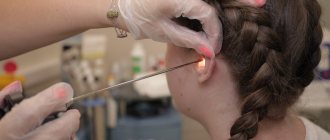
- surgical intervention: opening of the eardrum (paracentesis) is carried out if spontaneous rupture has not occurred.
All ENT doctor’s prescriptions must be followed in full: after all, following treatment recommendations is the key to a quick recovery.
Causes
The main pathogens that cause inflammation in the middle ear are: Streptococcuspneumoniae, Haemophilusinfluenzae, Moraxellacatarrhalis, Streptococcuspyogenes, Staphylococcusaureus. These microorganisms are also pathogens in other inflammatory diseases of the ear, nose and throat (sinusitis, pharyngitis, tonsillitis). Viral infection plays a major role in the occurrence of acute otitis media.
Viruses and bacteria, settling on the mucous membrane of the nose and nasopharynx, damage it. An inflammatory reaction is triggered - substances toxic to the mucous membrane are produced, blood vessels dilate, blood flow increases, and mucus production increases. As a result, nasal congestion, sneezing, and runny nose appear, which creates conditions for infection to penetrate through the auditory tube into the tympanic cavity.
What not to do during treatment
Some patients are overly self-confident and believe that a disease such as otitis media can be easily cured with the help of folk remedies and “grandmother’s” recipes. A wide variety of methods are used. This is a huge misconception!
The first mistake is that no foreign objects should be placed in the ear canal. Some are trying to use phytocandles, others, for example, geranium leaves. Such measures are fraught with the fact that leftover leaves may get stuck in the ear, which will provoke increased inflammation.
The second mistake is the use of heat and warming compresses for the purulent form of the disease. Some people replace compresses with a heating pad. At this stage of the disease, thermal heating will only increase the proliferation of bacteria.
The third mistake is trying to instill various oils or variations of alcohol into the ears. If during such treatment a perforation of the eardrum occurs, such instillations will not only cause pain, but will also cause scarring in the middle ear and eardrum.
Treatment of otitis media
If you have otitis media, treatment can only be prescribed by an otolaryngologist. Treatment of otitis media depends on the stage of the disease and the patient’s condition.
In acute eustachitis, treatment of otitis media is aimed at restoring the functions of the auditory tube. Sanitation of the paranasal sinuses, nose and nasopharynx is carried out in order to eliminate infection - rhinitis, sinuitis, etc.).
Vasoconstrictor nasal drops (otrivin, nazivin, etc.) are prescribed; in case of excessive mucous discharge from the nose, drugs with an astringent effect (collargol, protargol) are prescribed. Catheterization of the auditory tube is carried out using aqueous solutions of corticosteroids, and pneumomassage of the eardrums.
In the stage of acute catarrhal otitis media, catheterization of the auditory tube is carried out with the introduction of aqueous solutions of corticosteroids and antibiotics (penicillins, cephalosporins) into the cavity of the middle ear. Local anesthesia is prescribed (otipax drops, Anauran, Otinum). An intra-ear endaural microcompress according to Tsytovich is carried out: a cotton or gauze turunda soaked in a drug with an analgesic and dehydrating effect is inserted into the external auditory canal. Painkillers with an antipyretic effect (nurofen, solpadeine, etc.) are also prescribed. If there is no effect from symptomatic therapy, antibiotic therapy is prescribed within 48-72 hours.
Purulent otitis in the pre-perforated acute stage requires the same set of procedures as in the second stage, but supplemented with the following measures:
- prescription of penicillin antibiotics (amoxicillin, etc.), cephalosporins or macrolides;
- paracentesis (incision of the eardrum) when the eardrum appears to bulge.
It is important to prevent complications of the disease at this stage. After spontaneous opening of the eardrum or paracentesis, the disease progresses to the next stage.
The post-perforation stage of acute purulent otitis media involves the following treatment regimen:
- started antibacterial therapy continues;
- catheterization of the auditory tube is performed with the introduction of corticosteroids and antibiotics;
- a thorough toilet of the external auditory canal is carried out daily - cleaning it from purulent contents;
- transtympanic infusion of drops with an antibacterial and anti-edematous effect is prescribed (alcohol-based drops (otipax, 3% boric acid solution) are not used in this case).
In the scarring stage of AOM, spontaneous restoration of the integrity of the membrane occurs, and all functions of the ear are completely restored. However, this period requires mandatory observation by an otolaryngologist: there is a danger of chronic inflammation in the middle ear, its transition to a purulent form, or the development of an adhesive scar process in the tympanic cavity. It is also possible to develop mastoiditis.
1 Audiometry in MedicCity
2 Audiometry in MedicCity
3 Audiometry in MedicCity
In case of acute otitis media, timely contact with an otorhinolaryngologist is very important. The only measure to prevent complications is correct and timely diagnostic and treatment measures for otitis media. Sometimes the consequences of acute otitis media are adhesions in the tympanic cavity (adhesive otitis media), dry perforation in the eardrum (dry perforated otitis media), purulent perforation (chronic suppurative otitis media), etc. In addition, AOM can lead to such complications as such as mastoiditis, labyrinthitis, petrositis, meningitis, sepsis, venous sinus thrombosis, brain abscess and other life-threatening diseases of the patient.