What is lung lipoma
Lipoma belongs to a class of benign formations that grow from adipose tissue. Very rarely degenerates into malignant. There is an extremely low chance that this benign tumor will develop into liposarcoma, a form of cancer that arises from lipomas. It can rarely occur in children and people 50-60 years old.
Lung lipoma never becomes malignant. Like most benign tumors, it is surrounded by a membrane. It is represented by adipose tissue, which is formed from connective tissue.
Wen itching
When a lump itches and grows, it is important to immediately consult a specialist and determine the causes, since a benign tumor can develop into a malignant one.
During growth it does not cause pain. Causes concern when it grows and interferes with breathing. Common locations are the main and lobar bronchi.
Based on the tissue structure, this tumor is divided into 2 types: nodular and diffuse.
The nodular arrangement appears more often. The tumor is a lobular nodule of fat cells. The lobules are combined into a single dense formation.
The diffuse location of the tumor is the growth of fat cells that do not have a membrane. The growth occurs actively, sometimes leading to systemic pathology of adipose tissue.
Causes of wen appearance in children
It is believed that several factors are necessary for the formation of lipomas. Among them are hereditary causes, poor ecology, unhealthy emotional environment (stress), as well as unbalanced nutrition. However, the main factor, according to experts, is the general pathology of metabolic processes in adipose tissue. There are also versions that in older children the disease develops against the background of other diseases - liver problems, pathologies of the pancreas and thyroid gland. A typical wen in a child is a benign tumor. However, in extremely rare cases, there may also be wen in the form of malignant formations. They are called liposarcoma.
Causes of the occurrence and development of tumors:
- genetic predisposition;
- autoimmune hepatitis;
- Gilbert's disease;
- chronic gastroduodenitis;
- hereditary liver pathologies;
- improper metabolism and diseases of endocrine organs;
- hormonal disorders;
- blockage of the ducts of the sebaceous glands and others.
Causal factors of appearance
The basis for the formation of lung lipoma is considered to be a hereditary predisposition to this type of tumor. And its development is caused by some predisposing and particular factors that modern medicine identifies.
Predisposing factors:
- Viruses that involve the lungs in the infectious process. Their consequences become a trigger for the development of education.
- Inhalation of vapors of harmful substances that damage lung tissue.
- Smoking in all its forms.
- Excessive ultraviolet baths (natural/artificial).
- Consuming heavy metals with water, inhaling them through vapors.
Particular factors are represented by inflammatory diseases of the respiratory system, such as: chronic obstructive pulmonary disease, bronchial asthma, pneumonia, tuberculosis, bronchiectasis.
Inflammation of the bronchi significantly increases the possibility of failure of the cell program and the beginning of their spontaneous proliferation.
These factors significantly increase the possibility of lipoma. But without the presence of a hereditary factor, the rest are not significant.
Is treatment possible with folk remedies or without surgery?
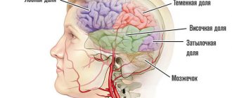
Practicing neurologists and surgeons most often choose observational tactics and advise paying attention to the cause of symptoms in the brain.
Need advice from an experienced doctor?
Get a doctor's consultation online. Ask your question right now.
Ask a free question
It is necessary to differentiate the symptoms; other concomitant diseases may be involved in their occurrence.
Traditional medicine resorts to folk methods and lifestyle adjustments. It is recommended to eliminate harmful factors and lead a healthy lifestyle. Traditional medicine offers several ways to combat lipomas with brain localization.
Burdock tincture. The roots need to be crushed and 1.5 cups of vodka added to 1 glass. Infuse for a month in a dark place. The tincture is taken 2 hours before meals, 1 tablespoon.
The most common and proven remedy for lipomas of any location is cinnamon. Daily dose 1-2 spoons.
The juice of beets, carrots, garlic and black radish are mixed and poured into a bottle. The mixture should be taken 2 dessert spoons after meals.
Chopped fresh celandine in a gauze bag with 200 g of sugar is placed at the bottom of a 3-liter jar, filled with warm whey. Leave for about 30 days in a dark and cool place. For consumption, filter and take 25 ml 30 minutes before meals. Be careful and follow the dosage.
Classification by location
The dependence on the position of the lipoma has given doctors grounds to divide them into several types. The main localization is represented by three types:
- Peripheral.
- Central.
- Mixed.
What does a wen look like on the body?
A fatty tissue (lipoma) is a benign neoplasm that almost never develops into a malignant tumor.
The tumor can occupy different locations. The most common 4 places to appear:
- In the area of the main bronchi.
- On the right at the corner of the heart and diaphragm.
- On the parasternal line, it is often designated in the diagnosis as a parasternal lung lipoma on the right.
The level of impact on a person’s lungs, the frequency of clinical manifestations, and the duration of the asymptomatic period depend on the location of the tumor in the lung. Parasternal lipomas develop for a long time without any manifestations or symptoms.
The superficial position of the tumor often allows it to grow into the bronchial tree. This is observed in diffuse type lipomas.
Classic lipoma is represented by adipose tissue. But there are formations that grow not only from adipose tissue. Fibrolipoma grows from connective and adipose tissue.
With myolipoma, pathologies of muscle tissue are associated. Angiolipoma is represented by adipose tissue, which is fed by a large vascular network. Myxolipoma is a formation that consists of fatty and mucous tissue.
University
- Oh, how did all this fit inside me? - exclaims the fragile 58-year-old Maria Antonovna (name changed - author's note), an employee of one of the capital's industrial enterprises. At the patient’s request, the head of the RCTC, Anatoly Tatur, showed a photo of the remote education. 25x18x10 cm! Weight - 2 kg!
Just 6 years ago, fluorography showed a homogeneous darkening in Maria Antonovna’s right lung. An in-depth examination - CT scan of the chest - revealed a tumor in the mediastinum. Doctors recommended removing it, but the woman decided to postpone the operation. After a couple of years, acute respiratory infections began to be tolerated worse - colds occurred with fever and took longer, which had not happened before. And recently I was very seriously ill with pneumonia. Repeated tomography revealed that the lipoma had grown, almost completely compressed the left lung, and the heart and other organs of the mediastinum had shifted to the healthy side.
As experts subsequently commented, only the patient’s right lung was fully functioning, so shortness of breath and palpitations occurred during physical exertion. Due to compression of the left lung by the tumor, gas exchange was disrupted, and in such conditions it is easier for infection to take hold...
After a consultation at the RCTC, Maria Antonovna agreed to the operation; she was hospitalized in the thoracic department of the 10th City Clinical Hospital. When analyzing CT data, doctors found that the “leg” of the giant lipoma, through which the tumor received nutrition, was localized in the anterosuperior mediastinum on the aortic arch, and its base lay on the diaphragm.
The choice of intervention technique was discussed by the council. At first they wanted to operate in a gentle way - thoracoscopically. However, the size of the lipoma was still too large. Manipulating thoracoscopic instruments in the cavity (the tumor occupied a significant volume of the pleural sac) was extremely difficult and even dangerous due to the risk of damage to large vessels of the mediastinum and the root of the lung. To remove such formations, it is ideal to have special equipment to destroy the structure of the tumor, and then simply aspirate it. We settled on a traditional anterolateral thoracotomy.
The intervention under endotracheal anesthesia lasted 2 hours. The surgeons carefully isolated the mediastinal “leg” of the tumor, preserving the capsule, and when the tumor was in a free cavity, they removed it. There were fears that after prolonged compression there would be difficulties with the lung. However, it was successfully dealt with while still on the operating table, quickly occupying the required volume of the pleural cavity, and negative consequences were avoided. The postoperative period proceeded well. On the 2nd and 4th days the drains were removed.
And on the 8th day, Maria Antonovna was discharged. Now she only needs outpatient monitoring.
...The woman gladly demonstrated to the correspondent how she could now take a deep breath, and then honestly summarized:
- Of course, the operation could not have been delayed...
A comment
When a banal lipoma becomes a non-trivial problem
Anatoly Tatur, head of the Republican Center for Thoracic Surgery, professor
1st Department of Surgical Diseases of BSMU, Doctor of Medical Sciences. sciences
Lipoma is the most common benign mesenchymal tumor. It can develop in all areas of the body where adipose tissue is present. Subcutaneous lipomas are usually small and do not manifest themselves for a long time. Patients become concerned when the tumor enlarges and a cosmetic defect develops.
Interventions to remove small subcutaneous lipomas are technically simple, so, as a rule, after consultation with an oncologist, they are trusted by novice general surgeons.
Among mediastinal neoplasms, lipomas account for only 2–3%. In the practice of a thoracic surgeon, the most common are abdominomediastinal, developing from preperitoneal tissue. It is extremely rare to deal with cervical-mediastinal ones.
10 years ago we treated a 45-year-old woman with a lipoma in the left neck area 16127 cm. The tumor compressed the trachea and esophagus and was located from the angle of the lower jaw to the aortic arch.
In the general surgical hospitals where the patient was consulted, they did not undertake the removal of such a formation, because, according to MRI, the carotid artery, internal jugular vein and vagus nerve passed through its thickness. The patient was operated on in our center using a combined left-sided cervicotomy and superior longitudinal-transverse sternotomy approach.
The tumor was isolated within the capsule, and after its longitudinal dissection and isolation of elements of the neurovascular bundle from the tumor tissue, it was removed. On histological examination, the formation had the same structure as ordinary subcutaneous lipomas. We shared our observation experience at a meeting of the Minsk Society of Surgeons.
Cases of giant mediastinal lipomas with intrapleural growth are also extremely rare. They increase slowly, remain asymptomatic for a long time, and are usually a radiological finding. The medical literature describes clinical observations of lipomas up to 3.5 kg. If they are large in size, they can cause serious hemodynamic and respiratory disorders due to compression of the structures of the mediastinum, lung and diaphragm.
In the diagnosis of mediastinal lipoma, the leading place today belongs to CT and MRI. Regardless of the location, there is a risk of malignancy, in particular degeneration into sarcoma (increases as the formation increases). Such tumors must be promptly disposed of in a specialized hospital. When they are still small, it is possible to perform minimally invasive video thoracoscopic intervention. Elena Kleshchenok Photos by the author and from the personal archive of A. Tatura Medical Bulletin , December 10, 2015
Symptoms
The development of symptoms occurs along with the passage of the tumor through certain stages of its growth and development.
At the first stage, the tumor does not have any effect on the lung. She is still small. The capsule does not allow it to grow quickly.
The second stage will be marked by compression of the bronchial tree, a rapidly growing formation. A person feels heaviness while breathing. Notes the appearance of shortness of breath. Feeling of tightness on one side of the chest. Possible chest pain.
Why can our articles be trusted?
We make health information clear, accessible and relevant.
- All articles are checked by practicing doctors.
- We take scientific literature and the latest research as a basis.
- We publish detailed articles that answer all questions.
At the third stage, narrowing of the main bronchi is possible. The cough may be mixed with blood and viscous sputum. Pulmonary hemorrhage may occur, which requires emergency care.
Injuries to the bronchial tubes by tumors lead to infection. Symptoms of fever appear. Intoxication of the body increases, urgent medical attention is required.
The clinical picture accompanying the development of formation is not the basis for making a diagnosis. Many respiratory system diseases are accompanied by the same symptoms.
By appearance
Based on how the tumor looks, lipomas can be divided into:
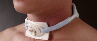
- Ring-shaped (location – neck, similar to a necklace);
- Tree-like (located inside the joints);
- Capsule-shaped (with a dense shell and clear edges);
- Diffuse (without capsule, clear boundaries);
- Ossified or petritic (when calcium deposits form inside);
- Pedicled (with a pedicle where the vessel and nerve are located);
- Dense;
- Soft.
The reasons may be as follows:
- Neutrophic;
- Lack of nutrition to the nerves in a certain area.
Lipoma contains fat cells of different sizes. Between the simple fat cells there are rudimentary ones, which affect the growth of the former.
Lipomas grow slowly. With frequent trauma, there is a risk of lipoma malignancy.
When localized superficially, the lipoma looks like a painless, mobile formation ranging in size from 0.5 to several centimeters. Lipoma can be either dense or dough-like. It does not hurt when touched, although it can compress nervous and vascular tissues. If it is located in a vulnerable place, then pus and inflammatory processes may appear from friction.
Most often, this tumor grows on the head, face, neck and waist, back, chest, but can also occur on the legs and abdomen.
The patient will complain of breathing problems if the lipoma is on the neck.
Typically complaints are cosmetic and aesthetic in nature.
During examination, a specialist doctor determines a lipoma by its location (if it is located in a place where adipose tissue accumulates), soft or compacted consistency, mobility, painlessness on palpation, tissue retraction when the skin is stretched.
Next, the puncture is taken for study.
They may prescribe ultrasound, x-rays, electroradiography of soft tissues, contrast x-rays, magnetic resonance and computed tomography.
The doctor must distinguish a lipoma from an atheroma.
You should not treat the problem yourself. After consultation with a surgeon, surgical treatment is prescribed, if required.
Prostate adenoma
Diagnosis criteria
Lipoma is not diagnosed by clinical manifestations. The pulmonologist, having noted the clinical picture, will definitely refer the patient to a plain chest radiograph (CHX). The X-ray results clearly show the approximate location of the tumor.
Having suspected a lipoma, doctors will send the patient for further diagnostics. Usually, when it is detected, a bronchoscopy with a biopsy of tumor tissue is performed. Bronchoscopy allows you to determine the exact location of the lipoma, and the results of a biopsy will tell you about the nature of the tumor.
Doctors will make the final diagnosis after the results of the biopsy. Without tissue examination, a pulmonologist or oncologist cannot confirm or refute the diagnosis. Before this, the detected tumor is called a neoplasm.
Bronchoscopy with biopsy reveals centrally located tumors. They are found in large bronchi. If the tumor is located on the periphery, it does not manifest itself for a long time. It is discovered accidentally during routine preventive examinations.
Diagnostics
A dermatovenerologist can recognize a wen visually, by its appearance.
Differential diagnosis can also be used. It is carried out for benign or malignant formations on the skin. We mentioned earlier that occasionally wen can be malignant. Then they are called not lipomas, but liposarcomas. To verify the diagnosis, ultrasound and computed tomography are performed.
Ultrasound examination helps to see the wen as a hypoechoic formation with a capsule, which is located among the subcutaneous fat layer or between the fibers of muscle tissue. A CT scan is needed to determine the structure of the tumor. For example, it is determined how a tumor absorbs X-ray rays in comparison with the tissues located around the tumor.
Research should be carried out only in a professional medical institution with good equipment. Then the diagnosis will be the most accurate and correct.
Treatment methods
Treatment for lipoma is required if its symptoms rapidly increase due to tumor growth. Getting rid of the tumor is possible only through surgery. Removal of the tumor is carried out using one of three methods possible for intervention on it.
Self-medication is dangerous with complications!
Attention
Despite the fact that our articles are based on trusted sources and have been tested by practicing doctors, the same symptoms can be signs of different diseases, and the disease may not proceed according to the textbook.
Pros of seeing a doctor:
- Only a specialist will prescribe suitable medications.
- Recovery will be easier and faster.
- The doctor will monitor the course of the disease and help avoid complications.
find a doctor
Do not try to treat yourself - consult a specialist.
Operation
The first method is represented by classic surgery on the chest. Used in emergency situations.
Indications for use are bleeding from the bronchi or narrowing of the bronchi, which leads to respiratory failure. The operation involves removing the capsule, which consists of healthy cells.
Liposuction
Used in the early stages of development of a fatty lung tumor. A lipoaspirator is used. The skin is incised 0.5 cm and work is carried out through this incision. Tumors larger than 3 cm cannot be removed by this method.
Laser removal
The lipoma is cauterized with a special laser. The operation is painless. Does not leave any marks on the skin after completion. There are no side effects or complications after the intervention.
All three methods, despite interfering with the internal environment of the body, are minimally invasive. This significantly reduces the risk of postoperative infection.
Surgical method for removing wen
If a child’s wen is too large, what to do? Usually in this case surgery is needed.
Surgical intervention may be prescribed in several cases:
- if the tumor is larger than 5 or 7 centimeters;
- if the lipoma grows very quickly;
- if the tumor develops rapidly in an infant in the first year of life;
- if there are white wen on the child's face.
During surgery, when it is performed on a small child, only general anesthesia is used. If the child is an older child or a teenager, then a local anesthetic is injected subcutaneously. General anesthesia is also used when operating on a lipoma on the face of a child.
The operation is performed on an outpatient basis if the lipoma is small. For newborns and children of the first year of life, surgery is performed only inpatiently. Surgery is also performed in a hospital if a tumor of significant size needs to be removed.
Before the lipoma removal procedure, certain types of tests are performed. This is a general blood and urine test. The blood is also tested for clotting and its group is determined. In addition, sometimes a test is prescribed to check for syphilis and/or HIV, etc.
Complications of unoperated lipoma
A lipoma that is not removed in time will grow and begin to compress the lung. After some time, complications will follow. Some can be fatal:
- Impaired outflow of mucus from the bronchial tree due to compression of the bronchi by a tumor.
- Difficulty in ventilation of the lungs: development of infections inside the lungs (bronchitis, pneumonia, tuberculosis) or oxygen starvation due to insufficient breathing.
- stenosis , which leads to oxygen starvation or suffocation, requires emergency assistance.
- Pulmonary hemorrhage, a condition that can lead to death, requires emergency surgical treatment.
Prevention of the development of complications is a regular x-ray or fluorographic examination of the chest.
2. Causes of benign tumors
The reasons why benign lung tumors appear are poorly understood. But in general, they often appear after health problems such as:
Inflammatory processes caused by infection:
- Fungal infections – histoplasmosis, coccidioidomycosis, cryptococcosis, aspergillosis;
- Tuberculosis
- Lung abscess
- Pneumonia
Inflammation not associated with infection:
- Rheumatoid arthritis;
- Wegener's granulomatosis;
- Sarcoidosis.
- Congenital pathologies such as lung cyst and others.
Visit our Oncology page
Prevention
The basis of prevention is represented by increasing the body’s immune forces and reducing the impact of negative factors on the lungs.
It is necessary to stop smoking. Tobacco smoke has a very negative effect on lung tissue. It is necessary to reduce the consumption of alcoholic beverages, which undermine the immune system.
Regular exercise will ensure your lungs have adequate ventilation. Medical examinations twice a year will ensure early diagnosis of all neoplasms.
Timely treatment of respiratory system infections will eliminate them as a factor in the occurrence of lipoma.
Lung lipoma is a relatively harmless and rare tumor. The chance of its occurrence in the lungs tends to zero. But if it has formed, removal with timely diagnosis is not difficult. A favorable prognosis allows the patient to forget about this disease forever after all procedures.
Localization of wen
Where do children usually develop wen? They can also occur on internal organs, but this is rare. Tumors grow under the skin, in muscle tissue, and in places above the bone tissue. Although lipoma is called a wen, it itself, as a rule, is located where there is almost no fat. Most often, a child has only one wen. However, if more than one of them is detected, then a diagnosis of lipomatosis is made.
Most often, formations are localized on:
- head and neck;
- face and back;
- shoulders and legs.
Therapeutic procedures
When a small wen is diagnosed with no discomfort and other associated symptoms, there is no need for urgent treatment. Doctors usually choose the tactic of monitoring the tumor. If signs of compression or disruption of the respiratory system appear, the patient is prescribed a course of therapy or removal.
The presence of serious complications and consequences with the development of tissue necrosis requires urgent removal of the lipoma using the following methods:
- Surgical excision of the tumor using a scalpel is required when diagnosing internal bleeding in the bronchial cavity or lungs. The method is also used in the presence of compression of the bronchial ducts. Surgery is considered effective, since the pseudocapsule formed by healthy cells and the lipoma capsule are removed. The risk of relapse is minimized.
- Liposuction is used to remove small tumors formed on the surface of the organ. The disadvantage of the procedure is the inability to remove the pseudocapsule, which increases the risk of relapse.
- Laser therapy is gaining popularity due to the lack of side effects and rapid recovery after the lipoma excision procedure. The operation lasts 15-20 minutes. The patient is discharged home almost immediately. Monitoring of the restoration of operated tissues is carried out on an outpatient basis.
The surgical method of treating wen is considered effective due to the complete removal of the tumor and the presence of a minimal chance of recurrence.
Together with the traditional course of therapy, it is allowed to use traditional medicine, but only after consultation with the attending physician and under medical supervision. Alternative medicine recipes can stop the active growth of fat formation and prevent the development of serious complications.
Traditional medicine recipes are used at the initial stage of wen formation or when diagnosing a small lump. The following means are considered the most popular:
- Consume 2 grams of ground cinnamon daily. You can simply eat it and drink it with water or add it to tea, coffee and ready-made dishes.
- Mix 1:1 juices of golden mustache, aloe and Kalanchoe with the addition of water (in a ratio of 1:3). Thoroughly rub the resulting mixture into the sore spot until the wen disappears completely.
- A decoction of Caucasian hellebore has a resolving effect on lipoma. Pour boiling water over the leaves and leave for 12 hours. Then take 1 tsp every day. within 3 months.
- Mix pine pollen and honey 1:1 and take 1 tbsp daily. after meal. It is recommended to drink it with an infusion of oregano or other herbal tea.
The use of traditional medicine helps prevent the formation of large formations in the lung tissues. Also, giving up bad habits will help avoid dangerous diseases.