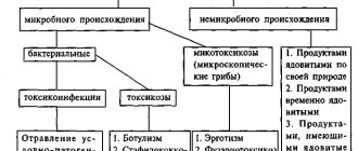
Issues regarding bleeding will never lose their relevance. After all, no matter how medicine learns to deal with them, there will still be unresolved issues in some cases. This is especially important in relation to massive blood loss, in which it is extremely important to instantly recognize specific types of bleeding, which will allow the correct assistance to be provided. And although, at first glance, there is nothing complicated about this, even experienced doctors in some critical situations can make mistakes, getting lost at the sight of a large amount of blood. Therefore, every person is obliged to know what a specific type of bleeding looks like, and what extent of measures should be provided in this case.
General classification
The division of bleeding into different types is of great expediency due to the ease of determining treatment tactics at different stages of medical care. Wherever she finds herself, all doctors know her clear algorithm. This approach minimizes the time spent and minimizes the amount of blood loss. People who are not involved in medicine should also know the main features and possible types of bleeding in order to help themselves or their loved ones if necessary.
The classification is given in table form.
| External bleeding (bleeding with direct contact with the external environment) | Internal bleeding (the spilled blood does not have direct contact with the environment) |
|
|
| According to the amount of blood loss during any bleeding | |
| |
How to help a patient with major blood loss
If a person has lost a large amount of blood, all measures must be taken to prevent vascular shock before the ambulance arrives. To do this, you should free the patient's chest and throat from clothing, give access to fresh air, and avoid crowding. The person should lie on his back, arms and legs raised, head slightly lowered. This position of the body will ensure proper blood supply to the brain, which will help maintain its activity. If there are no injuries to the abdominal area and the patient is conscious, it is recommended to give him water or weak sweet tea. In case of loss of consciousness and respiratory arrest, it is necessary to perform resuscitation measures in the form of cardiac massage and artificial respiration.
If there is significant blood loss, the patient must ensure correct body position
Capillary bleeding
The most common type of external bleeding is capillary. They occur with any traumatic injury that disrupts the integrity of the skin. They manifest themselves as a low-intensity, uniform flow of blood from the wound due to damage to the capillaries (the smallest vessels of the body). They rarely lead to severe blood loss, since in most cases they stop on their own. They present no difficulties either for diagnosis or for treatment. The exception is extensive superficial wounds, in which prolonged neglect of medical care can cause large blood loss.
Causes
There are two main causes of bleeding: as a result of injury and due to internal pathological processes, that is, they are traumatic and atraumatic (or pathological).
Traumatic
They arise as a result of exposure to traumatic factors that exceed the characteristics of the strength of blood vessels. In this case, mechanical damage to the vascular wall occurs. This is the most common cause of bleeding.
Atraumatic
They can begin without any provoking factor. Occurs in the following cases:
- during pathological processes occurring in the body: ulceration, necrosis, destruction of the vascular wall, for example, during the disintegration of a tumor, inflammation, peritonitis and others;
- with increased permeability of the vessel wall at the microscopic level, which can happen in diseases such as hemorrhagic vasculitis, vitamin C deficiency, scarlet fever, uremia, sepsis and others.
The bleeding process largely depends on the state of the coagulation system. Disturbances in its functioning themselves cannot cause bleeding, but they significantly worsen the situation. If a small vessel is damaged and the hemostatic system is functioning normally, significant blood loss does not occur and the blood quickly stops. If, for example, the process of thrombus formation in the body is disrupted, then even a minor injury can result in death from blood loss. An example of a disease in which the hemostasis process is impaired is hemophilia.
Venous bleeding
Venous bleeding occurs with superficial and deep wounds of any size, in which the integrity of the saphenous or intermuscular veins is disrupted. In this case, quite intense bleeding occurs. The following symptoms can clinically recognize venous bleeding:
- Dark blood;
- The bleeding is very heavy, like a constant flow of blood from the wound;
- It decreases when the area below the wound is pressed.
Venous bleeding is extremely dangerous if medical assistance is not provided in a timely manner. In this case, massive blood loss occurs in a short time, up to a state of shock. They rarely stop on their own, so stopping them should not be neglected. Superficial veins bleed less intensely, while damage to deep veins causes profuse bleeding.
Differences between arterial (a) and venous (b) bleeding
About the types of hemorrhagic conditions
In medicine, the internal type of hemorrhage is not classified in detail. Based on the cause of internal bleeding, there are the following types:
- Mechanical type. This condition occurs in the event of trauma to the vascular tissues through which blood flows. Occurs due to injury (often occurs in men) or due to surgery.
- The arrosive type of hemorrhage is considered to be a consequence of necrosis affecting vascular tissue or if newly formed structures grow and disintegrate.
- Diapedetic type. In this condition, the vascular tissue is not destroyed, but due to various pathological processes (hemorrhagic vasculitic changes, phosphorus poisoning and many other processes), the capillary network becomes highly permeable.
According to the type of vascular tissue there are:
- Arterial type, where destruction of the arterial vessel is diagnosed.
- Venous type, in which the veins are damaged.
- Capillary type. The blood medium spreads evenly from the capillary vessels. If blood pours out from different organs, then this is a parenchymal type of hemorrhage.
- Mixed type. Occurs with destructive changes in venous, arterial and capillary vessels.
If we take into account localization, then there are:
- Gastric and intestinal types of hemorrhages. Possible due to ulcerative processes in the stomach and duodenum, gastritis changes, inflammation of the intestines, also if the mucous membrane is cracked and there are newly formed structures. If esophageal hemorrhages are observed, the cause is liver dysfunction. In case of intestinal hemorrhages or intraperitoneal bleeding, the cause is hemorrhoidal changes or rectal fissures.
- Hemorrhages localized in the cavity spaces of the pleura due to torn vascular tissues between the ribs (hemothorax). This condition occurs when there is a closed injury to the chest.
- Hemorrhages that flow into the pericardium (pericardial sac), which compresses the heart muscle (hemipericardium). If this condition is ignored, it can manifest itself as heart failure followed by death. Such hemorrhage can occur with a mechanically damaged chest in the front, or surgical intervention on the heart muscle.
- Intra-articular hemorrhages (hemarthrosis). This type of bleeding often occurs when joint tissues are damaged (usually the knee joints).
According to the location where blood accumulates, there are:
- Hemorrhages in cavities. They are divided into bleeding of the abdominal cavity (if the abdominal area, chest and various organs are injured), pleural membrane (if the ribs are broken), and the skull.
- Hemorrhages within tissue structures, where blood accumulates in deep tissues that disintegrate, causing hematomas.
According to the amount of blood loss there are:
- Mild hemorrhages, where total blood loss is no more than 15% of the total blood circulation.
- Moderately severe hemorrhagic conditions, where blood is lost 20% of the total blood circulation.
- Severe bleeding, where the patient has lost approximately 1.5 liters of blood
- Massively manifested hemorrhagic condition. The total volume of blood circulation is reduced by more than 30%.
- Fatal blood loss occurs when the total circulating blood volume decreases above 60%.
Hemorrhages can occur:
- Obviously. After a certain time, the blood comes out into the external environment through the patient’s natural type of orifices.
- Is hidden. There are no main symptomatic manifestations; perhaps they are mild.
Given the time period, there are:
- Primary identified blood loss. They signal themselves after the vascular wall is ruptured.
- Secondary detected hemorrhages. They are observed a certain time after the traumatic factor that provoked them. This type of hemorrhage is divided into early, where manifestations occur after a period of 1 to 3 days due to the fact that an embolus has come out of damaged vascular tissue or the suture has been incorrectly applied. Late, secondarily detected hemorrhages appear three days after the vessel is damaged as a result of infection joining the wound.
Arterial bleeding
Given the deep location of the arteries in the tissues, their damage is the least common. The most common causes are knife, gunshot and mine-explosive wounds. In everyday life, these can be puncture wounds from thin and narrow objects. Clinically, arterial bleeding can be suspected by the following signs:
- Bright red blood;
- Flows out in the form of a pulsating stream;
- Very intense;
- Does not decrease with normal pressure on the wound or tissues above and below it;
- The localization of the wound corresponds to the projection of the course of large arteries.
Typically, arterial bleeding is very intense and quickly leads to massive blood loss and shock. If a complete rupture of an artery occurs, then in just one minute you can lose almost the entire volume of circulating blood. Therefore, such bleeding requires immediate assistance.
Wound tamponade
Emergency care for a person with bleeding in the head or neck area, when a tourniquet cannot be applied, is to pack the wound. To do this, use a sterile bandage or cotton wool; if you don’t have them at hand, you can take napkins. The tampon is pressed tightly to the wound site and a bandage is applied. The bandage should be made tight to avoid blood leakage. This method can be used in case of damage to the vessels of the upper and lower extremities. To do this, the wounded arm or leg must be raised to drain the blood, a tampon placed in the area of the affected artery, and a tight bandage applied to secure it.
Important! Despite the fact that the methods described above are only temporary help for the patient, and the patient still requires treatment in the hospital, clear actions of the helper can save a person’s life.
Internal bleeding
Unlike external bleeding, in which it is impossible not to notice their symptoms, internal bleeding is more insidious. After all, recognizing them is not so easy. They usually manifest themselves when there is already quite a lot of blood loss. Therefore, it is extremely important to know all the possible signs of this dangerous condition. These include:
- General weakness and drowsiness;
- Discomfort or pain in the abdomen;
- Unmotivated decrease in blood pressure;
- Frequent pulse;
- Pale skin;
- The appearance of pain in one of the halves of the neck, which occurs in a horizontal position and decreases in a vertical position (Vanka-Vstanka’s symptom).
The occurrence of internal bleeding is preceded by closed or penetrating wounds of the abdomen, lower back, rib fractures, stab or gunshot injuries. In this case, damage to internal organs occurs, which causes a violation of the integrity of blood vessels and bleeding. The result is an accumulation of blood in the abdominal cavity, chest, and saturation of the damaged organ or visceral fatty tissue (hematoma).
Such bleeding can progress at lightning speed, but can also increase over several days after the injury. It all depends on their intensity and the extent of damage to the injured organ. Usually the spleen is affected, less often the liver. With a one-stage rupture, bleeding occurs immediately; with a two-stage rupture, an intraorgan hematoma first appears, which ruptures after a few days, causing an immediate worsening of the patient’s condition.
Health care
After the victim has been taken to the hospital, his treatment is carried out by a general or vascular surgeon. The choice of therapeutic tactics always depends on the diameter of the artery damage and the location of the wound. To completely stop bleeding in a hospital, the following methods are used:
- plastic surgery of the damaged artery, that is, its suturing;
- tightly bandaging the wound. This method is used for minor damage;
- arterial replacement. The tactic involves replacing damaged artery walls with the patient’s own tissues; it is used for extensive lesions.
To prevent wound infection, local and general antibacterial therapy is carried out.
Arterial bleeding is one of the most dangerous. Lack of treatment leads to large blood losses and death of the patient. The quick reaction of others and competent emergency care ensure the safety of the victim’s life and prevent serious consequences.
Gastrointestinal bleeding
If you understand thoroughly, this type of bleeding cannot be classified unambiguously. After all, blood flows into the lumen of the gastrointestinal tract, but at the same time it comes into contact with air. But this is not as important as detecting the symptoms of such a condition. After all, the patient’s life sometimes depends on timeliness. Signs of gastrointestinal bleeding include:
- General weakness and dizziness;
- Frequent pulse and low blood pressure;
- Pale skin;
- Vomiting blood or brown matter;
- Thin, bloody or thick black stool.
Gastrointestinal bleeding occurs with peptic ulcers, tumor diseases, various necrotic processes in the mucous membrane of the digestive tract and some other diseases. Therefore, people with such a pathology must be aware of the possibility of bleeding and, if they occur, be sure to seek medical help.
Educational video on first aid for bleeding:
Pressure of arteries
In order not to get confused in case of bleeding and quickly find a suitable place to press the vessel, you should remember the following points of the body.
- Inguinal fold – in case of leakage from the thigh vessel.
- Popliteal region - with bleeding from the artery of the leg.
- The axillary region, the inner side of the biceps muscle - for the damaged arm vessel.
- The area on the neck (inner edge of the sternoclavicular muscle) - when flowing from the carotid artery.
- Supraclavicular surface - to stop bleeding from the subclavian vessel.
What to do if you are bleeding
Therapeutic tactics must be differentiated and depend on the specific type of bleeding. There is a general scope of activities that must be carried out in any type. All specific manipulations are purposeful, since their incorrect implementation can cause harm to the patient. General measures to help with bleeding include:
- Place the victim in a horizontal position;
- Monitor consciousness, pulse and blood pressure;
- Rinse the bleeding wound with hydrogen peroxide and apply a clean pressure bandage;
- If possible, apply ice to the source of bleeding;
- Transport the patient to the nearest facility.
The listed measures will never harm, regardless of the source and characteristics of the bleeding.
Differentiated tactics are presented in table form.
| Type of bleeding | Scope of necessary activities | |
| First aid (temporary stop of bleeding) | Specialized medical care (final stop of bleeding) | |
| Capillary |
| Stitching the wound if necessary. |
| Venous |
|
|
| Arterial |
| Suturing or prosthetic replacement of the damaged artery with further suturing of the wound. |
| Internal and gastrointestinal bleeding | General measures for bleeding, typical for the prehospital stage. |
|
Rules for applying a tourniquet
A tourniquet should be applied exclusively for arterial bleeding. Its erroneous use for venous bleeding will lead to their intensification. Characteristics of correct application of a tourniquet:
- It is applied to the affected segment no lower than 20 cm from the wound. It can be higher. A lower location is permissible only if it is impossible to perform the classic manual;
- A fabric bandage is placed on the skin under the tourniquet;
- A special hemostatic tourniquet or improvised materials that replace it can be used as a tourniquet;
- The first circular tours of the tourniquet around the limb are less tight. After them, tighter tours should be applied;
- After a properly applied tourniquet, bleeding decreases. If this does not happen, this indicates either that it was applied incorrectly or that there is no indication for this;
- The permissible duration of holding the tourniquet on a limb should not exceed 2 hours in the summer and 1-1.5 hours in the winter;
- If it is impossible to apply a tourniquet (neck wounds, high wounds of the shoulder and thigh), it is replaced by other methods of stopping bleeding: external digital pressure on the pulsating vessel above the wound or directly in it.
Correct application of a tourniquet as one of the ways to stop arterial bleeding
Only strict adherence to algorithms for determining the type of bleeding and step-by-step implementation of therapeutic measures can really effectively help in the fight against this problem. Remember the general rules and then you can protect yourself and your loved ones from dangerous complications of any bleeding.
Instructions for performing digital compression of the wound
The actions of the person providing assistance must be quick and concentrated. You must not panic, you must concentrate and strictly follow the following instructions:
- if the exact location of the wound is not clear, it should be pressed with your palms, this will help determine the source of bleeding;
- if necessary, remove clothing by tearing it or cutting it with scissors;
- It is recommended to squeeze the artery with your thumb or covering hand;
- You cannot remove your hand until the victim is bandaged;
- If you hold your fingers for a long time, cramps and numbness may develop; in such a situation, you should replace it with the other hand.
If the situation allows, before squeezing the wound with your fingers, it is recommended to wash them with soap or treat them with an antiseptic, this will avoid infection of the wound and the development of complications. This advice does not apply to situations where the main arteries are damaged and minutes are counting.