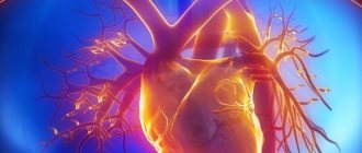
It branches like a tree, first into large branches (trunks), then into smaller branches and twigs, and is conventionally divided into several parts or sections:
- 1. The ascending aorta is the area from the aortic valve to the brachiocephalic trunk.
- 2. The aortic arch is a short section from which all the vessels supplying the arms and head (brachiocephalic arteries) depart. They anatomically form an arch connecting the ascending and descending aorta.
- 3. The descending (thoracic) aorta begins from the mouth of the left subclavian artery and continues to the diaphragm.
- 4. Below the diaphragm and before the bifurcation of the aorta (bifurcation) is the abdominal aorta.
Dividing the aorta into sections is very important for assessing risk and choosing optimal treatment tactics in patients with aortic aneurysms.
An aortic aneurysm is an area of local expansion.
What is an aneurysm
A thoracic aortic aneurysm is an abnormal bulge on the walls of the artery, formed due to intense blood pressure on a weak area of the vessel. An aneurysm can be:
- fusiform - has a convex shape, protrudes from both sides of the aorta;
- saccular - a rounded formation that protrudes from one side of the vessel.
A small aneurysm does not pose a health threat. However, if it is present, you must be observed by a doctor so as not to miss the moment of disease progression.
Classification of the disease
Pathology is divided into 3 types depending on the extent and location:
- In the ascending aorta with advancement along the arch. Occurs in 50% of the total number of recorded cases of the disease.
- In the ascending aorta. Diagnosed in 35% of cases.
- In the descending part of the aorta with progression down/up along the arch. Occurs in 15% of cases.
Stretching of the vessel occurs quickly - it does not require weeks. The process can be compared to inflating a balloon, which expands several times when filled with air.
Depending on the duration of the process, an aneurysm can be:
- acute – 1-2 days have occurred since the formation of aortic dissection;
- subacute – the endothelial defect appeared 2-4 weeks ago;
- chronic – the aneurysm formed 4 or more weeks ago.
Some patients ignore the symptoms of the disease and do not see a doctor for years. But the fact that the thoracic aortic aneurysm does not rupture is pure chance and luck. This can happen at any time, and in 90% of cases the person will die.
Symptoms of pathology
Symptoms of a thoracic aortic aneurysm differ depending on the location of the formation. In some cases, a person does not feel any discomfort at all, which greatly complicates the timely diagnosis of the disease.
Aneurysm of the ascending aorta is accompanied by:
- Acute pain in the heart area. This is due to the pressure of the damaged vessel on nearby organs and tissues;
- Shortness of breath, significantly increasing over time. It is problematic for a person with an aneurysm to walk long distances or climb stairs;
- Constant feeling of heartbeat. This is one of the most common complaints from patients. This phenomenon is associated with blood pressure on weakened vessel walls;
- Dizziness. May be accompanied by a severe headache that is not relieved by pills;
- Swelling of the face, upper half of the body. This phenomenon is due to the fact that the aneurysm compresses the superior vena cava.
Aneurysm of the aortic arch is characterized by the following symptoms:
- Swallowing problems. The swollen section of the vessel presses on the esophagus;
- Hoarse voice, periodic coughing. The main cause of the malaise is the pressure of the aneurysm on the recurrent nerve;
- Increased salivation. Occurs due to pressure from the damaged aorta on the vagus nerve;
- Breathing problems, shortness of breath. It manifests itself in the case of aneurysm pressure on the trachea and bronchi;
- Unilateral pneumonia. An aneurysm can put pressure on the root of the lung, preventing its normal ventilation. When an infection enters the body, in 94% of cases this condition develops into unilateral pneumonia.
An aneurysm of the descending aorta is accompanied by:
- Pain of unknown nature in the left arm, acute pain syndrome in the scapula area;
- Paresis, paralysis. It occurs due to the pressure of the aneurysm on the intercostal arteries, which disrupts the supply of oxygen to the spinal cord;
- Displacement of the vertebrae if the patient’s aneurysm is in a chronic condition;
- Painful sensations in the side, sternum, similar in nature to pain due to radiculitis/neuralgia.
Signs and symptoms of a ruptured aneurysm:
- Sudden, intense, and persistent pain in the abdomen, chest, or back
- Pain that radiates to the back or legs
- Sweating
- Sticky sweat
- Dizziness
- Low blood pressure
- Increased heart rate
- Loss of consciousness
- Dyspnea
- Weakness or paralysis on one side of the body, difficulty speaking, or other signs of a stroke.
Emergency cardiology deals with the treatment of such conditions.
Another complication of aortic aneurysms is the risk of blood clots. Small blood clots can develop in the area of the aortic aneurysm. If a blood clot breaks away from the inside wall of the aneurysm and blocks a blood vessel in another part of the body. This causes pain or blocks the flow of blood to organs and tissues, causing ischemia of the lower extremities, brain, and abdominal organs.
Reasons for development
A disease such as thoracic aortic aneurysm develops as a result of:
- Atherosclerosis. It is this pathology that in 90% of cases is the cause of aneurysm formation;
- Traumatic damage to a vessel. Occurs most often as a result of an accident, a fall from a great height, or a strong blow to the chest.
- Congenital pathologies. When a patient has a systemic connective tissue disease, the walls of blood vessels are in a chronically inflamed state.
- Complications after medical procedures. These include reconstructive operations on the aorta, cardiac catheterization and other interventions in the cardiovascular system.
- Infectious diseases. An aneurysm can develop in patients with tuberculosis, sepsis, syphilis, osteomyelitis, and pericarditis.
Previously, the pathology was diagnosed mainly in patients over 50 years of age. However, every year the disease becomes younger. Already today, young people aged 25-30 years often turn to doctors when an examination reveals a thoracic aneurysm.
conclusions
The creation of an aortic team in a separate center made it possible to obtain good reproducible results in the treatment of patients with various pathologies of the ascending aorta. The extent of surgical correction for pathology of the ascending aorta in itself is not a significant risk factor. Further improvement of the treatment of this pathology is associated with scientific developments at the intersection of various clinical and biomedical specialties (surgery, radiology, molecular biology and genetics). An urgent task is to create a unified register of patients with aortic diseases. Identification of new informative biochemical markers may affect the timing and extent of surgery in patients of this profile.
There is no conflict of interest.
Risk factors
The pathology has a number of risk factors, the presence of which significantly increases the likelihood of its development:
- Male gender. According to statistics, thoracic aneurysm is diagnosed 14 times more often in men than in women.
- Smoking. Specialists from the Moscow Regional Research Institute conducted mass screening diagnostics of patients with atherosclerosis and aneurysm. It turned out that 3/4 of the patients had a smoking history of more than 25 years.
- Age over 55 years. During this period, the walls of blood vessels stop producing collagen and elastin, as a result of which they become thinner.
- Hereditary predisposition. 14% of patients with thoracic aortic dissection have a similar pathology in close relatives.
- Insufficient physical activity. A sedentary lifestyle provokes blood stagnation in the extremities and puts a serious strain on the cardiovascular system.
- Obesity. Excess body weight negatively affects the functioning of the heart and vascular patency.
- Increased blood cholesterol levels. Scientists have long proven that the human body does not need cholesterol coming from outside to function properly - the liver produces it in sufficient quantities.
results
The Bentall-DeBono operation was most often performed - 177 (39.1%), the 2nd, 3rd and 4th places in terms of frequency of execution were taken by the combined operation of supracoronary VA replacement and AC replacement - 89 (19.6%), AC replacement in combination with VA repair - 67 (4.8%) and David I operation - 65 (14.3%). Supracoronary VA replacement operations were performed less frequently, either alone or in combination with AV repair. In 6 (1.3%) cases, the “elephant trunk” operation was performed, including 1 “frozen elephant trunk” operation using the E-Vita Open Plus hybrid device (Fig. 1).
Rice. 1. Surgical operations used in the treatment of aneurysms of the ascending aorta (n=453). 1. Bentall-DeBono - 177; 2. NP+PAK - 89; 3. PAK + PLAO - 67; 4. David - 65; 5. N.P. Ao - 33; 6. NP+PLAK - 9; 7. plao - 7; 8. “Elephant trunk” - 6.
David's operations had the longest duration of extracorporeal circulation (ECC) and myocardial anoxia (Fig. 2). In the immediate postoperative period, 13 (2.9%) patients underwent resternotomy due to bleeding. In 4 (0.9%) patients, fenestration and drainage of the pericardial cavity were required during their hospital stay. In 11 (2.4%) patients, severe manifestations of cardiovascular failure (CVF) were observed intraoperatively and in the early postoperative period, in 6 cases requiring the use of mechanical circulatory support (3 - intra-aortic balloon counterpulsator, 3 - left ventricular bypass). Complications of the immediate postoperative period are shown in Fig. 3. Hospital mortality was 2.6% (12 patients). The causes of unfavorable outcomes were acute heart failure (4 patients), cardiac arrhythmias (2), acute cerebrovascular accident (ACVA) (2), multiorgan failure (2) and uncontrolled intraoperative bleeding (2) with initial severe infectious damage to the structures of the VoA. There was no strict dependence of the complicated course of the immediate postoperative period on the volume of surgical correction.
Rice. 2. Parameters of extracorporeal circulation.
Rice. 3. Complications of the immediate postoperative period.
From January 1, 2006 to October 1, 2015, we performed 68 operations of valve-sparing aortic root replacement using the David I method for aneurysms and aortic root dissections. Of the 47 patients, 69% were men, 4 (5.9%) patients were operated on due to VA dissection. Hospital mortality was 2 (2.9%) patients, causes: acute CHF - 1 and fatal stroke - 1. In the immediate postoperative period, 7 patients underwent resternotomy for bleeding. When assessing the degree of aortic insufficiency (AI) in the immediate postoperative period, the vast majority of patients had grade 0-1 regurgitation; no severe AN was detected. In the long-term postoperative period, in the group of patients who underwent David's operations using the Valsalva aortic root prosthesis, no more than grade I AN was detected; in the group of patients with linear aortic prostheses, grade 0-I AN was observed in 76% of cases, grade II AN was observed in 16%, and severe AN was observed in 8% of cases. Of the last subgroup, 3 patients were reoperated (AC replacement was performed), 2 are under intensive observation. When studying predictors of relapse of AN in the long-term postoperative period, we identified factors such as the initial diameter of the fibrous ring (FC) of the AC (correlation coefficient with the degree of AN in the long-term postoperative period r
=0.55), position of the point of coaptation of the AC leaflets (
r
=–0.778), as well as the presence of asymmetrical prolapse of the AC leaflets (
r
=0.818).
Potential Complications
The most common complication of the disease is rupture of a thoracic aortic aneurysm. In this case, blood flows freely into the chest, abdominal cavity, pericardium, and esophagus. This condition requires emergency hospitalization and surgery. A ruptured aneurysm in the thoracic aorta without medical assistance will invariably lead to the death of the patient.
Other complications of the pathology are:
- Compression, erosion of adjacent structures;
- Thromboembolism;
- Coronary artery occlusion.
These conditions are deadly. The aneurysm itself is a favorable environment for the formation of blood clots, which under pressure from the bloodstream can break off at any time. If a blood clot enters the lungs, heart or brain, the patient has a more than 90% chance of death. That is why it is so important to consult a doctor at the first symptoms of the disease, who will conduct a diagnosis and select the correct treatment tactics.
Diagnostics
The first thing a patient needs to do is make an appointment with a therapist. He will conduct an examination, collect anamnesis and issue a referral to a specialist.
In 50% of cases, pathology is discovered accidentally during an X-ray examination of the lungs. In 70% of cases, the aneurysm is audible with a phonendoscope and manifests itself in the form of characteristic noise. The problem can also be identified during an ultrasound examination of the heart.
Specific methods for diagnosing an aneurysm are:
- Magnetic resonance imaging;
- CT scan;
- angiography.
Based on the data obtained during the examination, the doctor decides on the most appropriate treatment method.
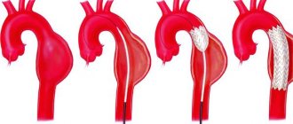
Surgery
It involves removing the damaged area of the aneurysm and replacing it with an artificial blood vessel - an aortic prosthesis. It is installed forever. The rehabilitation period after surgery ranges from 7 days to 1 month, depending on the patient’s age and the presence of concomitant diseases.
It is important to understand that aortic replacement does not guarantee the recurrence of an aneurysm in another part of the vessel. Therefore, it is necessary to strictly follow the doctor’s recommendations regarding nutrition and lifestyle.
The main indications for surgical treatment of an aneurysm are:
- Large size. The average is more than 5.5 cm in diameter. However, here everything depends on the initial diameter of the aorta. If the patient’s normal size is 3-3.5 cm, then surgery is indicated in case of aneurysm formation larger than 6.5-7 cm;
- Rapid growth - the damaged area of the vessel expands by more than 0.5 cm annually;
- An aneurysm provokes compression of the bronchi, the formation of an aortoesophageal/aortobronchial fistula;
- The vessel was stretched due to traumatic injury.
Material and methods
For the period from January 1, 2006 to October 1, 2015 in the Northwestern Federal Medical Research Center named after. V.A. Almazov performed 570 operations for aneurysms and dissections of the VoA. During the specified period, 453 operations were performed for AVOA without dissection - 339 (74.8%) men. Of the entire cohort of patients, 36.9% were patients with bicuspid aortic valve (BAV), 7.3% had hereditary connective tissue disorders, mainly Marfan syndrome, but the majority of patients (55.8%) belonged to the group of non-syndromic non-familial diseases GA . After previously performed interventions on the AC and VA due to the formed aneurysmal expansion of the VA, 24 (5.3%) patients were re-operated.
All patients underwent standard preoperative examination. The main stage of the operation was performed under conditions of moderate hypothermia (32-33 °C). Myocardial protection was carried out using retrograde intermittent isothermal blood cardioplegia or antegrade selective pharmacocold cardioplegia with Custodiol solution. Longitudinal median sternotomy was used as access in all cases. After cross-clamping of the aorta and cardioplegia, a longitudinal aortotomy was performed, the condition of the aortic leaflets, and the thickness of the aortic walls in different sections were assessed. Depending on the data obtained, a decision was made on the type of surgical correction. When aneurysmal expansion spread to the aortic arch (AA), prosthetics of the lower part of the AA (hemiarch repair) or the entire AA was performed (isolated AA prosthesis or “elephant trunk” operation, in 1 case - “frozen elephant trunk” using a hybrid device E-Vita Open Plus). In this case, the connection of the cardiopulmonary bypass apparatus (ACB) was carried out according to the right axillary/femoral artery–right atrium scheme. The surgical stage on DA took place under conditions of hypothermia 27-28 °C and antegrade mono- or bigemispheral cerebral perfusion under the control of continuous cerebral oximetry (Fore-Sight, CASMED). After reperfusion, removal of the clamp from the aorta and warming, the AIC was stopped and control transesophageal echocardiography (TEE) was performed. The wound was sutured tightly in layers, leaving drainage. If the immediate postoperative period was favorable, the patient was extubated several hours after the end of the operation. If the course was favorable, on the 7th day after surgery the patient was transferred to the cardiology department, where he stayed for 1 to 3 weeks until discharge.
The research results were processed using the computer program SPSS (Statistical Package for the Social Sciences) Statistica 22.0 (IBM). The significance level was assumed to be p =
0.05.
Quantitative data were tested for normality of distribution using the Shapiro-Wilk test, taking into account group sizes, and for homogeneity of variance using Levene's test. Indicators corresponding to the parameters of the normal distribution were described in the following form: arithmetic mean ( M
) ± standard deviation (
SD
). Indicators that did not correspond to the parameters of a normal distribution (non-parametric) were described as the median (25–75th percentile). Comparison of samples according to indicators corresponding to the criteria of normal distribution and sample homogeneity was carried out using one-way analysis of variance (ANOVA). To compare groups according to indicators that did not meet the criteria of normal distribution and homogeneity of variance, the nonparametric Spearman test was used when comparing two independent samples.
Endovascular intervention
If the shape and location of the aneurysm are not critical, the endoprosthesis replacement technique is used. The operation proceeds as follows:
- a puncture is made on the patient’s thigh, through which a conductor (narrow silicone tube) is inserted into the aorta;
- A vascular prosthesis is inserted through a conductor into the damaged aorta, which is fixed to the normal parts of the vessel above and below the location of the aneurysm.
The intervention is classified as minimally invasive. It does not require abdominal access, making the operation much easier for the patient to tolerate. The rehabilitation period is 2-3 days. During the day after the operation, the patient is under the supervision of a doctor.
Systematization of manifestations of aortic aneurysm
Currently, there is no unified approach to systematizing the manifestations of peritoneal aortic aneurysm. Most often, doctors and authors of medical works use the method of A. Pokrovsky and R. Ermolyuk, according to which aneurysms are divided:
- by etiology
– acquired (non-inflammatory or inflammatory) and congenital; - according to morphology
- into false (traumatic origin), true and stratifying; - by shape
- diffusion and saccular; - according to the course of the clinical process
- into diseases with an uncomplicated course, complicated and dissecting; - by type and location
- on the proximal segment of the peritoneal aorta, on the infrarenal section, as well as with total damage to the entire section of the abdominal aorta.
According to medical statistics, up to 95% of aneurysms are localized in the infrarenal region.
Forecast
If an aneurysm is detected, the patient needs constant monitoring by doctors. Even after surgery, it is necessary to undergo regular examinations to monitor the condition of the blood vessels. Today, no intervention provides a 100% guarantee that the aneurysm will not reappear in another part of the aorta. Therefore, you should take responsibility for your own health and not neglect regular visits to a specialist.
Statistics on aneurysm operations say that the mortality rate of patients six months after surgery and aortic replacement is 10-15%. Within 10 years from the date of surgery, this figure increases to 40%. This is due to the fact that most patients have concomitant chronic diseases.
In case of endovascular intervention, the prognosis is more favorable. The probability of complications and re-formation of an aneurysm is only 10%, while patient death occurs in only 2% of cases.
Medical observation
For small aneurysms, your doctor may recommend medical monitoring, which includes regular testing to make sure the aneurysm is not getting larger, as well as treatment for underlying conditions that may be contributing to the aneurysm's formation or enlargement.
Your doctor will give you regular tests to determine the size of the aneurysm. Once an aneurysm is diagnosed, you will have an echocardiogram in 6 months, as well as follow-up imaging tests.
If you have high blood pressure or have plaque in your arteries, it is likely that your doctor will prescribe medications to lower your blood pressure and reduce your risk of developing aneurysm complications. Such drugs include:
- Beta blockers. Beta blockers lower blood pressure by slowing your heart rate.
- Angiotensin II receptor blockers. Your doctor may also prescribe these drugs if beta blockers do not lower your blood pressure enough. These medications are recommended for people with Marfan syndrome, even if they do not have high blood pressure.
- Statins. These drugs for the treatment of atherosclerosis of the aorta can lower cholesterol levels, leading to a decrease in plaque deposits in the arteries and a reduced risk of developing aneurysm complications.
If you smoke or chew tobacco, it is important that you quit this bad habit. Tobacco use can worsen the aneurysm.
Prevention
There are no specific measures to prevent such pathology as thoracic aortic aneurysm. Doctors give general recommendations:
- Complete cessation of smoking, including hookah and electronic cigarettes.
- Complete abstinence from alcohol (the maximum allowed is 100g of medium-strength alcohol during the holidays).
- Regular exercise. However, physical activity should be moderate. It is recommended to contact a rehabilitation therapist and exercise therapy specialist, who will select appropriate exercises based on the patient’s diagnosis, the presence of concomitant diseases and the current state of health.
- Control of factors that can provoke a rise in blood pressure. These include stress, kidney pathologies, and prolonged exposure to the open air during the warm season.
- Control of atherosclerosis. This pathology requires treatment, because it can lead to other serious complications in addition to the aneurysm.
- Immediately consult a doctor in case of the slightest suspicion of malfunctions in the cardiovascular system, gastrointestinal tract or lungs. It is necessary to respond to the body's signals to prevent serious complications.
If an aortic aneurysm is already present, prevention consists of preventing the development of complications of the pathology:
- Taking anticoagulants prescribed by a specialist. Such drugs are not cheap, but they are vital. They prevent the formation of blood clots in the lumen of the aneurysm.
- Optimal physical activity. If the patient works in a position that involves heavy lifting or other physical labor, it is necessary to think about changing professions. Excessive force can cause rupture of the aortic wall.
- Control of hypertension. It is necessary to take all medications prescribed by the doctor to avoid increasing blood flow pressure on the thinned wall of the aneurysm. The patient must be aware that it can rupture at any moment.
- Control over psychological state. In medical practice, there have been cases where aortic rupture was caused by a minor stressful situation.
In order to monitor the condition of the cardiovascular system, it is necessary to visit a cardiologist annually. And do this not “for show,” but undergo a comprehensive examination. This way you can detect developing diseases in the early stages, which greatly increases the likelihood of successful treatment without incisions and pain. Take care of your health!
Still have questions about thoracic aortic aneurysm?
Free consultation with AngioClinic specialists
Author
Salmina Daria Vladimirovna
Geneticist. Graduated from the Chelyabinsk State Medical Academy. She completed an internship at the Northwestern State Medical University named after I.I. Mechnikov.
Abdominal aortic aneurysm
Abdominal aortic aneurysm is a chronic degenerative disease with life-threatening complications. An abdominal aortic aneurysm is defined as an increase in its diameter by more than 50% compared to the norm or a local bulging of its wall. Under the pressure of blood flowing through this vessel, the dilatation or bulging of the aorta may progress. The diameter of a normal aorta in the abdominal region is approximately 2 cm. However, at the site of an aneurysm, the aorta can be dilated to 7 cm or more.
Why is an aortic aneurysm dangerous?
An aortic aneurysm poses a major health risk as it can rupture. A ruptured aneurysm can cause massive internal bleeding, which in turn leads to shock or death.
An abdominal aortic aneurysm can cause other serious health problems. Blood clots (thrombi) often form in the aneurysm sac or parts of the aneurysm tear off, which move with the blood flow along the branches of the aorta to the internal organs and limbs. If one of the blood vessels becomes blocked, it can cause severe pain and lead to organ death or loss of a lower limb. Fortunately, if an aortic aneurysm is diagnosed early, treatment can be timely, safe and effective.