The main purpose of ultrasound of the abdominal aorta is to identify an aneurysm or atherosclerotic lesion of the aorta and damage to its large branches: splenic, hepatic, renal and mesenteric arteries. Ultrasound of the abdominal aorta is a non-invasive diagnostic method that has high value in identifying many vascular diseases. The results of ultrasound are very dependent on the examination technology, the quality of the equipment and the qualifications of the specialist. Today, the low price of ultrasound of the aorta and abdominal organs, the simplicity and safety of the study make it indispensable for the primary detection of abdominal aortic aneurysm.
The innovative vascular center specializes in vascular surgery, so we strive to diagnose vascular pathology at the highest level. Our center has excellent stationary and mobile ultrasound machines that allow us to study in detail the structure of the aortic wall and its branches, as well as identify aneurysms. The specialists of our medical center are dedicated to working specifically with vascular pathology, therefore they identify it much better than general ultrasound doctors.
What it is
We should start by defining the aorta. The aorta is the largest vessel in our body, and accordingly its importance is high. An aneurysm is an enlargement of a blood vessel. An aortic aneurysm can form in the thoracic or abdominal regions. Moreover, aneurysm of the abdominal aorta is much more common. This condition of the vessel may not cause any harm for a long time, however, an aneurysm is very dangerous due to the unpredictable course of expansion and the threat of rupture of the vessel walls when they become thinner - and this condition already poses a threat to life due to severe internal bleeding. In addition, blood clots can form at the site of vessel expansion due to changes in blood flow - the danger of this condition is the risk of the blood clot breaking off and clogging a smaller vessel.
How is an ultrasound of the abdominal aorta and its branches performed?
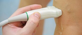
The principle of ultrasound scanning of the abdominal cavity is to process the ultrasound signal reflected from the organs. This allows you to evaluate the structure and size of this organ. Ultrasound examination of the abdominal aorta is not dangerous and does not cause any discomfort. The diagnostic process itself includes only a few steps:
- In the ultrasound diagnostic room, the patient sits on the couch to the right of the doctor and opens his stomach.
- The doctor lubricates a special sensor and the patient’s abdomen with a transparent gel, which reduces tissue resistance and helps the ultrasound wave penetrate inside.
- The specialist then slowly moves the sensor along the abdominal wall and records the results of the observations.
The capabilities of ultrasound devices allow not only to assess the size, shape and structure of the vessel wall, but also to study the characteristics of blood flow. This is achieved using Doppler ultrasound and color Doppler mapping. Dopplerography is based on recording the ultrasonic signal from moving red blood cells and changing the speed of this signal depending on the approach or distance of these particles to the sensor. The speed of blood flow, determined by Doppler ultrasound, is an indicator of the patency of blood vessels below the study site or in areas of narrowing of the vessel. In narrowed places it is significantly strengthened; if there is an obstacle to the outflow of blood below the test site, then the speed decreases.
Causes of aortic aneurysm in the abdominal region
Experts identify several reasons for the formation of an abdominal aortic aneurysm:
- heredity (it is worth undergoing additional examinations if one of your close relatives had such a diagnosis);
- trauma or surgery on the aorta;
- atherosclerosis (approx. link to article);
There are also risk factors for aneurysm formation:
- smoking,
- excess weight,
- high blood pressure,
- The disease is most often encountered by men over 50 years of age.
Quitting smoking and maintaining a normal weight remain factors that you can get rid of, protecting yourself from the risk of not only an aneurysm, but also other serious diseases.
Aortic aneurysm - symptoms and treatment
Treatment of aortic aneurysms is aimed at preventing its rupture. It can be conservative or surgical.
Conservative treatment
The main purpose of conservative therapy is to reduce blood pressure and the force of heart contraction, as well as the correction of concomitant diseases, such as coronary heart disease, diabetes, kidney disease, etc. [1]
Surgery
Modern surgical techniques for treating aortic aneurysms are divided into endovascular and traditional surgical interventions.
The endovascular method is the remote implantation of special tubular prostheses (stent grafts) into the affected area of the aorta from the inside through a catheter installed in the femoral artery. This method of treatment is performed under local or regional anesthesia, it is low-traumatic, low-painful and reduces hospital stay, and also reduces mortality. Increasingly, in the practice of vascular surgery, minimally invasive endovascular technologies are used in the treatment of aneurysms of the thoracic and abdominal aorta, although it is still too early to discount open surgical interventions [4].
Major operations for thoracic aneurysms are among the most difficult in medicine. They are performed in specialized cardiac surgery centers with the heart turned off during artificial circulation. The technology of the operation consists of replacing the dilated section of the aorta with a special tubular prosthesis, which is sewn into place of the aneurysmal sac [5][10]. The surgeon makes an incision to provide access to the aneurysm, excises the affected part and replaces it with a prosthesis. But in some cases, for example after injury, the aorta can be sutured end to end without the use of a prosthesis.
Abdominal aortic aneurysms with a diameter of more than 5.5 cm in men and 5.2 cm in women have a high risk of rupture, and therefore require immediate consultation with a vascular surgeon to determine indications for surgical treatment. It is performed in the vascular departments with access through the anterior abdominal wall (laparotomy) and also aims to replace the affected segment of the aorta with a prosthesis. Some medical centers have mastered laparoscopic treatment of aneurysms, which does not require incisions [4][7].
During surgical treatment of such severe diseases (as with other therapeutic and diagnostic interventions), there is a possibility of complications. It is also worth noting that not all regional, regional and republican medical centers have specialists who work on open heart surgery.
Symptoms of an abdominal aortic aneurysm
Most often, an aneurysm is accidentally diagnosed during an examination for other reasons of concern. The dilation of the vessel usually does not manifest itself in any way, but an aneurysm should be excluded during an examination by a doctor if you experience a dull, aching pain in the chest, under the ribs, or feel pulsation in the abdomen.
If the aneurysm is not detected in a timely manner, there is a risk of its rupture. This condition manifests itself in sharp, severe pain in the abdominal area, a dramatic drop in blood pressure, and fainting. In such a situation, it is necessary to immediately call an ambulance: aortic rupture is life-threatening.
Aneurysms of the abdominal and thoracic aorta
The aorta is the largest artery in the human body, through which oxygenated blood flows from the left ventricle. Branches extend from it to various internal organs and tissues, and at the level of the pelvis it is divided into two iliac arteries, which supply blood to the lower extremities. The aorta can be divided into thoracic and abdominal sections, located in the corresponding cavities. Sometimes, under the influence of various factors, the wall of this vessel becomes thinner, and due to blood pressure, a protrusion (aneurysm) is formed. Since various complications can develop, patients need to receive medical attention.
Etiology
An abdominal aortic aneurysm in most cases occurs against the background of atherosclerotic damage to its wall, which is observed in more than 80% of cases. Much less often, the cause of thinning of the muscle layer is inflammatory diseases (tuberculosis, rheumatism, syphilis), accompanied by vascular damage. Nonspecific aortoarteritis occurs against the background of other infectious diseases.
Congenital pathology of the connective tissue structure (fibromuscular dysplasia, Marfan syndrome) also plays a large role in the development of this pathological process.
Due to the development of vascular surgery in recent decades, aneurysms are increasingly caused by operations on the abdominal aorta:
- angioplasty, when using a special balloon to increase the diameter of the artery;
- removal of blood clots and atherosclerotic plaques along with the inner membrane (endarterectomy);
- thinning of the wall at the junction during aortofemoral replacement.
However, in most cases of complications of surgical intervention, a false aneurysm is formed, the walls of which are made not of the vessel, but of the surrounding tissues. Risk factors for the formation of an abdominal aneurysm are smoking, obesity, male gender, family history (the presence of similar diseases in close relatives) and hypertension.
Manifestations and complications
Symptoms of an aneurysm may be absent for quite a long time and appear only with a significant increase in its size, or with the development of various complications.
In the first case, patients complain of a feeling of pulsation in the abdomen, bulging of the anterior abdominal wall in a horizontal position. This is especially true for thin people. The pain syndrome is usually mistaken for renal or hepatic colic. In addition, dysfunction of internal organs caused by external compression may be observed:
- aneurysm dissection. When an aneurysm dissects, a false tract is formed, through which there is no blood flow;
- digestive processes are disrupted (heartburn, deterioration of peristalsis, constipation);
- from the excretory system, hematuria and urinary retention often develop;
- when exposed to vessels leading to the testicles, varicocele or ischemia may develop;
- when nerve roots and plexuses are compressed, symptoms of sciatica and radiculitis appear, characterized by pain in the lower back and lower extremities.
One of the most common complications is dissection of the aortic wall with the formation of a second lumen, which is usually blind, that is, it lacks adequate blood flow. As a rule, this is a continuation of a defect that arose at the level of the thoracic region.
A dissecting aneurysm is characterized by the following symptoms:
- Deterioration of blood flow to various internal organs. In this case, their function is disrupted, for example, when the mesenteric arteries are damaged, intestinal necrosis develops, and the renal arteries develop acute renal failure. If the blood supply to the lower extremities is impaired, intermittent claudication occurs.
- The pain is usually quite intense and is associated with an effect on sensitive nerve receptors located in the vessel wall and in organs that have been subjected to ischemia. It is localized in the anterior abdominal wall, but can also radiate to the back.
- Neurological disorders may appear in the form of paresis and paralysis, as well as peripheral polyneuropathy.
Rupture of an abdominal aortic aneurysm is accompanied by a sudden decrease in high blood pressure, sweating, pain in the abdomen and lumbar region, signs of motor agitation and weakness appear. If emergency measures are not taken, the patient dies within a short time from massive hemorrhage into the abdominal cavity.
Aneurysms of the thoracic aorta usually develop as a result of degenerative changes in its wall and are more often classified as true aneurysms. According to etiology, aneurysms are divided into: 1) non-inflammatory (atherosclerotic, traumatic, postoperative, iatrogenic); 2) inflammatory (syphilitic, rheumatic, with nonspecific aortoarteritis, mycotic); 3) congenital (with Marfan syndrome, cystic medianecrosis, coarctation of the aorta, congenital tortuosity of the aortic arch). Based on localization, they are distinguished: 1) aneurysms of the sinuses of Valsalva; 2) aneurysms of the sinuses of Valsalva and the ascending aorta; 3) aneurysm of the ascending aorta; 4) aneurysms of the ascending aorta and aortic arch; 5) aneurysms of the aortic arch; 6) aneurysms of the aortic arch and descending aorta; 7) aneurysm of the descending aorta; thoracoabdominal aneurysms. By type, aneurysms are divided into true, false and dissecting, and by shape - into saccular and diffuse (spindle-shaped).
Thoracic aortic aneurysms are observed predominantly in men. The clinical picture is poor. Sometimes an aneurysm is an incidental finding. Symptoms depend on the location, size and extent of the aneurysm. The leading symptom in 1/3 of patients is pain behind the sternum, in the neck, back, and between the shoulder blades. In another 1/3 of patients, the leading symptom is shortness of breath, stridor, and dry cough. Almost half of the patients have arterial hypertension. Sometimes hoarseness, bradycardia, Horner's symptom, and ischemic disorders of the brain are observed. Vascular murmurs are detected in 75% of patients.
Diagnosis of aneurysms at the prehospital stage is difficult. 40-50% of patients are hospitalized with an erroneous diagnosis. An accurate topical diagnosis is provided by the x-ray method, which reveals the expansion of the mediastinal shadow, calcification of the aneurysm wall, and deviation of the esophagus when it is contrasted with barium. The most valuable research methods are computed tomography and magnetic resonance imaging, which help identify the location and size of the aneurysm. Aortography confirms the diagnosis. Differential diagnosis includes tumors and mediastinal cysts.
The prognosis for untreated aneurysms is unfavorable. Within three years after diagnosis, 37.7% die, after 5 years - 54% of patients. Causes of death: rupture of aneurysm (35-40%), heart failure (20-36%), compression of the tracheobronchial tree, pneumonia (14-20%).
Diagnostics
Due to the absence of clear symptoms and gradual expansion of the aorta, most often the suspicion of an aneurysm appears to the doctor during an examination for another reason. The diagnosis is confirmed using ultrasound or CT scan of the abdominal cavity, angiography - which examination is suitable for a particular patient is decided by the attending physician. Examinations will provide information about the size of the aneurysm, the presence of a blood clot in this location, and the condition of the vessel walls.
In many patients, the identified dilatation of the aorta does not require surgical intervention by a specialist for a long time - the main task will be observation. In this case, the patient will be given a list of recommendations so as not to worsen the condition of the blood vessels (first of all, quitting smoking, if such an addiction is present), and will also be offered to undergo periodic examinations to monitor the condition of the aorta. But in any case, the doctor must exclude the risk of aortic rupture at the site of expansion. For this purpose, examination data is used: the width of the aorta and the rate of its expansion over time. If there is a threat of rupture, the doctor may decide to perform an operation, and this will require additional tests of blood, blood vessels, and urine to prepare for the operation.
Preparing for the study
To successfully perform an ultrasound of the abdominal cavity, preparation is necessary aimed at reducing the amount of gases in the intestines that interfere with seeing the retroperitoneal organs, including the aorta. The preparation rules for examining the abdominal aorta and its branches using ultrasound are simple:
- 2 days before the examination, exclude legumes, potatoes, cabbage, melon, dairy products, soda and all foods high in carbohydrates from the diet. These products may cause increased gas formation.
- On the eve of the ultrasound, start taking medications to improve bowel function. For these purposes, taking espumisan or regular activated carbon is suitable.
- It is advisable to skip dinner the evening before the procedure, and breakfast in the morning.
Treatment
As already noted, the most dangerous complication of an aortic aneurysm is the threat of its rupture and internal bleeding. If the patient did not consult a doctor and the aneurysm was not detected in a timely manner, then in the event of a rupture, the only option would be open surgery: the surgeon will remove the affected area of the aorta and install a prosthesis in its place. This method of emergency treatment of an aneurysm has contraindications; in addition, the operation is a serious intervention with associated risks and a long rehabilitation period (up to three months). Therefore, it is so important to contact a specialist if you are at risk and feel periodic pain and/or throbbing in the abdominal area.
Once an aneurysm is diagnosed, its further expansion is difficult to predict; regular monitoring by an experienced specialist, lifestyle adjustments, and control of risk factors are required. When the attending physician realizes that there is a risk of rupture, an alternative to emergency open surgery will be the planned installation of a stent graft. The installation is minimally invasive, i.e. the stent is inserted through the vessel, advanced to the site of the aneurysm and secured there. The structure of the stent resembles a vessel; it is fixed with one edge above the expansion of the aorta, and with the other below. After the operation, blood will flow through the stent, and the resulting cavity between it and the aortic wall will decrease over time. The stent graft is inserted under local anesthesia and requires only a couple of days of recovery.
Of course, any surgical intervention carries its own risks, which is why it is so important to choose a good medical center with experienced cardiovascular surgeons. Understanding the danger of aortic aneurysm as a disease, not only doctors in the cardiology department, but also in other departments of our center, when conducting routine examinations or examinations due to other diseases, monitor the patient’s condition and analyze the entire volume of data received: if a problem with the aorta is suspected, the patient within one center will be transferred for consultation with a cardiologist. For our patients, we try to organize the most convenient logic for staying in our center and consulting with specialists in order to avoid double examinations and unnecessary manipulations. Comprehensive patient management is one of the main principles of our work. And the quality of surgical and conservative care is maintained thanks to a team of experienced specialists, provided with the necessary diagnostic and treatment facilities.
You can sign up for a consultation using a special form on the website or by phone.
