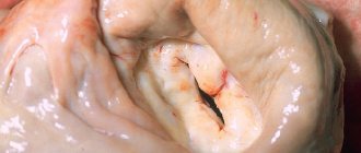
Prolactinoma is a benign formation that develops in the anterior lobe of the pituitary gland and produces the hormone prolactin in high concentrations. According to statistics, this hormonally active tumor develops much more often in women of reproductive age. The size of microprolactinoma is usually about 2-3 mm. The diameter of macroprolactinoma exceeds 1 cm.
As a rule, macroprolactinomas are detected in male patients, microprolactinomas are detected in female patients.
Prolactinoma: causes, symptoms, treatment, consequences
Prolactinoma
is one of the most common neoplasms of the anterior pituitary gland, the size of which most often ranges from 1-3 mm (
pituitary microadenoma, microprolactinoma
), less often - more than 10 mm (
pituitary macroadenoma, macroprolactinoma
). This type of adenoma rarely degenerates into a malignant neoplasm. Being a hormonally active tumor of the pituitary gland, prolactinoma promotes increased production of prolactin and an increase in its level in the blood serum.
Prolactin is a natural hormone that promotes milk production in postpartum women. Along with luteinizing and follicle-stimulating hormones, prolactin also regulates sexual and reproductive functions in the body of men and women. Excessive levels of prolactin in the blood have a suppressive effect on the production of female sex hormones - estrogens, which leads to the development of hypogonadism, menstrual irregularities and infertility. Prolactinoma in men contributes to the development of gynecomastia, decreased libido and erection due to dysregulation of the production of the male sex hormone - testosterone and decreased sperm activity due to hyperprolactinemia.
A third of pituitary tumors are prolactinomas, and in women of fertile age this type of tumor occurs 8-10 times more often than in men.
Radical methods of treating pituitary tumors
Due to the effectiveness of drug treatment for prolactinomas, surgery and radiation therapy are rarely used. Only a small proportion of patients with macroprolactinomas whose tumor size does not decrease with drug treatment may require surgery, especially if there is no improvement in vision. It should be noted that this operation is currently performed through a small incision near the sinuses, the so-called transsphenoidal approach. If a large prolactinoma steadily decreases in size as a result of taking pills, then this use continues in the future.
Sometimes experts recommend radiation therapy, which allows you to stop taking the medication. The effect of radiation develops gradually and is fully manifested only after a few years, so radiation therapy is not prescribed to young women who want to become pregnant (it is these women who predominate among patients with prolactinomas). For microprolactinomas, selective transsphenoidal adenomectomy is most often performed, but in 20-50% of patients, within 5 years after surgery, the tumor recurs and hyperprolactinemia resumes. With macroprolactinomas, even a short-term initial improvement after surgery occurs in only 10-30% of patients.
When carrying out radiation therapy or surgical treatment, pituitary insufficiency may develop, as a result of which secondary adrenal insufficiency and hypothyroidism develop and replacement therapy is required - glucocorticoids in the presence of adrenal insufficiency, L-thyroxine in the presence of thyroid insufficiency (hypothyroidism) and, possibly, sex hormones (estrogens for women and testosterone for men) as replacement therapy.
Causes and classification of prolactinomas
The reasons for the development of prolactinomas remain poorly understood to date. In a certain group of patients, a hereditary nature of tumor development was noted. Another group of patients with prolactinoma has genetic disorders in the form of a hereditary disease called multiple endocrine neoplasia, which is characterized by excessive production of hormones by the parathyroid and pancreas glands, as well as the pituitary gland. Geneticists do not stop searching for the gene responsible for the occurrence of prolactinoma.
Prolactinomas are classified
Depending on the size and location of the tumor relative to the pituitary fossa in the sella turcica, there are two groups:
— intrasellar prolactinomas
— the diameter of which does not exceed 10 mm, are located inside the sella turcica;
— extrasellar prolactinomas
- exceeding 10 mm in diameter and extending beyond the boundaries of the sella turcica.
Depending on the size of the prolactinoma, the clinical signs of the disease and the subsequent choice of treatment method will differ.
(Hyperprolactinemia)
Prolactinoma - hyperprolactinemia syndrome (HS) is both a manifestation of an independent hypothalamic-pituitary disease and one of the most common syndromes in various endocrine, somatic, and neuroreflex disorders.
It has been established that prolactinoma accompanies every third case of female infertility. Most often, GP occurs in young women 25-40 years old, much less often in men of the same age. Cases of hyperprolactinemia have been described in adolescents and the elderly.
Symptoms of prolactinoma
Prolactinoma is characterized by a dysfunctional disorder of the reproductive system, neurological signs, psycho-emotional disorders and changes in the endocrine system.
Prolactinoma in women is often presented as a microadenoma and manifests itself as menstrual irregularities
in the form of oligomenorrhea, opsomenorrhea or complete absence of menstrual flow. In addition, in the presence of menstruation, patients do not ovulate, and the luteal phase is shortened. The first menstruation usually comes late, and the subsequent cycle is irregular.
Most patients usually turn to a gynecologist due to a number of unsuccessful attempts to become pregnant.
.
A gynecological examination can reveal hypoplastic changes in the uterus
.
In one out of five women, the main complaint is the spontaneous release of fluid resembling milk from the mammary glands, not associated with childbirth - galactorrhea
. Moreover, the severity of galactorrhea can vary from single drops when pressing on the mammary gland to heavy discharge. However, this symptom may not appear even with a significant concentration of prolactin in the blood.
Due to disruption of the production of estrogen and progesterone, mastopathy
and reduction of mammary glands.
In addition, an increase in the level of prolactin in the blood leads to the leaching of calcium salts from bone tissue and the development of osteoporosis
, leading to frequent fractures due to brittle bones.
A decrease in the amount of estrogen contributes to the accumulation of fluid in the body and weight gain
.
In men, prolactinoma
manifests itself in the form of infertility, decreased libido and potency, testicular atrophy, the development of gynecomastia (enlarged mammary glands) and, in rare cases, galactorrhea.
If the tumor compresses the surrounding brain tissue, in particular the diaphragm of the sella turcica, patients complain of constant headaches
.
When the tumor grows beyond the sella turcica and compresses the optic nerves, there is a narrowing of the visual fields, double vision, drooping eyelids, and when the optic chiasm is compressed, complete blindness occurs. Psychoemotional disorders
that arise during the development of prolactinoma manifest themselves in the form of depression, irritability, emotional instability, narrowing of interests, apathy, increased anxiety, memory and attention impairment, and sometimes in the form of social maladjustment and autism.
Sometimes a clear clinical sign of prolactinoma is an acute hemorrhagic infarction of the pituitary gland, which is characterized by a sudden acute headache accompanied by nausea and vomiting, in some cases, loss of consciousness and a number of meningeal symptoms.
Diagnosis of prolactinoma
The most informative research method for identifying prolactinomas is magnetic resonance imaging of the pituitary gland
with a contrast agent, which allows you to determine the outlines of microadenomas, their location inside or outside the sella turcica.
When diagnosing macroadenomas, it is recommended to use computed tomography
of the sella region, in which the bone structures are clearly visible.
Laboratory research methods consist primarily of determining the level of prolactin in the blood, and the study is recommended to be carried out three times to exclude random fluctuations in the level of the hormone due to stress or other predisposing factors. Prolactinoma is characterized by a prolactin level in the blood exceeding 200 ng/ml.
Previously, to diagnose prolactinoma, stimulation tests with thyrotropin-releasing hormone were used, after the introduction of which into the body, in the first 20-30 minutes, the production of prolactin normally increased, doubling the initial level, and in patients with prolactinoma, the level of prolactin after stimulation did not change. Currently, this diagnostic method is not used due to its low reliability.
Prevention
The main direction of preventing the occurrence of pituitary adenomas is to minimize the impact on the body of provoking factors (brain injuries, neuroinfections). And prevention of relapses in the presence of a history of prolactinomas is constant monitoring and follow-up of such patients with MRI or CT scanning over time.
Our center’s specialists are always ready to find an approach to each patient and choose the most appropriate treatment tactics in each individual case, taking into account the nuances of the person’s health condition.
Treatment of prolactinoma
The main goals when drawing up a treatment plan for prolactinoma are to reduce the concentration of prolactin in the blood, influence the tumor by reducing it and preventing further growth, as well as corrective measures to combat infertility, hypogonadism, disorders of the visual organs and the skeletal system.
If a pituitary tumor is detected for the first time, there are signs of rapid growth, surgery is contemplated, or the disease is detected during pregnancy, patients should be hospitalized.
Conservative treatment of prolactinoma involves the use of dopamine agonists - cabergoline, bromocriptine, abergin, which affect the level of prolactin in the blood, allowing to restore menstrual function and influence the size of the tumor.
In cases where the use of medications does not bring the expected effect and the clinical signs of the disease progress, surgical treatment is resorted to, which involves removing the tumor transsphenoidally (through the nasal sinuses). If patients have contraindications to surgery due to the presence of severe concomitant pathology, or patients refuse surgery, radiation is used to treat prolactinoma.
After surgical or radiation exposure, patients take medication for a long time, sometimes even throughout their lives. Control images of magnetic resonance imaging are recommended to be carried out once a year, and prolactin levels in the blood are examined twice a year.
Doctor's opinion
Prognosis for prolactinoma depends on its size, clinical symptoms, as well as the timeliness of therapy initiated.
According to statistical data, when observing patients for five years after surgical treatment, relapse of the disease occurs in approximately 20-50% of cases. — Osina Ekaterina Aleksandrovna Reproductologist, obstetrician-gynecologist, Ph.D.
As for drug therapy, with its proper and long-term administration, regression and even complete disappearance of the adenoma is observed.
Prolactinoma and pregnancy
If, while taking drugs that reduce prolactin levels in the blood, reproductive function is restored and pregnancy occurs, dopamine agonists should be discontinued. In the first trimester of pregnancy, when the risk of spontaneous abortion in patients with prolactinoma is very high, natural progesterone is prescribed. Monitoring of a pregnant woman with prolactinoma by an ophthalmologist and neurologist throughout pregnancy is mandatory. Dopamine agonists are prescribed only when progressive tumor growth is noted. It is not advisable to perform magnetic resonance imaging during pregnancy, however, if there is no positive dynamics from the use of conservative treatment, it is necessary to decide on neurosurgical intervention.
A control magnetic resonance examination is recommended a couple of months after birth. There are no contraindications for breastfeeding, but in cases of tumor enlargement, suppression of milk production may be necessary.


