Hospitalization and treatment under the compulsory medical insurance quota. More details after viewing the pictures.
- Aneurysm clipping
A cerebral aneurysm is a local protrusion of the arterial walls. The disease often occurs without symptoms. As the brain aneurysm grows, a significant thinning of the walls of the formation occurs, which can lead to its rupture and the development of hemorrhagic stroke.
Depending on the shape, a cerebral aneurysm can be spindle-shaped or saccular. The exact type of disease can only be determined during a comprehensive diagnosis. Saccular aneurysm is much more common.
Causes of the disease
An aneurysm is a dangerous condition that primarily requires surgical treatment. Otherwise, complications will not be avoided. Experts believe that the disease is a consequence of developmental abnormalities that disrupt the structure of the vascular wall. Often, a cerebral aneurysm is combined with connective tissue dysplasia, polycystic kidney disease, and vascular disorders. The acquired form of the disease can be a consequence of traumatic brain injury. Also, a cerebral aneurysm occurs against the background of atherosclerosis and hypertension.
The vascular wall defect itself occurs gradually. Against the background of degenerative processes, tissue damage or underdevelopment, the area loses its former elasticity. Under the pressure of blood flow, the vascular wall begins to bulge. This is how a brain aneurysm forms. Most often, the formation occurs in the place where the arteries branch and there is the strongest pressure on the vessel wall.
What is the aorta
The aorta is the largest vessel in the human body, which carries blood from the heart to the organs and limbs. The upper section of the aorta runs inside the chest, this section is called the thoracic aorta . The lower part is in the abdominal cavity and is called the abdominal aorta . It delivers blood to the lower part of the body. In the lower abdomen, the abdominal aorta divides into two large vessels - the iliac arteries , which carry blood to the lower extremities.
The aortic wall consists of three layers: internal (intima), middle (media), external (adventitia).
Symptoms of an aneurysm
Two variants of the course of the disease can be distinguished: apoplexy and tumor-like. The characteristics of the clinical picture depend on the types of aneurysm. The tumor-like form of the disease is accompanied by an active course. In a short period of time, the aneurysm increases in size and compresses important anatomical structures of the brain.
A tumor-like aneurysm is most often localized in the area of the cavernous sinus or chiasm. When the chiasmal region is damaged, visual acuity decreases. Over a long period of time, optic nerve atrophy occurs. If a cerebral aneurysm is located in the cavernous sinus, paresis occurs and damage to the branches of the trigeminal nerve occurs. As the pathology develops, oculomotor disturbances occur and strabismus may appear.
Signs of a ruptured aneurysm
A sharp headache is the first symptom of a ruptured aneurysm. The pain syndrome is initially local in nature. But very quickly the headache spreads and becomes diffuse. In this case, patients note the appearance of nausea and repeated vomiting.
In the future, characteristic meningeal symptoms arise:
- stiffness of the neck muscles;
- Kernig's and Brudzinski's symptoms;
- loss of consciousness.
When a cerebral aneurysm ruptures, subarachnoid hemorrhage occurs. It may be accompanied by focal symptoms, which depend on the location of the formation. The most severe is hemorrhage into the ventricles, which often leads to the death of the patient.
With an aneurysm of the anterior cerebral artery, leg paresis develops, which is often combined with mental disorders. Damage to the middle cerebral artery leads to speech impairment and the development of hemiparesis.
If the pathology is localized in the area of the vertebrobasilar system, during the rupture of the aneurysm, swallowing disorders occur, nystagmus develops in combination with central paresis of the face and trigeminal nerve.
At the first sign of a possible aneurysm, you should immediately seek emergency medical help. It is important to maintain physical rest and avoid overexertion. Specialists will conduct an examination, diagnose, make an accurate diagnosis, and prescribe the necessary medications to reduce the risk of life-threatening complications.
Classification
The pathological process should be divided according to three bases: size, location and shape.
By size
Based on the criterion of the volume of altered tissue, the following types of pathological process can be distinguished:
- Miliary or microaneurysms. They represent the initial stage of the disorder or a full-fledged variety of it. They do not reach three millimeters in size. But they still pose a danger.
Therefore, it makes sense to check the patient every six months. If there is no dynamics as such, medications are prescribed, special living conditions and a diet are prescribed.
However, a microvariety may be a transitional variant. That is, as was said, the initial stage of the disorder. Then you cannot do without surgical correction.
- Small ones. Size up to 10 mm. They occur much more often. They pose immediate risks to the life and health of the patient. It is from this variety that the pathological process begins.
Treatment is strictly necessary. It is carried out under the supervision of a vascular neurosurgeon.
- Average. Up to 1.5 cm in diameter. These are serious entities. Enough physical stress is enough for the structure to rupture and lead to bleeding. Therapy is prescribed on an emergency basis. It is carried out strictly in stationary conditions.
- Large ones. 16-25 mm. Such forms are relatively rare. The size of the vascular formation leads to enormous risks of complications. The structure can rupture due to minor mechanical activity.
- Macroaneurysms. Over 2.5 cm in diameter. The danger lies not only in the possibility of rupture. Also, such a vascular formation compresses the surrounding tissues, leading to neurological deficits. Treatment is urgent, surgery is needed.
By localization
Regarding the second criterion, location:
- Anterior aneurysms. Located in the area of the parietal lobes. They are especially common. They do not pose great difficulties in terms of access, therefore the chances of recovery and a good course of the postoperative period are high.
- Medium varieties. Localized in the area of the parietal and temporal lobes. Access needs to be worked out as the issue is somewhat more complex.
- Localized in the area of the vertebrobasilar region. That is, closer to the occipital lobes.
- Located in the area of the internal carotid artery. It is difficult to reach such an aneurysm straight away. The correction method needs to be worked out. The task is not the easiest; it requires the participation of an experienced and qualified neurosurgeon.
The location must be clarified in order to carry out a competent and safe operation for the patient.
This classification is rather arbitrary, since an aneurysm can mature in almost any part of the brain.
By shape
The third basis for classification is the actual form of education. The following types of pathological structures are distinguished:
- Saccular. They occur most often. And account for more than 80% of the total number of situations. Externally they look like lateral wall protrusions of the artery, strictly in one direction. Such swelling became the basis for calling the aneurysm saccular.
- A fusiform change develops much less frequently. As the name suggests, the artery bulges in both directions. That is, there are no healthy, unaffected areas left.
A pathological process of this kind is much more dangerous. Because there is no restraining or compensating factor. Both sides of the vessel are involved in the breach and are at risk of rupture at any moment.
Doctors use all three classifications to describe the pathological process and plan treatment.
How is it diagnosed?
Since an arterial aneurysm can occur without significant symptoms, it is necessary to pay special attention to the first signs in women and men. A neurologist must diagnose the disease based on the examination, the diagnostic results obtained and the study of cerebrospinal fluid.
During a neurological examination, focal and meningeal symptoms are detected. Based on the location of complaints, it is possible to determine presumably where the pathological focus is located.
The neurologist obtains accurate information about the patient’s condition using the following studies:
- X-ray of the skull. A cerebral aneurysm is characterized by signs of bone tissue destruction.
- Computed tomography, magnetic resonance imaging of the brain.
- Angiography. Basic information about the location, shape and size of the brain aneurysm can be obtained.
- Lumbar puncture. The study reveals blood in the cerebrospinal fluid, which indicates the presence of hemorrhage.
Specialists carry out differential diagnosis of cerebral aneurysm. It is important to make an accurate diagnosis in a short time. This will avoid the adverse effects of a brain aneurysm. It is necessary to differentiate an aneurysm from tumor-like processes, cysts and abscesses.
Diagnostics
{banner_banstat10}
The examination is not particularly difficult. Neurosurgeons work with patients of this profile.
The list of events will be as follows:
- Questioning a person.
- Anamnesis collection.
- MRI.
- CT.
- Classic angiography, contrast-enhanced x-ray.
- EEG. To assess the degree of functional activity of the brain, to identify disturbances in the activity of cerebral structures.
Basically, this is enough to make a diagnosis. Additional measures are determined by a medical specialist.
Principles of treatment
Aneurysm treatment is carried out by a neurologist. The operation is performed only by a neurosurgeon. Surgical methods are aimed primarily at preventing aneurysm rupture, which can result in the death of the patient. Conservative therapy for this pathology is used to slow the progression of the disease.
Surgical treatment of a cerebral aneurysm involves two types of main operations: clipping and endovascular occlusion. Surgery prevents the aneurysm from rupturing. A radical method of treatment is the removal of arteriovenous malformation during a microsurgical operation.
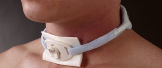
Aneurysm clipping
Aneurysm clipping is an open operation during which the neurosurgeon blocks blood flow in a certain area of the vessel and applies a clip. At the stage of preparation for surgery, specialists prescribe a procedure that allows them to stabilize the patient’s condition and minimize the risk of possible complications. Preventive examination makes it possible to identify possible disorders and diseases that can lead to adverse consequences of surgical intervention.
The operation is performed using modern microsurgical techniques through a small trepanation hole. During surgery, the neurosurgeon prevents rupture of the wall of the malformation.
After isolating the neck of the aneurysm, the specialist places a clip on it. Additional control with Doppler ultrasound allows you to assess the state of blood flow and ensure the effectiveness of the operation.
Treatment of an aneurysm using the clipping method is considered quite complex and should only be performed by an experienced neurosurgeon. A qualified specialist will do everything possible to ensure accurate and technically correct application of the clip. Otherwise, dangerous complications and a long rehabilitation period cannot be avoided.
The clip is applied to the neck of the aneurysm vessel. The accuracy of the neurosurgeon’s actions is confirmed by conducting high-quality diagnostics (Dopplerography) during the procedure.
Endovascular occlusion
During endovascular occlusion of an aneurysm, the neurosurgeon blocks the lumen of the dilated vessel with a special implant. The operation is used when clipping is not possible, for example, when the aneurysm is spindle-shaped. During surgery, a specialist inserts a catheter balloon through the femoral artery using angiographic control. It closes the lumen of the aneurysm. You can also use a microspiral to perform thrombosis. The choice remains with the attending physician. The microspiral in the cavity of the affected vascular area forms blood clots, which clog the lumen of the vessel and shut off the aneurysms from the blood circulation.
Taking medications
The main method of conservative treatment of an aneurysm is taking medications. The patient is prescribed a complex of drugs:
- drugs to lower cholesterol and prevent atherosclerosis;
- painkillers;
- anticonvulsants;
- drugs to normalize blood pressure: amlodipine, atenolol, anaprilin, enalapril (consultation with a doctor is required, there are contraindications)
Drug therapy is also indicated in the postoperative period. It eliminates neurological symptoms and pain, and reduces the likelihood of disease relapse.
Rehabilitation after surgical treatment of an aneurysm
The rehabilitation period takes place under the supervision of specialists. Professionals do everything necessary to prevent the occurrence of postoperative complications. The patient remains in the intensive care unit for several days under the constant supervision of specialists. Afterwards, the patient is transferred to a general ward, where strict bed rest must be observed for some time. The return to a normal lifestyle occurs gradually.
If the patient's well-being worsens after surgery, transcranial Doppler ultrasound may be prescribed. Rehabilitation is aimed primarily at preventing vasospasm and increased blood pressure. Specialists also monitor the general condition of the cardiovascular system and, if necessary, prescribe osmodiuretics to eliminate swelling of the brain tissue. Additionally, anti-inflammatory therapy is carried out.
Rehabilitation after surgical treatment of an aneurysm necessarily includes a program to restore impaired body functions and further socialize the patient. The recovery period lasts at least 6 months. With the help of high-quality rehabilitation, it is possible, among other things, to eliminate the adverse consequences of surgical treatment.
Rehabilitation measures include physiotherapy, massage, and the implementation of an individually selected rehabilitation program. After endoscopic clipping, the patient can return to normal life within a few weeks.
Prevention of pathology
All preventative measures can be divided into actions to prevent the formation of an aneurysm and methods to prevent the rupture of vessel formations. As a rule, in practice these methods coincide, so general recommendations for disease prevention can be identified:
- monitoring blood pressure and reducing it in a timely manner;
- giving up bad habits - alcohol, smoking, drugs;
- maintaining proper nutrition;
- control of bad cholesterol in the blood;
- eliminating stress;
- timely treatment of cardiovascular diseases;
- refusal of intense physical activity.
It is important to note that even the unquestioning implementation of the above measures does not guarantee the complete elimination of pathology and its complications. The only way to completely get rid of an aneurysm is through surgery.
Forecast for life
The prognosis of the disease depends on the location of the vascular protrusion and its size. The initial condition of the patient also has a huge impact. The mortality rate in case of rupture of a cerebral aneurysm exceeds 30-50%. Surviving patients sometimes retain the consequences of the condition in the form of movement restrictions, cognitive impairment and a significant decrease in quality of life. Mortality after recurrent hemorrhage exceeds 70%.
Therefore, it is so important to carry out surgical treatment in time, turning to a qualified neurosurgeon. The operation is postponed only if there are certain medical indications.
Successful neurosurgical operations are carried out at the Burdenko Research Institute. Patients with signs of an aneurysm or suspected aneurysm are admitted here. At the Institute of Neurosurgery, it is possible to carry out comprehensive diagnostics using the latest technology and further treatment of identified diseases.
Types of aortic aneurysms
There are “true” and “false” aortic aneurysms. A true aneurysm develops due to the gradual weakening of all layers of the aortic wall. A false aneurysm is usually the result of trauma. It is formed from the connective tissue surrounding the aorta. The cavity of the false aneurysm is filled with blood through a crack in the aortic wall. The aortic walls themselves do not participate in the formation of an aneurysm.
Depending on the form there are:
- saccular aneurysm - expansion of the aortic cavity on only one side;
- spindle-shaped (fusiform) aneurysm - expansion of the aneurysm cavity on all sides;
- mixed aneurysm - a combination of saccular and fusiform forms.