Damage to the organs of vision, unfortunately, occurs quite often in everyday life. Traumatic eye injuries occupy a leading position among the causes of blindness and disability.
Most often, subconjunctival hemorrhages and minor injuries to the cornea, eyelids and conjunctiva occur. In this case, the most dangerous are hyphema, hemorrhages in the vitreous body and retina of the eye. Rarely encountered: corneal rupture, retinal injury, fundus fracture, damage to the optic nerve or tear duct, lens displacement.
Causes of eye injuries
Acute injuries often occur when glasses or masks, i.e., equipment that is supposed to protect, are damaged. Shards of broken glass or plastic cause many penetrating wounds to the eyeball, incised wounds to the eyelids and face. Acute injuries can also be caused by nails, tree branches, and in some sports, such as basketball, water polo, rugby, and wrestling.
However, most eye injuries are blunt (contusions). Any object traveling at high speed can cause blunt force trauma. If the object is large, part of the energy is absorbed by the surrounding tissues, which can also be damaged (up to a fracture of the nose, cheekbone).
However, even a large and relatively “soft” soccer ball can protrude deep into the orbit and injure the eye. The eyeball is quite elastic, and when it “restrains” the blow, the kinetic energy is transferred further to the thin walls of the orbit and the so-called. a fracture of the orbital floor, and part of the contents of the orbit moves into the maxillary cavity. Such damage leads to the “drowning” of the eyeball into the orbit and limits its mobility, which causes diplopia.
Injuries to the eyes and eyelids most often occur during sports competitions (13% of all cases). The most traumatic sports in this regard, according to Norwegian statistics: football (up to 35%), bandy (13%), squash (up to 11%), handball (up to 7%). In Scotland, football also leads (up to 33%), followed by squash (up to 30%), ice hockey (up to 10%), tennis (up to 10%) and badminton (up to 8%).
In the US, basketball takes first place, and paintball comes in second (up to 21%). Perhaps the real danger of these sports is not so high, but the whole point is their popularity.
If a moving (flying) object has a small diameter, then it is more likely to damage the eye. Impacts from such objects cause damage to the eyelids and cornea (depending on whether the eyelids were open or closed at the time of impact). The impact deforms the eyeball and, as a result, intraocular pressure increases. There is a posterior displacement of the iris and lens, up to their rupture. As a result of contusion, the back wall of the eye can also be injured. Such events last milliseconds, but leave a “trace” for life.
Tears in the iris along the edge of the pupil lead to the impossibility of its narrowing, and tears along the periphery lead to the formation of a false pupil. Damage to the nerves in the iris causes an inability to regulate the amount of light entering the eye.
Blunt trauma to the eye leads to hemorrhage into the anterior chamber of the eye (hyphema) and the vitreous body. Hyphema is immediately visible; it is a kind of marker of serious damage, and the organ of vision should be examined by an ophthalmologist.
Prevention
There are no specific rules for preventing eye contusion. Herbs can arise at any time. This is due to external factors and mechanical stress. You can protect yourself from this by following safety rules. In general, safety glasses should be used.
Sometimes blunt force trauma can be caused by another person. For example, an attacker. In this case, it is difficult to protect yourself from such a situation. Therefore, the only advice is to follow general safety rules. If any degree of contusion occurs, you should immediately consult a doctor.
Eyelid injury
Superficial wounds of the eyelids damage only the skin and muscle layer, while penetrating wounds damage all layers of the eyelid. Since the skin of the eyelids is characterized by great extensibility and looseness of the subcutaneous tissue, swelling and hemorrhages appear here very early. The skin of the eyelids becomes tense, and the color ranges from dark blue to purple. The swelling may spread to the eyelid of the other eye.
A small external eyelid wound may conceal massive internal damage that requires immediate attention from an ophthalmologist.
Based on the appearance of the wound, one cannot draw a conclusion about the degree of damage to deeper tissues. A small external eyelid wound may conceal massive internal damage that requires immediate attention from an ophthalmologist.
If the wound is located vertically, then its edges gape due to rupture of the transverse muscle fibers. When the eyelid is injured, subcutaneous emphysema can form. This indicates a violation of the integrity of the bones of the paranasal sinuses.
Minor eyelid wounds end favorably, but if the wound becomes infected, scarring and deformation of the eyelid are possible.
If eyelid wounds become infected, scars can form, which in turn leads to cicatricial eversion of the eyelid. If the muscle that lifts the upper eyelid is damaged, ptosis of traumatic origin may appear. If you suspect the introduction of a foreign body into the tissue of the eyelids, orbit or lacrimal organs, you need to take an x-ray of the orbit.
Treatment
First aid for a wound of the eyelid - the skin around the wound should be treated with an antiseptic (miramistin, ethacridine, picloxidine, boric acid), and if the wound is contaminated, it should be cleaned and rinsed with a solution of hydrogen peroxide, and then apply an aseptic bandage.
If the eyelid wound is small and horizontal, then it does not require surgical intervention. If the wound gapes, surgical help is necessary. If it is impossible to carry out primary surgical treatment in a timely manner, it must be carried out later - even after a few days and in the absence of signs of suppuration. When treating eyelid wounds, it is necessary to take care of the damaged tissues and prevent their excision. If the wounds of the eyelids are through, then suturing is used “in two levels”: “first” - sutures on the conjunctiva and cartilage, and “second” - sutures on the skin of the eyelid.
Traditional therapy
Before using decoctions and lotions, you should consult an ophthalmologist: in treatment it is important not to harm or aggravate the condition.
Iodine mesh
This method is widely used for many diseases. Iodine eliminates swelling and relieves pain from bruises. The mesh is applied to the hematoma under the eyes with severe swelling and bruising. The substance stops inflammatory processes in tissues and destroys pyogenic bacteria. The mucous membrane of the eyes cannot be treated with iodine, otherwise there will be a burn.
Salt compresses
A 10% salt compress, which is applied to the swollen tissues under the eyes or under the eyebrows, helps relieve swelling. Salt applications are not applied to the eyes. The compress should be kept for 10 minutes, then the skin should be gently washed.
Burdock leaves
An ancient folk remedy for bruises and swelling, eliminates pain. The sheet should be washed well in running water and dried. This sheet is applied to the bruised area, a plastic bag for insulation and a warm cloth are fixed on top. This method should not be used on a fresh bruise, as heat will cause swelling to spread. Immediately after the blow, ice or a cold compress is applied to stop the spread of the hematoma. Warming is done the next day if the bruise hurts a lot.
Cabbage leaves
This remedy is used to eliminate the effects of a bruise and relieve a hematoma. The fresh leaf is slightly beaten with a rolling pin until the juice appears and applied to the eye. To keep the sheet in place, it is fixed with a bandage or towel. As the cabbage leaf dries out, replace it with a new one.
Marigold
Calendula is one of the popular folk methods for treating many diseases. It is used as a local remedy - in applications, lotions, rinses, compresses. For half a cup of boiling water, take a tablespoon of dried flowers and infuse. This compress can be used to treat a damaged eye and the skin around the eye. The application is applied in a warm state and kept until it cools.
Foreign body of the conjunctiva
A foreign body of the conjunctiva (usually small particles of earth, coal, stone or metal, grains of sand, hairs of cereal plants) can remain on the surface or penetrate into it, violating the integrity of the epithelial cover, in the latter case an inflammatory infiltrate is formed.
In the clinic, symptoms of eye irritation predominate: photophobia, pain, blepharospasm and, naturally, the sensation of the presence of a foreign body. During examination or biomicroscopy, you can see a foreign body on or inside the conjunctiva.
A foreign body can move due to blinking reflex movements of the eye. Often it lingers in the groove of the eyelid, on its inner surface. Then, when the eyelid is everted, an inflammatory infiltrate with papillae in the center of which is a foreign body (hair of cereal plants, etc.) is discovered.
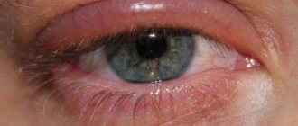
Often, damage to the conjunctiva leads to subconjunctival hemorrhage. This can happen due to trivial rubbing of the eye. A blood-red spot appears on the sclera. Despite the “terrible appearance,” such hemorrhage is not dangerous: it resolves on its own in two to three weeks and does not lead to visual impairment. But sometimes a perforation of the eyeball can be missed behind a bloody spot. Therefore, at the slightest suspicion, the victim should be sent for examination to an ophthalmologist.
Treatment
If it is located superficially, the foreign particle can be easily removed with a cotton swab soaked in a disinfectant solution. If penetration occurs deep into the tissues of the eye, you need to drip a solution of tetracaine (0.5%) into the eye and then remove it with a special needle or tweezers. If it is impossible to remove in this way, a section of the conjunctiva is excised along with the foreign particle.
Large fragments of coal, stone, glass, metal that cause severe irritation should be removed. And small particles of sand and stone that do not cause irritation do not need to be removed; they will “come out” on their own thanks to the blinking movements of the eye.
Injuries to the cornea of the eye
Most often, corneal injuries are caused by scratching with a fingernail or other foreign body, but more serious injuries, such as chemical burns, can also occur.
All corneal wounds are divided into linear and patchwork. They can be of various sizes and shapes. Clinical manifestations: lacrimation, photophobia, eye pain, blepharospasm. If an infection occurs, inflammatory infiltration of the wound edges can be detected.
To diagnose a non-perforated corneal wound, in addition to anamnestic and clinical data, the following method is used: drops of a fluorescein solution (1%) are instilled into the conjunctival sac, followed by rinsing with a sodium chloride solution (isotonic). The injured area of the cornea will turn yellow-green.
Treatment
Analgesics are not used for injury to the cornea of the eye, because this delays the healing process. Epithelization occurs within a few days without leaving a trace. If the damage was deep, it is possible that after healing there will be an area of cloudiness, which (sometimes) reduces visual acuity. In general, the prognosis for corneal injuries is favorable.
Foreign body in the cornea of the eye
Foreign particles can linger on the surface of the cornea or penetrate deep into the eye tissue (usually metal particles). This depends on the flight speed, the presence of sharp corners and teeth on the foreign body. Penetrating into the tissue of the eye, the foreign body disrupts the integrity and causes the development of an inflammatory infiltrate, visible upon examination in the form of a rim. A vascular pericorneal reaction appears.
The predominant symptoms in the clinic are: lacrimation, photophobia, a feeling of “mote in the eye,” blepharospasm, pain in the eye. Conjunctival injection is present. If it is impossible to remove, the foreign body of the cornea can gradually be rejected independently through demarcation inflammation. When chemicals are introduced, encystation and the development of purulent keratitis are possible.
Treatment
After pre-anesthetizing, you need to remove the foreign body from the cornea using a cotton swab soaked in boric acid (2%). For deeply located particles, a spear or grooved chisel is used. The smallest particles of coal, gunpowder, sand, and stone are often not removed unless they cause painful reactions. But bodies that can oxidize (steel, iron, copper, lead, etc.) must be removed, otherwise they will cause an inflammatory reaction with the formation of an infiltrate. Foreign bodies in the cornea can penetrate the anterior chamber of the eye, causing it to completely empty. In such cases, hospital treatment is mandatory.
The prognosis for superficially located foreign bodies of the cornea is favorable - they do not leave traces. But the extraction of foreign particles from the deep layers leads to clouding, which reduces visual acuity.
Forecast
The prognosis depends entirely on the degree of damage and the location of the injury to the eye structures. The first and second stages have a favorable prognosis in most cases. Difficult stages end disappointingly. The patient must be under the supervision of a doctor for at least 1 year.
During the examination, the doctor makes a diagnosis using ophthalmoscopy and tonometry to monitor intraocular pressure. If antihypertensive drugs do not have the desired effect, the patient undergoes surgery to eliminate glaucoma.
Hyphema
This is the name for hemorrhage in the anterior chamber of the eye. Hyphema can appear as a result of injury to the vessels of the iris. In this case, blood descends to the bottom of the anterior chamber of the eye. A bloody stripe is directly visible in the lower parts of the anterior chamber. The appearance of the hyphema changes depending on the change in the position of the victim's head. Rapid resorption of the hyphema is possible within one to two days, but sometimes the healing process is delayed.
If the size of the hyphema is small, visual acuity may not be affected. But no matter the size, the victim should be examined by an ophthalmologist.
Treatment
The goal of treating hyphema is to prevent bleeding, which can lead to glaucoma with cloudy corneas. The patient is prescribed bed rest for three to four days. Aminocaproic acid and antifibrinolytics are prescribed. In general, the prognosis is favorable.
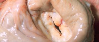
Lens displacement
Displacement of the lens occurs with blunt trauma to the eye. Here, damage to the ligament of Zinn occurs, unevenness of the anterior chamber appears, which leads to trembling of the iris (iridodonesis). The edge of the dislocated lens is visually determined. With ophthalmoscopy, we see two optic discs.
Displacement of the lens is combined with cataracts of traumatic origin, secondary glaucoma, and iridocyclitis. In the case of complete separation of the ligament of Zinn, the lens is dislocated and displaced into the anterior chamber of the eye, and sometimes into the vitreous body. At the same time, the anterior chamber of the eye deepens, the pupil narrows. In such cases, iridocyclitis and an acute attack of glaucoma suddenly appear.
The main symptom of lens displacement is iris trembling, or iridodonesis. Iris tremors can sometimes be seen with the naked eye. But it is better to observe this in the light of a slit lamp. But iris trembling may not always occur. In such cases, symptoms such as different depths of the anterior and posterior chambers of the eyes due to pronounced pressure, as well as movement of the vitreous anteriorly, to where the support of the lens is weakened, help to identify the displacement of the lens.
Lens subluxations most often occur in the upper inner quadrant. When the lens is dislocated, degenerative processes develop, followed by clouding, and in rare cases, its complete resorption.
Treatment of the eye after injury
Since with uncomplicated displacement of the lens, vision decreases insignificantly, no treatment is required. But in case of complications as a result of eye injury (lens opacification, secondary glaucoma), it is required to remove it and replace it with an artificial lens.
Hemorrhage into the vitreous body of the eye: symptoms and treatment
A blow to the vitreous area results in hemorrhage. Blood in the retrolental space of the vitreous expands it, and blood in the orbicular space leads to the formation of a specific rim (stripe) that surrounds the periphery of the lens behind.
Retrolental hemorrhage takes longer to resolve than orbicular hemorrhage. Sometimes minor hemorrhages may be invisible and are discovered later, when they “descend” to the lower part of the anterior chamber.
Hemophthalmos is understood as massive hemorrhage into the vitreous body, occupying a significant part of the latter.
Approximately on the third day after injury, the blood in the vitreous body undergoes a process of hemolysis with the loss of hemoglobin by red blood cells, as a result of which they become colorless and later disappear. And the hemoglobin of erythrocytes takes on the form of grains, which are subsequently absorbed by phagocytes. Hemosiderin is formed, which has a toxic effect on the retina. Sometimes the blood does not completely resolve and a blood clot is formed and replaced by connective tissue moorings. The clinical picture of hemophthalmos is dominated by loss of visual acuity from the state of light perception to complete blindness. Focal illumination and biomicroscopy make it possible to see behind the lens a dark brown granular, sometimes with a reddish tint, mass of blood that permeates the vitreous body. Ophthalmoscopy shows the absence of a fundus reflex. Later, when the blood clot dissolves, one can observe deformation of the vitreous with its liquefaction. Hemophthalmos must be distinguished from partial hemorrhage into the vitreous body, which quickly and completely resolves.
Hemophthalmos leads to the development of degenerative processes in the vitreous body.
Treatment
In case of vitreous hemorrhage, bed rest and a cold bandage are prescribed on the affected eye. Calcium preparations (tablets, eye drops and intramuscular injections), hemostatic agents (Vicasol) are used. To speed up the resorption of hemorrhage, heparin is used (subconjunctivally on days 1–2) and enzyme preparations and potassium iodide. Treatment of hemophthalmia: bed rest with the head end elevated. Use a binocular bandage for 2–3 days. Calcium chloride, pilocarpine 1% 2 times a day, glucose with ascorbic acid are used, and a solution of dicinone (12.5%) is injected subconjunctivally. Then, after 2-3 days, absorbable drugs are used: dionin, potassium iodide, lidase. Corticosteroids (under the conjunctiva) and fibrinolysin are also indicated. In the late period of treatment, ultrasound and physiotherapy help a lot. If there is no positive effect from therapy, then it is necessary to suction the vitreous, and sometimes excise part of it. The removed vitreous body is replaced with luronite (a hyaluronic acid preparation).
We must not forget that with somatic diseases (cardiovascular diseases, atherosclerosis, hypertension, blood diseases, endocrine pathology), the development of hemophthalmos is possible. But in these diseases, hemophthalmos occupies a small part of the vitreous body.
The prognosis depends on the area of hemorrhage. If the hemorrhage covers 1/8 of the vitreous body, it often resolves. With an area of 1/8-1/4 of the vitreous, moorings are formed, which leads to retinal detachment. In terms of restoring visual functions and preserving the organ in total hemophthalmia, when a blood clot occupies more than ¾ of the vitreous, the prognosis is unfavorable. Irreversible destructive changes occur in the vitreous body (organization of a blood clot, formation of adhesions). Tractional retinal detachment and atrophy of the eyeball may develop.
Diagnostics
First, the doctor asks the patient under what circumstances the injury occurred and how much time has passed since that moment. After examining the eyeball, sanitation is carried out - treating the tissues with a disinfectant solution.
In case of severe damage, hardware diagnostics are prescribed:
- biomicroscopy of the eye;
- ophthalmoscopy;
- gonioscopy;
- visometry;
- radiography of the facial part of the skull;
- MRI of the head;
- Ultrasound of the eye;
- non-contact tonometry.
Biomicroscopy is performed using a slit lamp. This is a quick, painless and effective method for diagnosing the eyeball. The ophthalmologist receives detailed information about the condition of the fundus, macula, retina, lens, corneal layer and other structures of the visual apparatus.
Ophthalmoscopy is an examination of the fundus of the eye using a beam of light that is directed onto the retina through the pupil. The ophthalmologist receives information about the condition of the retina, optic nerve and vascular system of the eyeball.
Gonioscopy - testing intraocular pressure, damage to the optic nerve, blockage of the tear ducts.
Visiometry is a test of visual acuity. Using the technique, you can determine the condition of the lens (presence of clouding), refractive error, pathology of the optic nerve and retina.
X-ray of the facial part of the skull is indicated for traumatic brain injuries that lead to eye contusion.
MRI of the brain shows the presence of internal hematomas and hemorrhages after receiving a strong blow to the head from a fall or other means. Sometimes, for a more accurate result, contrast is used - the introduction of a contrast liquid to highlight blood vessels and other structures.
Ultrasound of the eye is prescribed to clarify the condition of the visual apparatus after injury. The result gives an idea of the presence of hemorrhages, intraocular formations, the condition of the lens, and the nature of injury to the eyeball.
Non-contact tonometry gives an idea of the condition of the corneal layer. During the examination, a directed air flow is used.