This program can be prescribed only after consultation with a neurologist.
The regulation of all life processes depends on the functioning of the brain. Unfortunately, the most common cause of premature death is vascular disease. The final “blow” to the body is usually a stroke. In 90% of cases, patients who have suffered this disorder are unable to take care of themselves. There is practically no successful treatment for the consequences of a stroke, but there are effective prevention methods that improve cerebral circulation. Such measures will significantly reduce the risk of stroke.
Why does circulatory disturbance occur in the vessels of the brain?
In medical language, the deterioration of cerebral blood supply, which has become chronic, is called “dyscirculatory encephalopathy.” It causes various pathological processes in the central nervous system, often irreversible. According to doctors, this disorder is caused by atherosclerosis or arterial insufficiency. But there are also other fairly common reasons:
- constant stress;
- hypertension;
- chronic fatigue syndrome;
- scoliosis;
- constant excessive physical activity;
In addition, osteochondrosis sometimes provokes discirculatory encephalopathy. Sedentary work or lack of physical activity also significantly impedes the flow of blood through the vessels. Therefore, to improve blood circulation in the brain, it is advisable to do exercises in the morning and periodically warm up during the day.
Symptoms of poor circulation
The first signs are invisible to an uninformed person. However, over time, the progression of the pathology leads to discomfort and pain. Ignoring the following symptoms entails serious consequences and an advanced stage of the disease:
- chronic headache;
- causeless vomiting and nausea;
- pain in the eye area when moving them;
- causeless cramps and numbness of body parts;
- loss of consciousness;
- congestion and ringing in the ears;
- biased perception of reality, even mental disorders;
- chills, fever and pressure changes.
It is very important to recognize the signs of cerebrovascular accident in time. The success of treatment depends on this. The course of therapy is prescribed only by a specialist in this field. Treatment may be with oral medications or injectable medications. Prices for this type of procedure can be found below on the website.
Vasoactive drugs in the treatment of ischemic retinal diseases
Various diseases of the retina and optic nerve, caused by both general and local circulatory disorders, occupy a significant place among the causes of visual disability. Despite the undoubted successes in the treatment of vascular pathology of the eye, achieved in the last two decades, the number of patients with lesions of the optic nerve and retina continues to increase. This increase in the number of vascular diseases of the eye is directly related to the widespread prevalence of hypertension, atherosclerosis and diabetes,
which are practically impossible to cure. Existing treatment methods are aimed only at slowing down the development of the pathological process or stabilizing the functions of the affected organs. Ischemia is the most common pathological process that accompanies or causes ocular pathology.
As in other areas of practical medicine, in ophthalmology two main forms of the course of the ischemic process can be distinguished - acute and chronic
. This requires a different approach to the choice of treatment methods, as well as medications.
The use of vasoactive drugs as one of the links in the complex treatment of ischemic processes in the optic nerve and retina requires a careful and individual approach.
Drug treatment aimed at dilating blood vessels is often insufficient to improve blood circulation, and sometimes can even worsen ocular ischemia due to the development of “steal” syndrome. In this regard, it is necessary to increase perfusion, especially at the level of precapillaries and the arteriolar part of the capillary network.
The drugs most often used in the complex treatment of ischemic diseases are :
• improving blood circulation in the cerebral vascular system
(vinca preparations, pentoxifylline, nicergoline, cinnarizine);
• agents that dilate peripheral vessels and improve blood circulation at the capillary level
(xanthine nicotinate);
• vitamins
;
• means that improve redox and metabolic processes in the body
;
• biostimulants
;
• antioxidants, etc.
Drugs with a vasodilating effect are widely used to treat cardiovascular diseases. Vasodilators
divided into three main groups:
• direct acting vasodilators;
• calcium antagonists;
• blockers of postsynaptic a1-adrenergic receptors.
Among the direct acting vasodilators,
which in turn can be arterial, venous and arteriolar-venous, in practical ophthalmology venous vasodilators (nitroglycerin and other nitrates) were previously used for acute ischemia. Nitrates relax the smooth muscles of arteries and veins. But there is no accurate data yet that this improves the perfusion of the vessels of the eye. According to some data, under conditions of general redistribution of blood flow, the blood supply system of the eye may fall into the “stealing” zone. Doses of nitroglycerin that do not cause changes in systemic blood pressure (BP) often lead to dilation of arterioles in the face and neck, a rush of blood to the head and headache due to dilation of the meningeal vessels. According to M.M. Krasnov, a narrowing of the visual field and an increase in scotomas are often observed when taking nitrates.
The use of drugs acting through local factors that help reduce vascular tone and improve hydrodynamic parameters of the blood turned out to be more justified. Myotropic vasodilators have been used for many years to enhance peripheral circulation in areas where it is impaired due to acute or chronic vasoconstriction or spasm. Among the vasodilators currently most widely used in ophthalmology are nicotinic acid, xanthinol nicotinate, vinca preparations, and pentoxifylline.
. Using rheoophthalmography, a fairly high degree of vasodilating effect of the listed drugs on the vascular tract of the eye has been repeatedly proven.
Vinca preparations have a pronounced vasodilating effect on intraocular vessels.
, while they act anabolically, enhance the absorption of oxygen by brain cells and have an anti-aggregation effect. The therapeutic effect of vinca preparations develops gradually. In our clinic, vinca preparations were used for the treatment of acute (ischemic neuropathies, occlusion of the branches of the central retinal artery) and chronic (glaucomatous optic atrophy, dry form of senile retinal degeneration) ischemic diseases, 10 mg intravenously 1 time per day for 7–10 days . Upon discharge, the drug was prescribed 5 mg tablets 3 times a day for 2–3 months.
Many authors have noted the most pronounced vasodilatory effect when using drugs containing nicotinic acid
.
However, many authors consider the effectiveness of vasodilators in cerebrovascular insufficiency to be questionable. Myotropic vasodilators are capable of increasing cerebral blood flow under physiological conditions. With vascular pathology, which often accompanies ocular ischemia or is its cause, the affected areas of the vessels react less strongly to them. In addition, it is known that in poorly perfused areas, blood circulation can improve by increasing blood pressure. When using drugs of this group, especially intravenously, there is a drop in systemic blood pressure, which can lead to the development of “steal” syndrome.
For the same reasons, some ophthalmologists express doubts about the advisability of prescribing vasodilators for glaucoma, arguing that a decrease in systemic blood pressure can lead to a deterioration in the blood supply to the eye due to a decrease in perfusion pressure. However, most studies have found that blood supply to the eye improves under the influence of vasodilators when systemic blood pressure decreases by no more than 10% from the initial level. A more significant decrease in blood pressure leads to a deterioration in the blood supply to the eye in some patients.
Considering that the effect after
lasts
for 2–8 months
; courses of drug treatment must be carried out 2–3 times a year.
In addition, other methods of therapy have been developed based on improving the rheological parameters of blood (increasing the deformability of erythrocytes, reducing their aggregability, etc.). Many of the modern drugs used to treat ischemic conditions of the eye combine several of these properties simultaneously.
Quite widely in ophthalmology, purine derivatives are used to treat ischemic conditions of the posterior segment of the eye. Xanthinol nicotinate
reduces the level of fibrinogen in the blood and the aggregation ability of platelets, reduces blood viscosity. With long-term use of xanthinol, nicotinate helps lower blood levels of cholesterol and atherogenic lipoproteins. The drug can be used (as part of a complex treatment) intravenously (preferably by drip, 300 mg per 200 ml of saline once a day), intramuscularly (300 mg once a day) and parabulbarly (75 mg each) for 7–10 days, after which you should switch to the tablet form for 2–3 months.
Pentoxifylline is also a purine alkaloid.
. The classification of pentoxifylline into the group of vasodilators is very conditional, since it has a mild vasodilator effect, with almost no effect on hemodynamic parameters. The action of pentoxifylline is based on its effect on the hydrodynamic properties of blood. This drug acts on the rheological properties of the blood, on the one hand, and on tissue metabolism, on the other. In a hospital setting, it is advisable to prescribe pentoxifylline once a day intravenously, 5 ml of solution (100 mg) and/or parabulbar 10–15 mg for 7–10 days. After discharge from the hospital, the drug is prescribed at a dose of 400 mg 3 times a day for 2–3 months.
Another drug that deserves attention is picamilon, which consists of two biogenic components - GABA and nicotinic acid.
Among the many properties of picamilon, the most important are its antihypoxic, antioxidant, and vasodilating properties. In addition, it has been proven that picamilon reduces platelet aggregation ability and has anticoagulant and antiatherosclerotic effects.
The therapeutic activity of picamilon was studied in the treatment of compensated glaucoma, retinal pigmentary abiotrophy, ischemic neuropathy, and central chorioretinal dystrophy. According to a number of authors, after a course of treatment with picamilon (0.02 g 3 times a day orally), in 34-45% of patients, depending on the stage of the disease, there was an improvement in eye hemodynamics, an increase in light sensitivity, an expansion of the peripheral boundaries of the visual field, and a decrease in the area with a cat.
A relatively new and promising direction in vascular therapy, in our opinion, is the use of calcium antagonists for the treatment of acute and chronic vascular pathology of the posterior part of the eye. The most interesting for research is nimodipine
, which is so far the only drug from this group that has a cerebral effect. There are isolated reports of the use of nifedipine, isradipine, diltiazem and verapamil in tablet form in the treatment of open-angle compensated glaucoma. Studies have proven the positive effect of calcium channel blockers on ocular hemodynamics. A decrease in peripheral vascular resistance and an increase in perfusion pressure in intraocular vessels were noted. Visual functions stabilized or improved. In 6 patients, we successfully used nimodipine in the complex treatment of acute ischemia (occlusion of the branches of the central retinal artery and ischemic neuropathy).
It is necessary to say a few words about the methods of administering drugs.
Intravenous and parabulbar methods of administering vasoactive drugs are traditional in ophthalmology. However, a number of disadvantages of these methods reduce the effectiveness of the use of drugs. In particular, intravenous administration of vasodilators to a greater extent than local (retro- and parabulbar, subconjunctival) affects blood pressure, which increases the likelihood of deterioration of blood circulation in the eye due to a decrease in perfusion pressure. With parabulbar injections, the main part of the drug enters the orbital tissue, where there are many blood vessels and the drugs are quickly carried out by the blood from the eye area.
In 1991, A.P. Nesterov and S.N. Basinsky proposed a new method - the introduction of drugs into Tenon’s space using sub-Tenon implantation of a collagen infusion system
(SIKIS), which was mainly used to treat glaucomatous optic atrophy. In our studies, we compared the effectiveness of three methods of drug administration - intravenous, parabulbar and using SICIS. The effectiveness of vasoactive drugs was highest after the use of a collagen infusion system. The use of SICIS made it possible to achieve the maximum effect of drugs introduced into Tenon's space.
Thus, the use of vasoactive drugs in the complex treatment of ischemic diseases of the retina and optic nerve can be considered necessary.
Literature
1. Bunin A.Ya., Conde L.A. On the critical level of systemic blood pressure when prescribing vasodilators to patients with glaucoma // Vestn. ophthalmol. 1983; 3: 17-20.
1. Bunin A.Ya., Conde L.A. On the critical level of systemic blood pressure when prescribing vasodilators to patients with glaucoma // Vestn. ophthalmol. 1983; 3: 17-20.
2. Krasnov M.M. To analyze the features of intraocular hemodynamics and the possibility of therapeutic effects on it in glaucoma and blood supply deficiency. // Vestn. ophthalmol. 1989; 6: 36-43.
3. Metelitsa V.I. Cardiologist's Handbook of Clinical Pharmacology. M. 1987; 367 pp.
4. Egorov E.A., Svirin A.V., Puzakov V.P. The use of Cavinton in the complex treatment of glaucoma and some other eye diseases // New drugs used in ophthalmology. M. 1978; 62-4.
5. Katsnelson L.A., Mikhailova N.A., Gurtovaya E.E., Yakovlev A.A. Results of an experimental and clinical study of the drug trental//Vestn. ophthalmol. 1980; 1:41-3.
6. Tikhomirova N.A., Sukhorukova A.Yu. Local use of the drug “Cavinton” for vascular eye diseases. – In the book: Pathology of the fundus. Abstracts of conference reports. M. 1986; 50-1.
7. Konde L.A., Yakovlev A.A. On the mechanism of therapeutic action of a new drug complex aimed at stabilizing visual functions in patients with primary glaucoma // Pathophysiology and biochemistry of the eye. M. 1986; 25-30.
8. Konde L.A., Yakovlev A.A. Experience of using Cavinton in the treatment of glaucoma // Glaucoma. M. 1984; 59-61.
9. Gabrilyan E.S., Amroyan E.A., Akopov S.E. Physiology and pharmacology of the vascular wall. - Yerevan. 1987; 279 p.
10. Mashkovsky M.D. Medicines. M.: Medicine. 1993; 738 pp.
11. Ishchenko M.M., Dorogiy A.N. The effect of complamin on systemic and cerebral hemodynamics in patients with atherosclerosis and chronic cerebrovascular accident // Neuropathology and Psychiatry. 1985; 1: 17-20.
12. Stefanovich V. On the question of the biochemical mechanism of action of pentoxifylline // In collection. Clinical significance of the drug trental. M. 1977; 16-8.
13. Kruger A, Matulla B, Wolzt M. et al. Short-term oral pentoxifylline use increases choroidal blood flow in patients with age-related macular degeneration // Arch. Ophthalmol. 1998; 116 (1): 27-30.
14. Bondareva G.S., Umovist N.M. On the differentiated use of trental, complamin, solcoseryl and increased doses of vitamin B6 in the complex treatment of retinal dystrophy. Current issues in the pathology of the posterior part of the eye. Abstracts of reports. – Odessa. 1989; 107-8.
15. Davydova G.A., Kolomoitseva E.M., Eliseeva E.G., Pereverzina O.K. Results of the use of picamilon in some eye diseases // Picamilon in medical practice. M. 1997; 50-5.
16. Kolomoitseva E.M., Mukha A.I. On the effect of picamilon on the visual functions of patients with open-angle glaucoma // Picamilon in modern neurological and psychiatric practice / Proceedings of the Russian Conference. Moscow, November. 1994; 157-160.
17. Efimova M.N., Yakubova L.V. The influence of the calcium antagonist nifidipine on the visual functions of patients with primary glaucoma // Issues of ophthalmology. Omsk 1994; 137-9.
18. Efimova M.N., Yakubova L.V. Immediate results of the influence of systemic administration of calcium antagonists on the visual fields of patients with primary open-angle glaucoma with normalized pressure // Glaucoma. M. 1994; 82-4.
19. Yakubova L.V. The use of vasoselective calcium antagonists in the complex treatment of patients with primary open-angle glaucoma with normal intraocular pressure. Dissertation of a candidate of medical sciences. M. 1995; 146 p.
20. Brand L., Anderson K., Junegreen B. et al. Cerebrovascular and cerebral effects of nimodipini – an update // Acta Neurochir. 1988; 45 (1): 11-20.
21. Netland PA, Chaturvedi N., Dreyer EB Calcium channel blockers in the management of low-tension and open-angle glaucoma // Am. J. Ophthalmol. 1993; 115: 608-13.
22. Varno M., Pecori Giraldi J., Covelli GP et al. Effetti della nimodipina sulla sensibilita retinica nella neurotticopatia glaucomatosa // Bollettino di oculistica. 1990; 69 (5): 449-55.
23. Flammer J. Psychophysical mechanisms and treatment of vasospastic disorders in normal-tension glaucoma // Bull. Soc. Belge. Ophthalmol. 1992; 244: 129-34.
| Applications to the article |
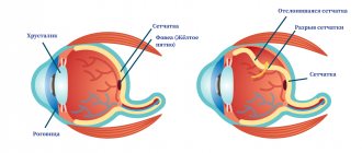
| “Flying floaters” before the eyes are not dangerous and do not require treatment, but their sudden appearance in a myopic eye can be a symptom of incipient retinal detachment and requires immediate examination by an ophthalmologist. |
| The maximum time for continuous work at a computer is 10–15 minutes for younger schoolchildren, 25–30 minutes for older students, and 3 hours a day for senior students. |
| The desirable medicinal effects of tricyclic antidepressants (amitriptyline, etc.) include tear hyposecretion, pupil dilation, accommodation paresis, increased IOP, and even an acute attack of angle-closure glaucoma. |
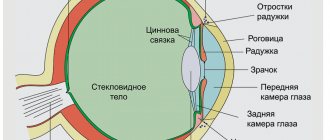
| Ischemia is the most common pathological process in ocular pathology |
| Administration of drugs using sub-Tenon implantation of a collagen infusion system can enhance and prolong their effect |
Droppers to improve cerebral circulation
Treatment is prescribed by a specialist only after a series of examinations. Injections and drips are considered one of the most effective methods of combating stroke. The action of the main part of vasodilator drugs is focused on improving blood flow to brain cells. Droppers to improve blood circulation in the brain are placed only in a hospital under the supervision of a doctor.
The pharmacological action of the drugs makes it possible to prevent oxygen starvation, which is one of the main causes of stroke. Injections also relieve nutritional deficiencies, improve energy metabolism in brain cells and normalize brain functions. The duration of the course depends on the patient’s condition and the stage of the disease.
Treatment regimens for retinal angiopathy
Drug therapy in the case of angiopathy and its vascular complications should be comprehensive. If necessary, treatment can be continued in courses. Before prescribing it, an ophthalmologist develops a specific, most effective regimen for using drugs. As a rule, these are the following appointments:
- Drug therapy aimed at activating blood circulation in the vessels of the eyes is necessary. This is a course treatment with Emoxipin, Vazonit, Mildronate, Arbiflex, Solcoseryl, Trenatal. They help activate local microcirculation processes. Another property of these drugs is improving the plasticity of red blood cells. This allows them to move faster through the small vessels of the eyes.
- Pentoxifylline and Curantil help prevent possible thrombus formation. Xanthiol nicotinate and nicotinic acid improve blood rheology.
- To reduce vascular permeability, ginkgo biloba and calcium dobesilate are prescribed.
- Actovegin injections are designed to provide better nutrition to the eye tissues. To improve metabolic processes in eye tissues, ATP and cocarboxylase are used.
- It is mandatory to take vitamin complexes to maintain eye health - Lutein-intensive, Anthocyan Forte. It is important to take ascorbic acid, which accelerates microcirculation processes in blood vessels and maintains visual acuity.
- For diabetic retinopathy, it is necessary to adhere to a special diet that can neutralize diabetes mellitus and improve blood flow in the retinal vessels. It is worth remembering that prohibited foods include any food rich in fast carbohydrates (baked sweets, sweet soda, etc.). The same applies to excessively high-calorie foods. Limiting salt is also important, because it helps normalize metabolism and improve recovery processes.
- Vigorous physical exercise, included in the daily routine to give energy to the muscular system, also improves the condition of the eye vessels.
- When treating angiopathy caused by hypertension, it is mandatory to lower blood pressure levels through medications and specially designed dietary adjustments. To do this, you should contact a cardiologist and nutritionist.
- It would not be superfluous to prescribe physiotherapeutic procedures. In the case of angiopathy, magnetic therapy courses and acupuncture can show good results.
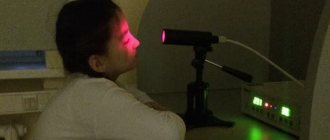
- Another physiotherapeutic method is “Sidorenko Glasses”. This device combines the effects of pneumomassage, infrasound, phonophoresis, and color therapy. The powerful effect on the retina of such a complex makes it possible to achieve good results in a short time.
- Treatment also helps with massage courses on the cervical spine.
Injections to improve cerebral circulation
In some cases, injections are given intramuscularly. These may be medications that affect metabolic functions in the nervous system and stimulate brain activity. They speed up the process of transmitting impulses through nerve cells, improve attention, memory and eliminate various brain lesions. Injections that improve cerebral circulation can be done at home. However, a prescription for drugs can only be written by the attending physician. Never buy medications yourself!
You can undergo an examination of the brain and its vessels and have an MRI of the brain with angiography done at the diagnostic and treatment center. Modern high-precision equipment will produce the most accurate results. The center’s doctors will study the diagnostic results and, if necessary, prescribe medications that improve cerebral circulation. You can find out the cost on the official website of the center, or by calling the contact phone numbers at the reception.
The program includes:
Intraocular injections
Intravitreal injections are the injection of a drug directly into the eye cavity (into the vitreous body). Such injections are performed for hemorrhages (partial hemophthalmia), wet form of macular degeneration, retinal edema, the appearance of newly formed vessels and other serious eye diseases.
In our clinic, intravitreal injections are performed by ophthalmologists with extensive experience in complex intraocular injections in sterile procedure rooms, in compliance with appropriate medical protocols. Such therapy for retinal pathologies, using effective medications, allows one to preserve visual function and also achieve a significant improvement in visual acuity.
Lucentis
The active component of the drug is ranibizumab, a substance that suppresses excessive angiogenesis (the formation of pathological blood vessels), which is characteristic of age-related macular degeneration. In addition, it helps relieve macular edema, normalizing the thickness of the retina. The drug quickly penetrates completely into the layers of the retina, reducing the size of the lesion, preventing possible hemorrhages and further progression of the growth of newly formed vessels with a pathological wall. More about Lucentis >>>
Eilea
The active substance of Eylea is aflibercept. Its action is aimed at inhibiting the progression of age-related macular degeneration (wet form). In addition, it has the ability to act effectively and safely in reducing visual acuity in the case of diabetic macular edema and edema caused by retinal vein occlusion.
The innovative drug Eylea has a long-lasting effect, so injections can be performed less frequently than with Lucentis, which is much more convenient for patients. More about Eylea >>>
Ozurdex
It is used in cases of macular edema caused by occlusion of the retinal veins. The drug is produced in the form of a thin implant in the form of a rod, which is inserted into the vitreous body. The implant contains the potent steroid dexamethasone, which begins to be released inside the vitreous in small portions. This innovative method of dosed delivery of a medication to the affected area significantly increases the duration of action of the hormone, which can last up to six months. Read more about Ozurdex >>>
Parabulbar and intravitreal injections are performed under sterile conditions