Make an appointment Inflammation of the prostate gland is a common urological disease that affects men of reproductive age. Bacterial prostatitis is an inflammation caused by pathogenic microflora. It enters the prostate gland from the environment or moves from other pathogenic foci through the circulation of lymph and blood. In the absence of timely treatment, the disease quickly passes from the acute form to the chronic phase. A urologist is involved in eliminating this pathology.
If you require treatment for your prostate, see a specialist immediately.
Features of diagnosis and treatment of chronic prostatitis - yesterday and today
The content of the article
Inflammation of the prostate gland (lat. Prostate) is a serious clinical problem, especially its chronic form. The hidden nature of the disease, of course, is perennial, and the many somatic and psychological complications make this disease an interdisciplinary problem in medicine.
Inflammation of the prostate gland
Despite significant progress in diagnosis, pharmacotherapy, physiotherapy and rehabilitation, treatment results are often unsatisfactory. Considering that almost every second man has had or has had prostatitis, the problem is of a social nature.
As a clinical manifestation, prostatitis appears in nineteenth-century literature. In the early years of this century, factors contributing to chronic prostatitis were reported to include alcohol abuse, frequent horse riding, colds, and sexual abuse. Some sexually transmitted diseases are also associated with the process, for example, gonococcal urethritis.
For many years, scientists incorrectly associated prostatitis solely with a bacterial infection. The currently noted high progress in microbiological, microscopic, histological and clinical diagnostics refutes these views and allows in many cases to clearly determine the cause of the disease, which is not always caused by bacteria.
Therefore, types of prostatitis were distinguished depending on the etiological factor or its absence. This served as the basis for the creation of the school in 1968. Meres and Staemi classification of prostatitis. In 1995, this system was modified based on the latest advances in modern medicine at the US National Institutes of Health.
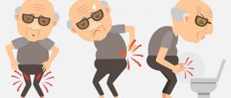
What is the difference between acute and chronic prostatitis?
The symptoms of acute prostatitis and its treatment do not cause serious problems, but the chronic disease is one of the most difficult and controversial both from the point of view of diagnosis and from the point of view of treatment. This situation appears to be due to the fact that it has long been believed that such a small organ as the prostate should not attract much scientific attention. After all, no one has yet been recognized as disabled due to chronic prostatitis!
However, recently chronic prostatitis has received a lot of attention, especially abroad. In the United States, it is estimated that a quarter of all men who see a doctor each year suffer from prostatitis.
Prostate diseases are associated with privacy, so the doctor must be very careful when talking with the patient. But most patients do not understand the essence of this disease, and therefore are dissatisfied with the treatment and the doctors themselves. They, in turn, try to send dissatisfied patients to another place. Many patients believe that this is an incurable disease, so there is no need for treatment, and they come to their senses when their symptoms worsen significantly.
Men are the most unbalanced and are concerned about the deterioration of potency as a result of untreated or poorly treated chronic prostatitis. This complication not only destroys family life, but also often changes the character of a man. Having lost the joy of life, such a patient becomes a social problem.
Features of the structure and functions of the prostate gland - why this organ is called the second heart
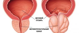
The anatomical relationship of the male reproductive system, the outflow of sperm and urine is due to the origin of most of these structures from a common rudiment - the endorenal duct. From it the epididymal ducts, vas deferens, ejaculatory ducts, seminal vesicles, ureters, and prostatic urethra are formed. Only the prostate gland and testes originate from other points. The prostate forms in the form of numerous vesicles from the posterior urethra, which develops from the urogenital sinus.
The prostate has the shape of a chestnut and, surrounding the urethra at the base of the bladder, lies on the urogenital diaphragm. The gland, covered on the outside with a thick connective tissue sac, is a tubular-alveolar organ with numerous smooth muscle fibers.
Anatomically, the organ is divided into right and left lobes, and clinically there are two lateral lobes and one middle lobe. The prostate is vascularized by branches of the inferior arteries of the bladder, the arteries of the internal genital organs and the inferior rectal arteries. Innervation comes from the fibers of the autonomic system from the pelvic plexus.
Pancreatic secretion makes up 15% of the sperm composition. The secretion contains a number of active enzymes with highly active proteolytic factors such as fibrinolysin and fibrinogenase. Prostate secretion components also include diastase, β-glucuronidase, fructose, transaminases and amino acids. The prostate is the main producer of citric acid and acid phosphatase in human sperm. The secretions also contain spermine and spermidine and an antimicrobial zinc-containing polypeptide. Immunoglobulins are synthesized locally.
The main task of the prostate gland and seminal vesicles, like Cooper's glands, is to create seminal fluid along with the secretion of the testicle and epididymis. It provides transportation and protects the metabolism and motor activity of sperm.
The mixing of these ingredients occurs during ejaculation. The first portion of ejaculate, which does not coagulate or liquefy immediately, contains products of the Cooper and prostate glands. The last part of the ejaculate liquefies very slowly, about 15-20 minutes, and contains products of the seminal vesicles, including fructose. The first fraction includes activators of lysozyme, alpha-amylase, spermine and plasminogen.
Seminal protein concentration is half that in plasma and is 4.0 grams%. This is mainly albumin, the level of which is 140 milligrams% of the quantitative ratio of the secretion of the prostate gland to the secretion of the seminal vesicles. Besides proteins and enzymes, there are also their coenzymes, which are metals such as zinc, magnesium, calcium and selenium.
Systemic humoral influence is mediated by cellular receptors specific for testosterone, FSH and LH. In the most general form, they set the pace and direction of many biological reactions.
Nutrition
This factor also plays a big role; a proper diet will help you recover faster from illness and reduce the risk of relapse. It is recommended to select food according to the following principles:
- products containing all the microelements necessary for the body;
- food should be well digestible and not heavy;
- foods high in fiber stimulate the digestion process and prevent intestinal dysfunction;
- Monitoring your drinking regime will help prevent dehydration.
The main task when creating a diet is to normalize intestinal function, since constipation is one of the causes of exacerbations of prostatitis.
The prostate has a defense mechanism
The prostate gland is physiologically protected from infection. Defense mechanisms include mechanical factors, antibacterial substances, cellular and humoral factors.
- Mechanical ones include: the length of the urethra, urination, ejaculate and the characteristic location of the canals.
- Antimicrobial substances are: prostate antimicrobial factor containing zinc and spermine, spermidine and lysozyme. The prostate contains the highest concentration of zinc in the body, which decreases with inflammation, leading to a local decrease in immunity.
- The cellular protection factor for prostate infection is the characteristic presence of leukocytes in urine, prostate secretions and semen. Increased phagocytosis in patients with infectious forms of prostatitis indicates a very important role of the cellular response in infections of the lower genitourinary system.
- In patients with prostate infections, titers of immunoglobulins in prostate secretions are elevated, most often IgA, IgG and IgM, and their levels are much higher than in blood serum.
Types of prostatitis and their distinctive characteristics
According to its course, prostatitis can be divided into acute and chronic. From the point of view of cytological and microbiological assessment of prostate secretion, the following can be distinguished:
- acute bacterial prostatitis;
- chronic bacterial prostatitis;
- chronic nonbacterial prostatitis;
- prostatodynia.
This classification was proposed by the scientist. Meares and Stamey in 1968
The classification of inflammation is an expanded form of the above section (Table 1). It specifically classifies non-bacterial chronic inflammation of the prostate as type III. This type of inflammation includes those whose symptoms last more than three months. Type III A is inflammatory and III B is non-inflammatory.
Table 1. Classification of prostatitis (according to NIH; US National Institutes of Health, 1995).
| Type I | Acute bacterial prostatitis (acute prostate infection) | |
| Type II | Chronic bacterial prostatitis (recurrent urinary tract infections, chronic prostate infection) | |
| Type III | Chronic nonbacterial prostatitis (chronic pelvic pain syndrome). — Pain and discomfort in the pelvis for at least three months with urination and sexual dysfunction — There are no signs of infection. | |
| III A. | Inflammatory, chronic pelvic pain syndrome. Increased leukocytosis of prostate secretions (VB 3). | |
| III B | Non-inflammatory chronic pelvic pain syndrome. Lack of leukocytes in prostate secretions (VB 3). | |
| Type IV | Asymptomatic prostatitis. Features of inflammation during prostate biopsy, sperm and prostate secretions (VB 3). No symptoms. | |
These two types of prostatitis are distinguished based on microscopic examination of prostate secretions. The presence and number of leukocytes are assessed as an indicator of inflammation.
In infectious forms of prostatitis, pathogenic microorganisms enter it through an ascending route. The common urinary and seminal tracts allow infection to spread from the posterior urethra into the gland.
Infectious prostatitis
Prostate infection usually occurs when the infection is transmitted from the urethra through urine. It mainly affects the posteroinferior and peripheral part of the prostate. The reason for this is the constant flow of urine stream at the prostate tubules. In the central, upper part of the urethra they pass perpendicularly or backward to the flow of urine.
Phimosis, a narrowing of the urethra or contraction of the external sphincter, contributes to urethral infections. As a result, pressure increases in the back of the urethra and urine flows out. The microbes are carried upward and carried in the urine into the prostatic tubules by backflow through the posterior urethra.
This mechanism can also cause chemical prostatitis. Thus, urates and creatinine can penetrate into the organ tissue. Increased levels of urate in the prostate can cause the formation of stones in the prostate gland, which contributes to the development of bacterial infection.
Bladder sphincter spasms may be a reaction to rectal pain associated with hemorrhoids or anal fissure (anogenital syndrome). Spasms of the external sphincter can be caused by neurosis and anxiety.
Inflammation mainly affects the lower peripheral part, in particular the clusters and ducts of the glands. Due to swelling or small deposits in the gland ducts that obstruct the flow of secretions, the infection is usually chronic.
Emergency care for urological diseases
Acute prostatitis is a nonspecific acute inflammation of the prostate gland.
Acute prostatitis develops as a result of a pathogenic infectious agent (E. coli, other Enterobacteria, Pseudomonas, Enterococcus and Staphylococcus) entering the prostate gland.
Ways of infection entering the prostate:
- canalicular - through the excretory ducts of the prostate (the source of infection is the posterior urethra);
- hematogenous - with congestion in the gland from hypothermia, constipation, prolonged sexual abstinence or excess, etc. (the source of infection is purulent foci in other organs);
- lymphogenous.
Acute prostatitis is divided into catarrhal, follicular, parenchymal.
The disease begins with frequent (5-7 times per night), painful at the end, difficult urination in small portions, with continued urge and pain in the perineum, which intensifies with defecation. In some cases, terminal hematuria appears, caused by the involvement of the mucous membrane of the neck and bottom of the bladder in the inflammatory process. Body temperature is high.
Possible complications of acute prostatitis:
- acute urinary retention;
- prostate abscess;
- paraprostatitis;
- phlebitis of the paraprostatic venous plexus.
Diagnostics
During the survey they find out:
- presence of chronic prostatitis;
- whether hypothermia occurred (swimming in cold water, etc.).
With prostatitis, urine reflux from the posterior urethra into the prostate occurs in response to increased intraurethral pressure.
A digital rectal examination reveals an enlarged, sharply painful prostate gland, sometimes with signs of fluctuation in one of the lobes. This study is performed very carefully; it requires sufficient experience from the doctor.
Laboratory diagnostics. A blood test (leukocytosis, band shift, increased ESR) gives grounds to judge the degree of the inflammatory process and purulent-inflammatory intoxication. Urinalysis reveals pyuria and bacteriuria, and it is especially important to examine the first portion of urine.
Acute prostatitis should be differentiated from various diseases that occur with fever, and primarily from paraproctitis. In differential diagnosis, the main role belongs to digital rectal examination.
Main directions of therapy
- Broad-spectrum antibiotics:
- aminoglycosides - gentamicin (gentamicin, gentamicin-K, gentina) 80 mg 2 times a day IM, amikacin (selemycin, amikin, amikozit) 500 mg 2 times a day IM;
- cephalosporins of the II, III generations - cefotaxime (Talcef, Ceftax) 1.0 g 2 times a day IM, ceftazidime (Cefadim, Fortum) 1 g 2 times a day IM), ceftriaxone (Medaxone, Oframax, Rocephin) 1 –2 g once a day IM and IV, cefoperazone (medocef, cefobid) 1–2 g 2 times a day IM, cefuroxime (Axetin, Zinnat, Supero) 750–1500 mg 3–4 times a day day IM and IV;
- parenteral fluoroquinolones (ciprofloxacin - medociprin, siflox, ciprolet) 50 ml of 0.2% solution 2 times a day i.v.), ofloxacin (geoflox, ofloxin) 200 mg in 5% glucose solution i.v., pefloxacin (abactal, perti) 250 ml in 5% glucose solution;
- in mild cases - fluoroquinolones orally: ciprofloxacin 0.5 g 2 times a day, ofloxacin 0.2 g 2 times a day, lomefloxacin (lomflox, maxaquin, xenaquin) 0.4 g 1 time a day, pefloxacin 400 mg 2 times a day day).The duration of antibacterial therapy should be at least 14 days.
- If there is no positive dynamics within 1–2 days, a cystostomy is performed to drain urine from the bladder (Fig. 1).
Clinical pharmacology of individual drugs
III generation cephalosporins (cefoperazone, cefotaxime, ceftazidime, ceftriaxone, etc.) are active against gram-positive and gram-negative bacteria. The drugs are prescribed 1–2 g 2 times a day parenterally.
Common therapy errors:
- Installation of a urethral catheter and other manipulations on the urethra - with the development of acute urinary retention, an emergency epicystostomy is necessary.
- Reducing the duration of antibacterial therapy increases the likelihood of recurrence of the disease and its transition to a chronic form.
Acute prostatitis, especially in elderly and senile people, as well as in the presence of severe hyperthermia with chills, requires urgent hospitalization in a urological hospital.
Analysis of clinical cases
Patient R., 19 years old. Complaints of frequent and painful urination, pain in the perineum, fever up to 38.5°C with chills. History: the day before I bathed in cold running water. In the evening of the same day, he noted an increase in temperature against the background of frequent and painful urination. On examination: the abdomen is soft and painless. Pasternatsky's symptom is negative on both sides. On digital rectal examination, the prostate is slightly enlarged in size, sharply painful, with areas of hardening and softening. Diagnosis: acute prostatitis. The patient was hospitalized in the urological hospital. Treatment: intensive antibacterial therapy.
Patient Z., 65 years old. Complaints of sharp pain in the anus and perineum, aggravated by defecation, frequent and painful urination, fever up to 39°C with chills. History: complaints appeared 5 days ago and grew progressively. Periodically, body temperature rose to 39.5°C with chills, then decreased, with increased sweating. On examination: the patient is pale, breathing heavily, heart rate 95 beats/min. Rectally, an enlarged prostate is detected, which protrudes into the intestinal lumen, is sharply painful and asymmetrical due to the right lobe, where fluctuation is determined. Diagnosis: acute prostatitis; prostate abscess. The patient was hospitalized in the urological hospital. Treatment: surgical drainage of the prostate abscess, antibacterial and detoxification therapy.
Acute epididymitis is an acute inflammation of the epididymis that can involve the testicle (epididymo-orchitis).
The causative agents of acute epididymitis are Chlamydia trachomatis, N. gonorrhoeae (in patients under 35 years of age) and E. coli, P. aeruginosa (in older patients).
Routes of infection:
- retrograde (from the urethra due to its bougienage, catheterization, endoscopic manipulation, etc.);
- from the prostate gland;
- hematogenous.
There are specific epididymitis (gonorrheal, chlamydial, trichomoniasis, tuberculous) and nonspecific.
The disease usually begins acutely, less often develops slowly. Fever appears, pain is localized in the testicular area, intensifying and radiating along the spermatic cord, due to swelling, the scrotum increases in size.
Possible complications of acute epididymitis:
- suppuration with the formation of an abscess of the epididymis and phlegmon of the scrotum;
- an increase in intoxication phenomena with the development of bacteriotoxic shock or urosepsis;
- fertility disorders, especially with a bilateral process.
Diagnostics
On examination, swelling and redness of the skin of the scrotum are noted. On palpation, the appendage is enlarged, compacted, and sharply painful. As the inflammatory process progresses and exudate accumulates in the membranes of the testicle, the testicle itself and the epididymis form a single conglomerate.
Laboratory diagnostics. A general blood test reveals leukocytosis and band shift with an increase in ESR; in some patients, leukocyturia and bacteriuria. Before starting antibiotic therapy, a urethral smear and midstream urine should be obtained for microbiological examination.
Acute epididymitis is differentiated:
- with torsion of the spermatic cord, in which the testicle is usually located above its normal position and is often turned transversely, the onset of the disease is sudden and is not accompanied by an increase in temperature;
- hydrocele;
- testicular injury;
- strangulated hernia.
Main directions of therapy
- Ice and a suspension are applied to the scrotum.
- Prescribe suppositories with diclofenac.
- Antibiotic therapy:
- young patients with mild epididymitis - fluoroquinolones (for example, ofloxacin 200 mg 2 times a day) or tetracyclines (for example, doxycycline - medomycin, tetradox, unidox solutab - 100 mg 2 times a day) orally;
- in severe cases and in older patients - aminoglycosides (amikacin, gentamicin) and fluoroquinolones parenterally (see acute prostatitis), then fluoroquinolones (for example, ciprofloxacin 500 mg 2 times a day) orally.
- When an epididymal abscess forms, surgical treatment is indicated.
The duration of the course of antibacterial therapy should be at least 14 days.
Common therapy errors:
- Discontinuation of antibacterial therapy after fever and pain have decreased.
- Continuation of antibiotic therapy as the only method of treatment despite the lack of effect for more than 3 days without additional diagnostics.
Hospitalization to a urological hospital is indicated for most patients with moderate to severe acute epididymitis, especially with signs of chronic prostatitis. Only young patients with mild epididymitis are treated outpatiently.
Analysis of clinical cases
Patient K., 25 years old. Complaints of sharp pain in the testicular area, fever up to 38.5°C. History: bruised scrotum a week ago while riding a bicycle. About 5 days ago I discovered a lump in the scrotum area, which was progressively increasing in size. The day before treatment, the temperature increased to 38°C. He suffers from focal pulmonary tuberculosis, for which he receives treatment irregularly. On examination: the right half of the scrotum is increased in size and moderately hyperemic. Palpation reveals an enlargement and thickening of the right testicular epididymis; examination is sharply painful. Diagnosis: acute right-sided epididymitis, possibly of tuberculous etiology. The patient was hospitalized in the urological hospital. Treatment: antibacterial therapy with broad-spectrum antibiotics. If the tuberculosis etiology of the process is confirmed, the patient is transferred to a specialized medical institution.
Patient D., 82 years old. Complaints of pain in the area of the right half of the scrotum, increased body temperature to 38°C, severe general weakness. History: 8 days ago there was pain in the right half of the scrotum and its enlargement. The pain progressively increased, weakness appeared, and the day before treatment the temperature was 38°C. On examination: the patient's condition is serious. The right half of the scrotum is significantly increased in size and is sharply painful on palpation. Palpation reveals a single conglomerate with foci of softening; it is impossible to differentiate the testicle and epididymis. Diagnosis: acute epididymo-orchitis; purulent melting of the testicle and epididymis. The patient was hospitalized in the urological hospital.
Acute urinary retention (AUR) is the accumulation of urine in the bladder due to the inability to urinate independently with a painful urge to urinate.
The causes of AUR can be different.
- Mechanical:
- benign hyperplasia and prostate cancer;
- acute prostatitis;
- sclerosis of the bladder neck;
- foreign body;
- stone and urethral rupture;
- neoplasm of the lower urinary tract;
- uterine prolapse.
- Diseases and damage to the central nervous system (tumor, injury, etc.).
- Reflex dysfunction of the bladder.
- Poisoning with psychoactive substances (hypnotics, narcotic analgesics).
Mechanical and dynamic mechanisms are involved in the pathogenesis of AUR.
In elderly men, in response to gradually increasing bladder outlet obstruction (mechanical factor), nervous regulation changes - the tone of the smooth muscle cells of the detrusor increases and the detrusor hypertrophies. The histomorphological structure of the bladder wall gradually changes: muscle elements are replaced by connective tissue, trabecularity develops. The volume of the bladder increases. The process enters the stage of decompensation - hypotension of detrusor smooth muscle cells develops (dynamic factor). In such a situation, any provoking factor (hypothermia, alcohol intake, spicy food, prolonged sitting position, constipation) causes venous stagnation in the pelvis, the veins of the bladder neck dilate, swelling of the prostate occurs, which, in turn, leads to deformation and compression of the prostatic parts of the urethra (mechanical component). Against the background of existing pathological changes in the detrusor, AUR develops.
Often, AUR in older people occurs after injection of atropine or its derivatives due to a decrease in detrusor tone, more often with an existing urological disease (for example, prostate adenoma).
Reflex AUR is more often observed after operations, especially in children, due to disruption of the nervous regulation of the detrusor and striated sphincter of the bladder. In addition, it can occur with trauma to the perineum, pelvis and lower extremities, with severe emotional shock, alcohol intoxication, fear, hysteria.
Clinical picture: the patient is restless, experiences severe pain in the suprapubic region, a painful urge to urinate, and a feeling of fullness in the lower abdomen.
In older men, AUR often becomes chronic and causes:
- inflammation in the urinary tract (infectious agents can be introduced during bladder catheterization);
- chronic cystitis and pyelonephritis;
- stone formation.
With a hypotrophied bladder wall, vesicoureteral-pelvic reflux develops, leading to bilateral urethrohydronephrosis and chronic renal failure.
Diagnostics
During the survey they find out:
- how the patient urinated before AUR;
- what color was the urine;
- whether he took drugs that promote AUR.
When examining patients with asthenic physique, the symptom of a “ball” in the suprapubic region is determined. Percussion - a dull sound above the bladder. Palpation is painful due to a strong urge to urinate.
Ultrasound allows you to determine the volume of urine in the bladder and the condition of the upper urinary tract.
Laboratory diagnostics. Clinical tests of blood and urine (obtained during catheterization) in new-onset AUR are usually normal.
In patients with severe cystitis, pyelonephritis, signs of inflammation are detected in a blood test, and an increase in leukocytes and fresh red blood cells in the urine.
In chronic renal failure, hemoglobin levels and the number of red blood cells in the blood are reduced, and the level of urea and creatinine in the blood serum is increased.
Acute urinary retention is differentiated from anuria: with anuria there is no urge to urinate, palpation of the suprapubic area is not painful.
With paradoxical ischuria, the bladder is full, the patient cannot urinate on his own, but urine is involuntarily released in drops. Once the urethral catheter releases urine, urine leakage stops until the bladder becomes full again.
Main directions of therapy
- Urgent emptying of the bladder by inserting an elastic catheter.
Contraindications to bladder catheterization:- acute urethritis and epididymitis (orchitis);
acute prostatitis and/or prostate abscess;
- urethral injury.
- If AUR lasts more than 2 days, a permanent urethral catheter is installed in the bladder.
- Antibiotics are prescribed to prevent inflammatory diseases of the scrotal organs and drugs from the α-blocker group (Table).
In this case, it is necessary to resort to puncture of the bladder, which is performed only in a urological or surgical hospital.
Drugs from the group of α-adrenergic blockers tamsulosin (omnic), doxazosin (zoxon, cardura, magurol), terazosin (setegis, hytrin, cornam), alfuzosin (dalfaz):
- relax the smooth muscle elements of the prostate (mechanical component of the prostate);
- improve blood microcirculation in the wall of the bladder, restoring the contractility of the detrusor (the dynamic component of the bladder).
Side effects of α-blockers:
- blocking α1-adrenergic receptors located in the stroma, prostate capsule, bladder neck, urethra;
- vascular dilatation;
- reduction of peripheral vascular resistance;
- decrease in blood pressure.
Common therapy errors:
- Self-medication, taking diuretics.
- Incorrect, inept catheterization and formation of false urethral passages. A metal catheter should not be used in the prehospital setting.
Urgent hospitalization to the urology department is indicated in the following cases:
- difficult first catheterization;
- urethrorrhagia, acute inflammation of the urethra, scrotal and prostate organs, urethral trauma;
- impossibility of inserting a catheter (you cannot make more than two attempts);
- unsuccessful repeated catheterizations of the bladder.
Analysis of clinical cases
Patient M., 77 years old. Complaints of sharp pain in the lower abdomen, inability to urinate independently. History: I took alcohol the day before. He did not urinate for about 12 hours. For 5 years he noted a deterioration in urination: a sluggish stream of urine, the need to strain when urinating, nocturia up to 2 times. I did not see a urologist. On examination: the patient is restless, holding his lower abdomen with his hands. The “ball” symptom is determined in the suprapubic region. Percussion - dull sound. On digital rectal examination: the prostate is enlarged 1.5 times, has a tight-elastic consistency, the median groove is smoothed. The rectal mucosa above the prostate is mobile. Diagnosis: benign prostatic hyperplasia, AUR. Treatment: catheterization of the bladder with a Nelaton catheter. It is recommended to take α-blockers, anti-inflammatory therapy, examination and observation by a urologist.
Patient Yu., 68 years old. Absence of urination for more than a day with the urge to urinate, no pain. History: three cases of acute urinary retention, which resolved after catheterization. During the last catheterization (6 months ago), the patient accidentally removed the catheter with an inflated balloon, after which he noted painful urination with blood at the beginning of urination. He has been suffering from benign prostatic hyperplasia for about 7 years. I took α-blockers, which had no effect in the last six months. On examination: the bladder is at the level of the navel, percussion - 10 cm above the womb. Palpation is sensitive, but does not cause sharp pain. On digital rectal examination: the prostate is enlarged 2–2.5 times, has a tight-elastic consistency, the median groove is smoothed. The rectal mucosa above the prostate gland is mobile. Diagnosis: benign prostatic hyperplasia, post-traumatic urethral stricture, AUR. The patient is indicated for hospitalization in a urological hospital to decide on the tactics of further treatment (if it is impossible to install a urethral catheter, perform a trocar cystostomy).
Patient T., 20 years old. Complaints about the inability to urinate independently, pain in the lower abdomen. History: the patient has not urinated for more than 20 hours. She experienced emotional stress, felt a strong urge to urinate, but was unable to visit the toilet, after which she was unable to urinate. On examination: the “ball” symptom is determined in the suprapubic region. Percussion reveals the bladder 8 cm above the pubis. Diagnosis: AUR of a neurogenic nature. Treatment: bladder catheterization. Further examination by a urologist and neurologist is recommended.
Anuria is the absence of urine in the bladder.
Risk factors for the development of prerenal anuria:
- decreased cardiac output (cardiogenic shock - cardiac infarction);
- systemic vasodilation (sepsis, neurogenic shock);
- hypovolemia and a sharp decrease in circulating blood volume: - blood loss; -plasma loss (with extensive burns); -dehydration (with vomiting, diarrhea, forced diuresis); -the emergence of a “third space” (with sequestration of fluid into the abdominal cavity - ascites, into the subcutaneous tissue - edema), etc.
Violation of general hemodynamics and circulation with a sharp depletion of renal blood circulation leads to renal ischemia; when it worsens, ischemic necrosis of the epithelium of the renal convoluted tubules occurs and prerenal anuria can develop into renal anuria.
Risk factors for the development of renal anuria:
- acute tubular necrosis, the most common causes of which can be:
- renal ischemia (with prolonged clamping of the renal artery, with thrombosis and thromboembolism of the renal vessels - intravascular block, renal hypoperfusion as a result of prolonged arterial hypotension - prerenal factor);
- nephrotoxic factors: - iodine-containing radiocontrast agents during angiography; - salts of heavy metals (lead, mercury, copper, barium, arsenic, gold); -antibiotics (aminoglycosides, amphotericin B); -organic solvents (glycols, dichloroethane, carbon tetrachloride); -uricuric crises - intrarenal occlusion of tubules by uric acid crystals during gout, chemotherapy for myelo- and lymphocytic leukemia, treatment with sulfonamides, etc.;
- acute and chronic end-stage renal failure due to glomerulonephritis, malignant arterial hypertension, hemorrhagic fever with renal syndrome, etc.
Postrenal anuria is an acute violation of the outflow of urine from the kidneys to the bladder, resulting from occlusion of the upper urinary tract on both sides.
Causes of postrenal anuria:
- urolithiasis, mainly in the form of ureteral stones;
- external compression of the urinary tract due to retroperitoneal fibrosis;
- cancer of the uterus, ovaries, etc.;
- obstruction of the ureter of a single kidney.
There are four forms of anuria:
- arenal anuria (renoprival) - with congenital aplasia of both kidneys, accidental or intentional removal of both kidneys or the only functioning one;
- prerenal anuria (hemodynamic) - caused by acute disruption of the blood supply to the kidneys;
- renal anuria (parenchymal) - caused by damage to the renal parenchyma;
- postrenal anuria (obstructive) - resulting from an acute violation of the outflow of urine.
Early symptoms of anuria are always associated with its cause.
With anuria the following are noted:
- disturbance of water-electrolyte metabolism, hyperkalemia;
- acid-base imbalance;
- damage to the central nervous system (uremic intoxication), Kussmaul breathing;
- increasing azotemia;
- uremic pulmonary edema;
- acute bacterial and non-bacterial inflammation of organs.
Possible complications of anuria:
- cardiovascular disorders (arrhythmias, pulmonary edema, pericarditis, hypertension);
- metabolic disorders (hyperuricemia, metabolic acidosis, hyperphosphatemia, hypocalcemia, hyperkalemia, hyponatremia);
- neurological disorders (convulsions, somnolence, coma);
- gastrointestinal disorders (nausea, vomiting);
- hematological disorders (normocytic normochromic anemia, disorders in the hemocoagulation system and vascular platelet insufficiency with the development of hemorrhagic rashes, ecchymosis, gastrointestinal bleeding);
- infectious complications (pneumonia, urinary tract infections, septicemia, wound infections).
Diagnostics
During the survey you need to find out:
- whether there was exposure to nephrotoxic factors;
- whether the patient has diseases leading to anuria (urolithiasis, prostate disease, gynecological diseases, heart disease, etc.);
- whether there were episodes of renal colic.
When examining a patient, you need to pay attention to:
- presence of free fluid;
- the presence of massive edema;
- skin turgor;
- condition of mucous membranes;
- muscle tone;
- presence or absence of neurological symptoms;
- the patient's consciousness.
The patient needs to measure blood pressure (if blood pressure is <70 mm Hg, prerenal anuria may develop).
Auscultation in the case of uremic pulmonary edema reveals moist rales of various sizes over the entire surface of the lungs.
Radiologically, anuria is characterized by multiple cloud-like infiltrates in both lungs; in other cases, edema of the airways of the lungs is localized in the hilar zones, forming homogeneous butterfly-type opacities, while the peripheral parts of the lungs are free from edema.
An ECG can reveal hyperkalemia by tall, narrow, pointed positive T waves, gradual shortening of the QT interval, with possible slowing of atrioventricular and intraventricular conduction and a tendency to sinus bradycardia.
Laboratory diagnostics. A biochemical blood test is performed and the following is assessed:
- level of creatinine, urea;
- ionogram;
- acid-base status of blood:
- level of creatinine and sodium in daily urine;
- osmolarity of urine and blood.
The leading role in identifying hyperkalemia and monitoring potassium levels belongs to biochemical monitoring.
If necessary (if the patient’s consciousness is confused, the memory is clouded, the explanations of the patient’s relatives are sufficient), the presence of urine in the bladder is determined by installing a urethral catheter.
Anuria is differentiated from acute urinary retention. For this:
- perform a detailed history taking;
- perform catheterization of the bladder with a catheter with a balloon (No. 14-16.18 according to Charrière);
- the catheter is left in the urinary tract to monitor the possible appearance of urine.
Catheterization is performed delicately, wearing sterile gloves, since any trauma to the urethra during infection can lead to resorptive fever and urethritis.
Main directions of therapy
Therapy directly depends on the cause and clinical manifestations of anuria.
In case of prerenal anuria resulting from cardiogenic shock or collapse, it is necessary:
- maintain cardiac activity;
- stabilize or even increase blood pressure;
- reopolyglucin, reoglukin 400–1000 ml intravenous drip, albumin 150–200 ml 10–20% solution intravenous drip, native or fresh frozen plasma 400 ml, glucose 400–800 ml 5% solution intravenous drip.
In case of postrenal anuria it is necessary:
- determine the level of urinary tract obstruction;
- restore their patency, especially in patients with sepsis.
In case of renal anuria due to poisoning, it is necessary:
- urgently rinse the stomach through a tube or give the patient to drink 2-3 liters of water, then induce vomiting, repeat the manipulation several times;
- if a toxic substance is clearly identified, administer antidotes;
- carry out detoxification therapy;
- correct acidosis and water-electrolyte balance.
The treatment algorithm is presented in Figure 2.
Clinical pharmacology of individual drugs
Reopolyglucin reduces the aggregation of blood cells, promotes the movement of fluid from tissues into the bloodstream, increases the volume of circulating blood, has a detoxification effect, improves the microcirculation system, and helps restore blood flow in small capillaries. It is necessary to administer 400–1000 ml intravenously.
Dopamine (dopmin, dopamine Solvay 50) increases myocardial contractility, increases renal blood flow and has a diuretic effect (at an infusion rate of 2–4 mcg/kg/min). 200 mg of dopamine is dissolved in 400 ml of 5% glucose solution (1 ml of solution contains 500 mcg, and 1 drop - 25 mcg of dopamine), for a patient weighing up to 70 kg, the infusion is carried out at a rate of 3 mcg/kg/min, more than 70 kg - 10 mcg/kg/min.
Mannitol increases osmotic pressure in the renal tubules, reduces water reabsorption, eliminates spasm of afferent arterioles and thus increases glomerular filtration. The drug also increases renal blood flow, but is ineffective in cases of already developed renal failure and tubular necrosis. Apply 10–20% mannitol solution intravenously by drip or slow stream at a dose of 1 g/kg. Mannitol is dissolved in 5% glucose solution.
Common therapy errors:
- The use of Lasix and osmotic diuretics for postrenal anuria.
- The administration of large amounts of fluid (more than 600–800 ml) is dangerous due to the high risk of developing severe extracellular hyperhydration.
For any form of anuria, emergency hospitalization to the urology department is indicated.
Analysis of clinical cases
Patient K., 68 years old. Complaints of lack of urine for 18 hours, chills. History: benign prostatic hyperplasia for 15 years, takes adrenergic blockers, but periodically experiences difficulty urinating, and in the last 24 hours he noted the inability to urinate independently. On examination: no signs of dehydration were detected, no pathology in the organs, the per rectum gland was significantly enlarged, densely elastic in consistency, the median sulcus was smoothed. A physical examination revealed no abnormalities; body temperature was 38.9°C. The patient was taken to the urology department. Treatment: a urethral catheter was inserted into the bladder cavity, 680 ml of urine was obtained, after which the catheter was clamped. Over the next 3 hours, 2000 ml of urine was obtained, in which a large number of gram-negative microorganisms were found, 15–20 leukocytes in the field of view. Antibacterial and anti-inflammatory therapy was prescribed, which led to an improvement in the condition. Over the next 9 hours, the patient excreted about 2500 ml of urine, and 1000 ml of saline was administered. Over the next 3 days, diuresis decreased to normal, serum creatinine was 14 mg/l.
E. B. Mazo , Doctor of Medical Sciences, Professor, Corresponding Member of the Russian Academy of Medical Sciences, Russian State Medical University, Moscow
Acute bacterial prostatitis
Type I most often develops with an ascending infection from the urethra, less often through the bloodstream or lymphatic route.
The main symptoms of acute prostate infection are:
- severe pain in the perineum, rectum, pubic joint;
- increase in body temperature;
- pain is accompanied by oliguria, pollakiuria and other dysuria;
- during urination there is a burning sensation in the urethra;
- There may be mucopurulent discharge from the urethra, and there may be purulent contents and blood in the seminal fluid and discharge.
Severe pain in the perineum with acute prostate infection
Diagnosis of acute bacterial prostatitis is not difficult. Laboratory tests of urine, glycemia and semen secretion show the presence of bacteria and leukocytes.
The main bacteriological test is still the four-sample test introduced in 1968. Mercom and Stamey.
Urine is cultured from the general and middle stream. Then a prostate massage is performed, after which prostate secretions are collected for microbiological examination. Finally, the patient urinates on the culture after prostate massage. Bacterial prostatitis can be diagnosed if the number of microorganisms found in 1 ml of urine after prostate massage and/or in 1 ml of prostate secretion is 10 times higher than the number of microorganisms cultured from 1 ml of urine obtained before the massage.
In addition, in the analyzed samples it is possible to estimate the number of leukocytes, characterized by different shapes of cell nuclei. For practical reasons, it is assumed that the presence of 10 white blood cells may indicate the presence of inflammation. The white blood cell count is a cytological predictor of infection severity.
Bacterial prostatitis is most often caused by gram-negative bacteria, the main representative of which is Escherichia coli. The cause of inflammation is also bacteria of the genus Klebsiella, Pseudomonas, Enterobacter, as well as gram-positive bacteria: Enterococcus faecalis, Staphylococcus aureus, Streptococcus Beta haemoliticus. Anaerobic bacteria are also an etiological factor. After identifying bacterial strains, an antibiogram is performed.
In the acute phase, prostate massage and instrumental examinations should not be used due to the possibility of sepsis.
On rectal examination, the organ is enlarged, the prostate is dense, tense, and very painful.
Treatment of acute inflammation consists of antibiotics, non-steroidal anti-inflammatory drugs, antipyretics, muscle relaxants, analgesics, alpha blockers and adequate fluid intake.
The effectiveness of antibiotic treatment depends on their bioavailability and ease of transfer from the blood to the tissues. It is recommended to use the complex: trimethoprim-sulfamethoxazole, erythromycin, fluoroquinolones. They include, but are not limited to: ciprofloxacin, enoxacin, enofloxacin, fleroxacin, lomefloxacin, norfloxacin, ofloxacin, pefloxacin, sporfloxacin, rufloxacin, temafloxacin, rosoxacin.
Treatment of prostatitis with antibiotics
With acute inflammation, a prostate abscess can develop, which sometimes breaks through and enters the urethra and rectum. A prostate abscess is the occurrence of untreated or poorly treated prostatitis. Main symptoms:
- worsening urination;
- urinary retention;
- pollakiuria;
- pain in the perineum, rectum, lower abdomen;
- hematuria;
- purulent discharge from the urethra.
Very often there is a high temperature. When diagnosing doubtful cases, TRUS, CT and NMR should be performed.
A possible prostate abscess should be removed by puncture with a thick needle. The procedure is performed through access from the perineum. If pus appears in the needle, the abscess is dissected under local anesthesia with a thin scalpel inserted along the needle. The abscess should be drained over several days. The second method of emptying is transurethral electroresection of the tissue covering it.
Very often, despite the treatment, the acute stage becomes chronic.
Clinical picture of acute prostatitis
With prostatitis, as with any other inflammatory process, patients complain of high temperature, poor health, and a feeling of weakness. The temperature reaction to inflammation of the prostate has a peculiarity: if a thermometer is placed under the arm, as is usually done here, then the numbers may be normal. But temperature measurements in the anus and groin always show results higher than normal.
Patients also complain of pain - pain in the groin, in the lumbar region, lower abdomen, less often in the scrotum, anus. They experience discomfort due to painful and frequent urination, and the urge to urinate at night. Sometimes pus is discharged from the urethra. Acute urinary retention may occur.
Chronic bacterial prostatitis
Chronic bacterial prostatitis - type II, acute inflammation may occur. May appear with gonorrhea, purulent nonspecific urethritis. Most often, the cause of pathology is bacteria that enter the prostate ascendingly through the urethra, through the blood and lymphatic route through the wall of the colon and through the return flow of urine into the prostate tissue.
CKD is most often caused by Gram-negative bacilli from the family Enterobakteriaceae, with Escherichia coli being the predominant organism. It can be assumed that the bacterial etiology is similar to that in the case of cystic fibrosis.
In the chronic form of GC, the clinical picture is less characteristic than in the acute form, and may be poorly perceptible or invisible.
Remissions, alternating with periods of exacerbation, can last up to several months. In addition to moderately severe pain, mucopurulent discharge from the urethra is sometimes observed. Very often the dominant symptom is dysuria. Infertility is also a common sign of chronic inflammation.
Dysuria
On rectal examination, the prostate is enlarged, normal, soft, slightly tender or tender.
In the diagnosis of chronic prostatitis, microscopic and microbiological examination of urine and prostate secretions is important.
These tests must be repeated due to different activity of the inflammatory process and changes in the patency of the ducts inside the glands. Urography with voiding cystourethrography, abdominal ultrasound, and transrectal ultrasound should be performed. A urodynamic examination may also be performed. PSA test results should also be taken into account in those patients who are at risk for prostate cancer. This applies to all other forms of inflammation in PD.
Female partners must also undergo microbiological diagnosis and treatment.
Chronic bacterial prostatitis is treated by administering antibacterial agents that reach high therapeutic concentrations in the prostate. These drugs should be used in accordance with the antibiogram. After the symptoms disappear, treatment should be continued for several days.
The duration of taking the drug is determined to be at least several weeks. In more severe cases, the course of treatment must be extended to several weeks. If symptoms persist or recur, the maintenance dose should be maintained for a longer period of time. At the end of treatment, repeated bacteriological studies should be carried out.
Great success in the treatment of bacterial prostatitis has been achieved with the use of quinolones. The pharmacokinetic properties of these drugs regarding tissue and GC secretion are very good. They reach very high therapeutic concentrations in the prostate. Additionally, antibacterial vaccines, autovaccines, and immunostimulants can be used.
During treatment, systematic kneading of purulent discharge by massaging the prostate gland may be useful.
It is advisable to eliminate the possible cause of inflammation, for example, phimosis, urethral strictures, prostate stones, and also eliminate painful conditions of the anus. This applies to hemorrhoids, anal fissures and excessive tension of the anal sphincters, both external and internal.
A complication of bacterial prostatitis can be swelling of the prostate and, as a result, urinary retention. Acute or chronic cystitis may occur. Inflammation of the sciatic nerves, joints, endocardium, iris, epididymis, testicles, as well as acute or chronic pyelonephritis may also occur.
Diagnostics
The disease has a characteristic clinical picture, so making a diagnosis is not difficult. But the urologist must differentiate this particular type of pathology from others, according to the classification. Before treating bacterial prostatitis, a full examination is carried out, which includes:
- patient interview;
- rectal examination of the prostate by palpation;
- general clinical and bacterial analysis of urine and blood;
- PCR (polymerase chain reaction) test to detect pathogen DNA and determine the stage of the disease;
- sampling and culture of urethral secretion to determine the severity of inflammation and study the internal flora.
Additional examination techniques may be required:
- PSA is a test for prostate-specific antigen to identify oncological processes in the prostate gland;
- TRUS – transabdominal ultrasound;
- spermogram – if infertility is suspected;
- urodynamics - to determine the source of disturbances in the urination process.
Signs of bacterial prostatitis play an important role. It is the clinical picture that is the main factor for the urologist in making a diagnosis and determining treatment.
Inflammatory prostatitis
Inflammatory prostatitis - type III A can be caused by the viruses Chlamydia trachomatis, Trichomans vaginalis, Mycoplasma hominis, Ureaplsama urealiticum, Candida albicans. The cause may also be chemical factors or autoimmune processes.
Similar to the bacterial form, it is necessary to examine the sexual partners of patients.
A PSA test should also be considered in patients at risk for prostate cancer.
PSA test
This form of inflammation is treated in the same way as the bacterial form. After determining the etiological factor, the inflammation is treated for six to twelve weeks. If necessary, treatment is extended with a maintenance dose up to several months.
Depending on the etiological factor, targeted drugs are used, for example, doxycycline, tetracycline, minocillin, erythromycin, and antifungal drugs. In addition, in the second stage of treatment, alpha-blockers, anti-inflammatory drugs and prostate massage are used for patients with urinary disorders. Transurethral thermotherapy and herbal medicine procedures can be performed. 5-alpha reductase inhibitors are also used.
Prevention
If there are symptoms of bacterial prostatitis in men, treatment will be more effective the sooner the patient consults a doctor. But measures can be taken to prevent pathology:
- do not overcool;
- observe the rules of personal hygiene;
- remember safe sex;
- avoid dubious and promiscuous intimate relationships;
- eat well;
- do not forget about physical activity;
- avoid prolonged sexual abstinence;
- control bowel function.
When patients present with characteristic symptoms, urologists at the Desir clinic strive to obtain the most complete picture of the ailment. Doctors not only conduct a full examination, but also try to cover all aspects of the development of pathology - from physical to psychological. Contact our specialists and make an appointment. We will make every effort to restore your men's health.
Non-inflammatory prostatitis
Non-inflammatory prostatitis - type III B (prostatodynia). This form of prostatitis most often occurs in middle-aged men.
Patients with prostatodynia report uncharacteristic pain in the perineum, lower abdomen, rectum, groin, and sacrum. These complaints are similar to those of patients with chronic inflammation of the prostate. Complaints of pain are not systematic; they alternate with periods of remission and are very disturbing. Additionally, nervousness and irritability may appear. Patients also report problems with urination and ejaculation.
Symptoms reported by patients are associated with irritation of the lower urinary tract and difficulty urinating. This is caused by hyperreflexia of the muscles of the urogenital diaphragm, smooth muscles of the prostate and urethral sphincters. Increased urethral resistance resulting from contraction of the external urethral sphincter forces urine to pass from the prostatic urethra into the prostatic tubules. The consequence of this phenomenon may be chemical irritation of the pancreas tissue.
In prostatodynia, the prostate is normal on rectal examination. Laboratory tests of urine, glucose and semen, as well as microbiological tests, show no changes. Therefore, urodynamic tests are performed, which usually confirm impaired relaxation of muscle fibers. You also need to do a PSA level test.
Despite negative microbiological tests, approximately four weeks of antibacterial treatment is recommended, the effects of which are usually unsatisfactory.
Prostatodynia is treated with drugs of three groups.
- Alpha adrenergic blockers. High doses of these drugs are preferred.
- Painkillers with amitriptyline.
- Muscle relaxants.
Painkillers are used for 14 days, which can be replaced with non-steroidal anti-inflammatory drugs, while reducing the dose of muscle relaxants. Treatment with alpha-blockers lasts more than three months. Physiotherapeutic procedures can be used. The help of a psychologist or psychiatrist is often necessary.
Treatment and prevention of prostatitis
Treatment of acute prostatitis is carried out with antibiotics (fluoroquinolines and cephalosporins, macrolides), alpha-blockers, non-steroidal anti-inflammatory drugs, neuromodulators.
Few antibiotics are able to penetrate the prostate gland; pathogens are immune to some drugs, so bacterial culture is necessary [4]. Conservative urological treatment may also include acupuncture, herbal medicine, remote shock wave therapy, thermal physiotherapeutic procedures (after acute inflammation), massage [4].
Prevention of prostatitis includes both medical procedures and the formation of healthy habits:
- use of barrier contraceptives;
- regular sexual activity in conditions of minimized risk of infection;
- physical activity;
- elimination of deficiency conditions - hypo- and avitaminosis, mineral deficiency;
- compliance with aseptic conditions and careful technique for performing invasive urological interventions;
- regular preventive examinations using laboratory tests.
Asymptomatic prostatitis
Asymptomatic prostatitis - type IV. The doctor recognizes this type of inflammation based on the results of a prostate biopsy, as well as examination of secretions and sperm. Patients do not report any symptoms, and a biopsy is performed when diagnosing, for example, prostatic hyperplasia or fertility problems.
Prostate biopsy
This form is treated by eliminating the hypothetical cause of the histopathological changes. However, visible inflammatory changes in prostate tissue are not an indication for antibiotic therapy.
Subacute inflammation of the prostate gland
The subacute form at the initial stage does not differ from the acute one. However, it is formed due to incomplete or interrupted treatment. At the same time, the patient’s vigilance is lulled by the fact that the most acute symptoms, such as fever, which most often disappear completely, disappear. But the remaining symptoms - dysuric disorders, disturbances in the intimate sphere, pain or discomfort in everyday life - remain, albeit with minimal manifestations. Gradually the patient gets used to not paying attention to them.
A constant, sluggish process gradually turns into a chronic one. Very often, any weakening of the immune system leads to an exacerbation of the process with the development of the clinical picture. The treatment of subacute prostatitis is based on:
- Antibiotic therapy with mandatory determination of the sensitivity of microorganisms.
- Painkillers, most often with a long period of action.
- Antispasmodics and drugs that improve urine flow. In this case, longer courses are necessary, since some of the changes become difficult to reverse.
- Locally acting drugs with activation of local immune and organotropic mechanisms of resistance. One of the most frequently prescribed drugs are preparations containing extract of prostate tissue.
In case of subacute prostatitis, it is extremely important to completely complete the course of treatment and conscientiously follow all the necessary recommendations. In this case, there is a chance to cure the disease and prevent it from becoming chronic, from which it will no longer be possible to get rid of it.