What is prostate cancer?
Prostate cancer is a malignant neoplasm that develops from prostate tissue.
The prostate gland, or prostate, is the male reproductive organ, similar in size to a chestnut. It is located under the bladder and covers the anterior part of the urethra.
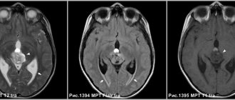
Rice. 1. MRI (T2-weighted images) picture of the lesion in the left lobe of the prostate gland.
The gold standard is transrectal biopsy.
A common method for taking biopsies from a man's prostate gland. Features of transrectal biopsy:
- It is carried out under ultrasound control through the rectum.
- The doctor makes 12 punctures with a needle with cutting ends, on which particles of tissue remain. They are preserved and sent for examination.
- Transrectal intervention causes minimal tissue trauma.
The disadvantage is that there is a possibility of obtaining an insufficient number of biopsies, which leads to the need for a repeat biopsy.
Types of prostate cancer
In 95% of cases, prostate cancer develops from the epithelial cells of the prostate glands (they are called “acini”), from which the peripheral part of the prostate gland is formed. This form of the disease is called acinar adenocarcinoma. In the remaining 5% of cases, the introductal variety is diagnosed, which is characterized by a more aggressive course.
An important characteristic of adenocarcinoma is the degree of its differentiation, which is revealed by histological examination of a biopsy or biomaterial obtained during surgery. Differentiation today is expressed by the Gleason scale, according to which a score is assigned: from 6 (the most favorable prognosis) to 10 (the most unfavorable option).
Stages
Oncologists distinguish four stages of prostate cancer.
- The size of the neoplasm is very small, so it cannot be felt and is not detected by ultrasound of the gland. Signs of the disease are detected only with the help of a special prostate antigen (PSA) test.
- The tumor increases in size, but does not extend beyond the prostate. It can be detected: with a digital examination, compactions are felt, which are detected by ultrasound. There are problems with urination and painful sensations.
- Malignant tissue affects the bladder, rectum and other nearby organs. An admixture of blood appears in the urine, and severe pain is felt when urinating.
- The tumor metastasizes to other organs: lymph nodes, liver, lungs, bone tissue. The patient's condition worsens, he feels a loss of strength, and excretory processes are disrupted.
Characteristic signs of the disease
The risk of developing prostate cancer increases with age: the average in this category of patients is 68 years. There are also risk factors, that is, things that increase the chance of developing cancer. Modern medicine has not identified reliable factors that lead to an increased risk of developing prostate cancer (any medications, diet, bad habits, poor environment, etc.). Therefore, the main factor remains age, as well as age-related hormonal imbalance (between estrogens and androgens).
Causes and risk factors
The only cause of prostate cancer is a malignant modification of cells that lose their original function and direct the generated energy to uncontrolled division. How the biological mechanism that triggers this process works has not yet been established. However, the main factors that increase the likelihood of developing a tumor are well known:
- age over 65 years – up to 75% of patients belong to this age group;
- hormonal imbalances associated with age-related changes in the body;
- unbalanced diet, excessively saturated with animal fats;
- inherited predisposition to the disease;
- the presence of pathologies of the prostate gland - adenoma, prostatitis, hyperplasia, adenosis;
- prolonged exposure to sunlight or exposure to artificial ultraviolet radiation;
- work in hazardous industries with prolonged exposure to carcinogenic compounds.
Symptoms, first signs
In the early stages, a malignant tumor usually does not manifest itself. In addition to adenocarcinoma, people with an increased risk of developing prostate cancer almost always have concomitant pathologies (prostatitis, prostate adenoma), and they can cause symptoms. The most common symptoms are:
- Frequent urination, including nocturia (that is, increased frequency of urination at night);
- difficulty urinating;
- feeling that the bladder is not completely emptied;
- pain when urinating;
- hematuria, that is, blood in the urine;
- hemospermia - during ejaculation, the presence of blood impurities in the semen;
- bone pain that appears when prostate cancer metastasizes to the skeleton.
Thus, the higher the stage, the greater the likelihood of symptoms. Most often, prostate cancer is detected during a preventive examination (it is recommended for all men over 40 years of age). Such an examination includes:
- Ultrasound of the prostate;
- digital rectal examination;
- determination of PSA (prostate-specific antigen).
PSA is a marker used for early detection of prostate cancer. It is sensitive and specific enough to suspect the presence of cancer at an early stage. In addition to PSA, its derivatives can be analyzed - prostate health index, PSA density, ratio of free PSA to total.
Diagnostics
Since it is extremely difficult to recognize the disease by symptoms, laboratory and instrumental diagnosis of prostate cancer, which includes a number of studies, is of utmost importance.
- Serum PSA test. A glycoprotein produced by cells of the epithelial tissue of the prostate serves as a specific marker that allows one to determine the tumor process of the prostate gland at an early stage.
- PSA analysis 3. Another biomarker that allows one to judge not only the presence of a malignant process, but also the size of the tumor.
- Ultrasound of the prostate. It is performed using a special sensor, which is inserted into the anus to detect compacted areas of the gland.
- Biopsy. The most accurate method for diagnosing cancerous tumors is to remove samples of tumor tissue using a thin, hollow needle.
- Histological analysis. The sample taken is examined under a microscope to study the cellular structure and determine the type of tumor.
- Cytological analysis. Microscopic examination of a tumor cell allows you to accurately diagnose cancer and determine its aggressiveness.
Attention!
You can receive free medical care at JSC “Medicine” (clinic of Academician Roitberg) under the program of State guarantees of compulsory medical insurance (Compulsory health insurance) and high-tech medical care.
To find out more, please call +7, or you can read more details here...
Diagnostic methods
The basis for diagnosing prostate cancer is prostate biopsy, in other words, morphological verification.
Indications for biopsy:
- PSA level is higher than normal. It should be noted that the upper limit of normal (4 ng/ml) can be lowered for relatively young men (age 40-50 years) to 2-2.5 ng/ml.
- Suspicious focal changes (hypoechoic foci) detected by ultrasound (or TRUS) or MRI. It is now recommended to do a biopsy after an MRI rather than before because it improves the interpretation of changes. Important: it is better to do MRI in a specialized institution, whose specialists have the necessary experience, and whose diagnostic equipment is specialized software (that is, multiparametric MRI).
- Focal changes detected during digital examination.
Important! If the PSA level is below the upper acceptable limit, this does not always mean that prostate cancer is absent. Approximately 25% of morbidity cases occur against the background of normal values of this indicator. Therefore, the decision on the need for a biopsy should be made after a comprehensive examination, which includes all types of diagnostics.
Prostate biopsy options:
- Standard, or transrectal multifocal. This biopsy is usually done on an outpatient basis. It is performed through the rectum; during the procedure, at least 6 biopsies are taken (preferably 10-12). The disadvantage of this type of biopsy is the likelihood of missing prostate cancer if it is small in size and localized in certain areas of the prostate gland.
- Perineal. It is usually carried out using an extended technique (saturation procedure). During this process I take much more biopsy samples – from 20. Such a biopsy is indicated for those who have undergone standard biopsies, but they did not reveal prostate cancer, while the risk of developing the disease remains. Another indication: planning organ-preserving treatment (focal therapy, brachytherapy). The disadvantages of the technique are the need to provide the patient with spinal anesthesia, the use of specialized equipment, and inpatient conditions. However, it is precisely this biopsy that makes it possible to most accurately identify the nature of pathological changes.
- Fusion. This is a modern type of prostate biopsy that uses modern equipment and MRI data taken in advance. The widespread use of this technique is now limited due to the lack of necessary equipment in medical institutions.
Rice. 2 A., 2 B. Fusion biopsy. Master class at the National Medical Research Center of Oncology named after. N.N. Petrova
Palpation of the prostate gland through the rectum for prostate cancer
Due to the proximity of the prostate to the rectum, a urologist can perform a digital rectal examination. A simple method allows you to detect the presence of a tumor, since it differs from other tissues in its immobility and increased density. In most cases, tumors are located in the peripheral zone, so the doctor can feel them through the rectum.
A healthy gland has an elastic consistency, symmetrical contours and reacts when pressed. During palpation, the doctor pays attention to:
- asymmetry and blurred contours;
- surface roughness;
- immobility when pressed;
- the presence of a dense tumor protruding into the intestinal lumen.
Digital examination is the primary diagnostic method, which must be carried out when examining a man. The result is supported by laboratory and instrumental studies.
Stages of prostate cancer
Staging of prostate cancer and determination of the risk group for recurrence after possible therapy are carried out after histological verification of the disease.
Staging with the standard approach involves osteoscintigraphy and MRI of the pelvic organs. Magnetic resonance imaging is needed to identify the degree of local spread of the process in the prostate area (germination into the seminal vesicles, growth of the tumor beyond the gland capsule), and also to determine whether there is damage to the regional lymph nodes.
Rice. 3. Ways of spread of prostate cancer to the pelvic lymph nodes.
If necessary, an additional CT scan of the chest or abdominal cavity is performed.
The purpose of osteoscintigraphy is to identify possible tumor damage to the skeletal bones.
Additional studies may be prescribed - radiography (sighted), ultrasound, uroflowmetry.
The risk group is determined based on the PSA level before the start of therapy, the Gleason score according to biopsy data, and the clinical stage of the disease. The risk group can be low, intermediate or high. Its determination is extremely important in order to choose the optimal treatment method.
At-risk groups
| Low | Intermediate | High |
| cT2a or less | st2v - st2s | sT3a |
| PSA up to 10 ng/ml | PSA from 10 to 20 ng/ml | PSA more than 20 ng/ml |
| Gleason sum 6 | Gleason sum 7 | Gleason sum 8-10 |
Signs of prostate cancer and disease
Symptoms of prostate cancer resemble those of hypertrophy - enlargement of the prostate as a result of benign growth of the organ. However, the tumor develops in the peripheral part of the gland, as opposed to a benign growth, so it produces symptoms quite late and has a long asymptomatic period.
In addition, symptoms of prostatitis are common among many older men. They are usually associated with inflammation of this gland and are also similar to signs of cancer.
Symptoms of prostate cancer include difficulty urinating due to pressure on the urethra caused by an enlarged prostate gland.
A diseased prostate may cause problems with urination, such as:
- Frequent urination (pollakiuria);
- Having to go to the toilet several times at night (nocturia);
- Despite the urge to the bladder, urine flows out in a small amount, in a weak stream;
- Sudden urge to urinate. Appear quickly, even soon after emptying the bladder, forcing the patient to quickly visit the toilet;
- Urinary incontinence - the patient cannot stop the flow of urine, especially often during stress or coughing. Patients may also complain of spontaneous leakage of urine after finishing urination;
- Difficulty starting to urinate. Despite a full bladder, the patient must concentrate on urinating and it takes a long time for urine to start flowing from the urethra;
- Patients also complain of problems stopping urination, which has already begun.
Frequent urination
A weak, intermittent stream of urine is also a symptom of prostate cancer.
Treatment methods
According to the results of the multicenter prospective randomized study ProtecT (2016), radiation therapy and surgical treatment demonstrate early antitumor efficacy and provide reliable disease control in the majority (more than 90%) of prostate cancer patients with low and intermediate risk of disease recurrence. Currently, the decisive factor when choosing antitumor treatment in this category of patients is the safety of therapy and reducing the risk of complications.
Let's consider the main types of therapy: surgical treatment, brachytherapy, stereotactic radiation, combined radiation therapy.
Hormone-dependent malignant prostate tumor - life expectancy
An increase in testosterone levels in a man's body can lead to the development of hormone-dependent prostate cancer. The survival prognosis for this form of cancer is negative. The tumor progresses rapidly; once metastases appear, life expectancy for prostate cancer of this type is no more than 3-4 years. If prostate cancer is detected, a survival prognosis is made after a complete examination of the patient and diagnosis.
The Yusupov Hospital provides comprehensive diagnostics of prostate cancer. The type of malignant disease and the stage of tumor development are determined. Diagnosis of the disease is carried out using various research methods:
- PSA test. A blood test is performed to check for tumor markers of prostate cancer. This analysis allows you to detect a malignant tumor at the first stage of development. Every year, the test is prescribed to men who have a hereditary predisposition to prostate cancer.
- The patient is examined by a urologist or oncologist. The doctor performs rectal palpation, determining the presence of a formation, its location, and size.
- Transrectal ultrasound of the prostate gland is prescribed.
- To determine the extent of tumor invasion into neighboring tissues, the presence of metastases in regional or distant lymph nodes and organs, the doctor refers the patient to MRI, CT or PET-CT studies.
- After the studies, a biopsy of prostate tissue affected by the tumor is prescribed.
Depending on the test results, age, and health status of the patient, the oncologist prescribes treatment. The hospital's oncology department uses innovative methods for treating prostate cancer. You can make an appointment with a doctor by phone.
Surgical intervention
RP, or radical prostatectomy, is a surgical procedure to remove the prostate gland, as well as surrounding tissue and lymph nodes. During this operation with the gland, the seminal vesicles and a section of the urethral canal are removed as a single block.
Rice. 4. PET-CT images of patient M. with damage to the pelvic lymph nodes
RPE varies according to the type of access and degree of invasiveness:
- Open. There are two main types of access: perineal and retropubic.
The retrotropic approach involves an incision in the lower abdomen through which the prostate and local tissue are removed.
The perineal technique is an open method in which a small incision is made in the area between the anus and the musculocutaneous sac, that is, the scrotum. The technique allows you to remove the prostate, but when using it, it is also impossible to remove unfavorable tissues and nodes located near the gland. If, after perineal surgery, cancer cells are found in the pelvic organs, an additional lymphadenectomy will be necessary. Nowadays the perineal technique is used extremely rarely.
- Laparoscopic. Main approaches: through the preperitoneal space or abdominal cavity. To perform the operation, several small incisions are made on the front wall of the abdomen. Through them, special manipulators are inserted into the preperitoneal space or abdominal cavity and the prostate gland, pelvic fat, and regional lymph nodes are removed.
The laparoscopic technique is the most gentle. The doctor has access to the affected organ through a small incision in the lower abdomen. A camera and all the instruments the surgeon needs are inserted into it. The camera displays an image of the pelvic organs on the screen, thanks to which the doctor fully controls the process and the patient receives a minimum of harm. With this method, blood loss is minimized, foreign organs are almost not injured, erectile function is partially or completely preserved, etc.
Let's also consider the most common complications that can arise after prostate surgery:
- Urinary incontinence. This complication occurs in 95% of cases immediately after removing a special catheter from the patient’s bladder. Further, in 45% of cases, this complication resolves 6 months after removal of the prostate cancer. In 15% of cases, incontinence persists for up to 1 year.
- Loss of erectile function - complete or partial. Doctors are able to significantly reduce this complication by performing laparoscopic prostatectomy. This technique minimizes damage to the neural stem cells of the pelvic organs. If erectile dysfunction is observed after surgery, the patient is prescribed a course of drug therapy and external medications that dilate blood vessels.
Prostate cancer treatment and chances of recovery
For localized forms of prostate cancer (T1-2N0M0), there is a high probability of permanent cure after radical prostatectomy (removal of the prostate) and/or a course of radiation therapy. Surgical treatment of prostate cancer in Germany using Da Vinci technology will completely remove the tumor, preserving the nerves and vessels that supply the anatomical structures. When performing robot-assisted surgery, the probability of maintaining potency and urinary continence exceeds 90%.
If the cancer has already spread to the organs surrounding the prostate, patients with stage ≥T3a disease may be offered radical prostatectomy (RP) combined with lymphadenectomy (removal of the affected lymph nodes). In this case, robot-assisted surgery using Da Vinci technology provides high precision surgical intervention and a good functional result in the long term; maintaining potency is not a priority.
At stages T3b - T4, when metastases are detected in distant organs, radical tumor removal is not always possible. Therefore, it is so important to consult a doctor at the first signs of prostate cancer, when surgical treatment is still possible and, ideally, can be performed using nerve-sparing technology.
Brachytherapy
Brachytherapy is the introduction of radiation sources into tissues. This technique is the “youngest” among the methods of treating prostate cancer. Today this is one of the most popular methods of prostate irradiation, providing very high dose selectivity. The main feature of brachytherapy is that the prostate is irradiated from the inside - the radiation source is introduced directly into it. This method makes it possible to use high doses (100-140 Gy or more), while avoiding the high risk of radiation damage to tissues not susceptible to cancer.
The rapid growth in the clinical use of brachytherapy, compared with surgical interventions, is due to its high effectiveness, which is comparable to prostatectomy, with a much lower incidence of complications.
There are 2 types of brachytherapy, depending on the method of introducing the radiation source into the gland and its power:
- high-power, which is characterized by a short-term introduction of a high-power radiation source into the tissue;
- low-power – a low-power source is installed for the entire duration of treatment.
During low-power brachytherapy, a radiation source is implanted into the prostate tissue and remains there until it completely disintegrates. For a long time, this type of brachytherapy was used most often for prostate cancer. The most commonly used isotope of radioactive iodine, i.e. I125, is used for therapy.
According to numerous studies, low-power brachytherapy does not provide very high radiation accuracy. This is explained by a shift in the radiation source, a change in the shape and size of the prostate, and damage to adjacent healthy organs. In view of this, the low-power technique is indicated mainly for patients with the very initial stages, when the tumor is small and does not extend beyond the gland. This type of brachytherapy has other significant disadvantages. The first is the high frequency of complications arising from the urinary tract; acute urinary retention may even occur and the need for epicystostomy, that is, the formation of a suprapubic vesical fistula, for a long time. The complications are based on swelling of the prostate gland due to the fact that several hundred grains (foreign bodies) remain in it. In addition, radioactive grains, if they remain in the body for a long time, are sources of radiation that pose a certain danger to other people. Because of this, the patient’s contact with family is limited (close contact with small children is prohibited).
Rice. 5. High-power (high-dose) brachytherapy
The most modern technique of interstitial therapy is high-power brachytherapy. Radiation sources are automatically loaded and retrieved. This radiation therapy has a fundamental advantage - high precision irradiation, achieved by introducing needles under the control of a special ultrasound machine. At the same time, doses are automatically calculated and the radiation treatment plan can be quickly adjusted. The radiation source is in the patient’s body temporarily, so the level of complications is the lowest compared to all radical methods of treating prostate cancer, including a low-dose type of brachytherapy.
The technological features of the technique make it possible to offer it to most patients, regardless of the size of the malignant neoplasm and its spread beyond the prostate. In addition, high-power brachytherapy is the “gold standard” for combined treatment, that is, simultaneous use with external irradiation in patients with unfavorable characteristics of the tumor.
The biggest disadvantage of the high-power technique is the high requirements regarding the qualifications of medical personnel, as well as the need to use high-tech equipment. This explains the low prevalence of the method in Russia.
Contraindications to brachytherapy are divided into general and urological. The most common urological contraindications are serious disorders of the urination process:
- IPSS (urinary quality questionnaire index) more than 20;
- the volume of residual urine is more than 50 ml;
- the highest urination rate recorded during uroflowmetry is up to 10 ml/sec;
- transurethral resection of soft tissue of the prostate less than 9 months before the intended brachytherapy.
It should be noted that the large volume of the prostate, which is important for low-dose brachytherapy (50-60 cm3), almost does not limit the possibilities of treatment in the high-power technique.
General contraindications:
- distant metastases;
- malignant tumors, infections and inflammations of the bladder;
- malignant tumors, infections and inflammations of the rectum;
- intolerance to anesthesia;
- absence of the rectum due to previous operations.
These contraindications apply not only to brachytherapy, but also to other methods of radiotherapy for prostate cancer.
Which treatment method is more effective?
The effectiveness of the surgical and drug methods is the same, however, the advantage of drug hormone therapy is the possibility of intermittent treatment and its cancellation, which is not possible after removal of the testicles.
Hormone therapy for prostate cancer can be used alone or in combination with other treatments, and can also be used to treat relapse of the disease.
You should know that hormone therapy does not cure prostate cancer, but it can slow down its growth, reduce the size of the tumor and alleviate the symptoms caused by the tumor!
Stereotactic irradiation
STRT (stereotactic radiation therapy) is a high-precision technique for treating prostate cancer lesions with high doses of ionizing radiation.
Rice. 6. Stereotactic beam accelerator
Today, STLT for prostate cancer is implemented by several main methods, each of which has its own characteristics, pros and cons:
- Proton irradiation. The main advantage is the presence of a Bragg peak, which provides a high dosage gradient. However, this technique is more labor-intensive and costs an order of magnitude more when compared with photon radiation therapy (including the CyberKnife device and STLT performed on a linear accelerator).
- Cyber-Knife (installation of a cyber-knife) has a significant advantage, which consists in an almost unlimited number of directions of the radiation beam. This makes it possible to accurately replicate the geometry of the tumor. The disadvantages include: the duration of the session is up to 40-50 minutes (during this time, the likelihood of patient displacement and the risk of changing the relative position and geometry of the pelvic organs increases), as well as the low uniformity of dosage distribution in the lesion.
- STDT on a linear accelerator using RapidArc and VMAT technology is characterized by a short session duration (4-6 minutes), comfort for the patient and uniform dosage distribution at the site of the disease.
Comparative characteristics of prostate CTLT techniques
| Characteristic | Proton | On the device there is a cyber knife | Using ViMAT and RapidArc methods |
| Price | + | ++ | +++ |
| Duration of irradiation session | ++ | + | +++ |
| Radiation exposure to affected organs | +++ | ++ | ++ |
STLT is used when the patient can be classified as a low or intermediate risk group, provided that the malignant process has not spread beyond the prostate: during instrumental examinations, no data were obtained about damage to regional lymph nodes, there are no MRI signs that the process has spread beyond the capsule glands.
It must be said that of all the radical methods of treating prostate cancer, the SBRT technique is the only non-invasive one and is often chosen if it is impossible to treat the patient with other methods due to the presence of contraindications.
Stages of conducting STLT:
- Radio-opaque gold markers are implanted into the prostate tissue under ultrasound or TRUS guidance.
- After 2-3 days, CT and MRI topometric studies are performed. For the purpose of topometric preparation, a CT simulator with a wide aperture is used; this is necessary in order to accurately contour the organs and plan irradiation.
- After CT and MRI, the results obtained are sent to the planning system of the complex for STLT. CT and MRI images are combined in the planning system by comparing the localization of markers implanted at the first stage.
- When dosimetric planning is completed, radiation therapy can be carried out, consisting of 5 sessions (1 time per day).
The entire course of treatment, including preparation, lasts 7-8 days and can be carried out on an outpatient basis.
Can prevention prevent disease?
There is no reliable evidence that a diet low in animal fats can prevent or reduce the severity of the disease. You may have read about the benefits of consuming foods rich in selenium or vitamin E for cancer prevention. The Selenium and Vitamin E Cancer Prevention Trial did not support this hypothesis.
Conscientious doctors do not talk about prevention, but about early detection of prostate cancer. Early diagnosis will allow you to undergo treatment without compromising the quality of life and remove the tumor, preserving potency and urinary continence.
Combined radiation therapy
Concomitant irradiation is advisable for patients who are at high risk of prostate cancer recurrence. This is due to the risk that regional lymph nodes are affected. This treatment involves comfortable remote irradiation of the pelvic area for 1.5 months: 1 session per day for 5 days with a break on weekends, a total of 25 sessions. Irradiation is carried out using a linear accelerator; an additional dose is delivered to the prostate in the form of 2 sessions of high-power brachytherapy or several sessions of stereotactic irradiation.
MRI of the pelvis with contrast
Magnetic resonance imaging is an accurate diagnostic method that is used to clarify the results of ultrasound examination. To determine the nature of the tumor, an MRI is performed with a statement - a drug with paramagnetic substances is injected into the vein, which makes images of tissues and blood vessels clearer. A malignant tumor quickly accumulates contrast, after which it is easily recognized in the image. Multiparametric MRI is safe for the body; the administered substances do not cause allergic reactions. Tomography allows you to determine the location of tumors and the extent of damage to surrounding tissues. MRI is prescribed in preparation for fusion biopsy.
Suspicious lesions are classified according to the Pirads scale:
- 1-2 points – no cancer.
- 3-4 points – possible oncology.
- 5 points – high risk of cancer.
Recurrence of prostate cancer with radiation therapy
Radiation therapy is one of the most effective and modern methods of treating patients with prostate cancer. However, after its use, relapses are possible.
The earliest method for diagnosing a possible relapse of prostate cancer is to determine the PSA level, which also serves as the basis for the use of more expensive and complex diagnostic methods (osteoscintigraphy, MRI, PET).
Rice. 7. PET-CT image of a prostate lesion in patient V.
After radiation, your PSA level should decrease compared to your baseline level. The dynamics of decline and the minimum indicator are of great importance for assessing the effectiveness of therapy. It must be remembered that the PSA level must be determined each time in the same laboratory, since the test systems have different sensitivities, so there may be significant discrepancies in the indicators if biomaterial is taken from the same patient, but examined in different laboratories.
The generally accepted definition of relapse after treatment is an increase in PSA levels of more than 2 ng/ml relative to its lowest value. The value of the lowest PSA level is individual and depends largely on the initial clinical indicators of the tumor (degree of malignancy, local extent of the process).
When determining PSA levels after radiation, there are 3 important factors to consider:
- Ionizing radiation destroys tumor cells in the prostate. At the first stage, this may be accompanied by a temporary increase in PSA compared to the initial value. Therefore, a control analysis after irradiation should be done no earlier than 3 months after its completion. Further determinations of PSA levels are carried out once every 3 months for several years.
- The process of lowering PSA after irradiation can take a long time, in contrast to radical prostatectomy: by the end of the first month after surgery, the PSA level usually reaches its lowest value. This is explained by the fact that the preserved prostate gland continues to produce a small amount of PSA. Cases have been described in medicine where, after irradiation, the PSA level gradually decreased over 5 years of observation.
- Up to 30% of patients following radiation may experience periods of elevated PSA during follow-up. This phenomenon is called “benign relapse” or “biochemical jump.” The marker level increases very slightly (up to a few tenths of ng/ml) and temporarily. This process is based on the same idea that the prostate persists and produces a small amount of PSA. This is usually observed in patients with adenoma and a large volume of the gland.
Questions and answers
How long do you live with prostate cancer?
About 80% of men treated for prostate cancer live more than 10 years. In the absence of treatment, life expectancy depends on the stage of the disease and the aggressiveness of the tumor, and death becomes inevitable within several years.
What does prostate cancer look like?
There are no external signs of a tumor, so it is impossible to independently detect the presence of cancer pathology. Initial symptoms resemble those of prostatitis or prostate adenoma. For diagnosis, you need to contact a urologist or oncologist.
Is there a cure for prostate cancer?
Currently, malignant prostate tumors can be successfully cured in most cases if they are detected in a timely manner. If any signs of deterioration in urinary function appear, you should visit a urologist as soon as possible and undergo a quality examination.
Attention! You can cure this disease for free and receive medical care at JSC "Medicine" (clinic of Academician Roitberg) under the State Guarantees program of Compulsory Medical Insurance (Compulsory Medical Insurance) and High-Tech Medical Care. To find out more, please call +7(495) 775-73-60, or on the VMP page for compulsory medical insurance
Forecast
Survival rates determine the effectiveness of not only radiation, but also other radical methods of treating prostate cancer. According to numerous studies, the main factor influencing patient survival after radiation is the amount of dose delivered to the prostate gland. According to today's ideas, the dose should be from 72 Gy.
Table of dependence of 5-year survival without relapse on the total focal dose that was delivered to the prostate
| Total focal dose indicator, Gy | Percentage of 5-year relapse-free survival |
| 65 | 48-52% |
| 70 | 57-60% |
| 80 | 82-84% |
| 90 | 91-95% |
Outdated irradiation techniques did not allow the prostate to be exposed to the indicated doses without a strong negative effect on nearby organs. This explains the relatively small number of patients who chose radiation as a treatment option.
Modern techniques make it possible to deliver doses to the prostate gland that exceed 90 Gy. The level of radiation complications is low. Therefore, equal or superior survival rates are provided compared to surgery.
Who is at risk?
First of all, these are men over 45 years of age with a genetic predisposition to the disease. It has been confirmed that hereditary factors play a key role in the development of clinically significant forms of prostate cancer. If a father has been diagnosed with prostate cancer, the risk for his son increases by at least 2 times. Detection of the disease in two or more close relatives (for example, father and brother) increases the risk by 5 to 11 times.
Cases of breast or ovarian cancer in a mother and/or sister are another reason to pay closer attention to your health. Mutations in the BRCA1/2 genes greatly increase the risk of aggressive, early-onset breast and prostate cancer. For men aged 50–70 years without an unfavorable family history, arterial hypertension and waist circumference >102 cm have a statistically significant effect on the increase in the risk of prostate cancer. A body mass index (BMI) of 32.5 worsens the prognosis of treatment success by 30%.
What treatment is prescribed in such cases?
The decision on treatment methods is made based on the patient’s complaints, general health, age and existing diseases. Drug therapy typically includes alpha-blockers, alpha-5 reductase inhibitors, low-dose phosphodiesterase 5 inhibitors (PDE5i), and combinations thereof.
For patients who have not responded to drug treatment, surgery may be recommended:
- minimally invasive endoscopic laser surgery;
- TUR-prostatectomy;
- robot-assisted isolated transvesical prostatectomy;
- open prostatectomy with severe enlargement of the gland.
Assessing the risk of lymph node metastasis
| Risk group | PC stage | PSA, ng/ml | Sum of Gleason scores | Probability of metastasis to lymph nodes | Relapse-free survival for 10 years |
| Short. All factors | T1–T2a | < 10 | 2 — 6 | < 5 % | 70 – 90 % |
| Intermediate. one of the factors | T2b–T2c | 10 — 20 | 7 | 5 – 15 % | 60 – 75 % |
| High one of the factors | T3a | > 20 | 8 — 10 | 16 – 49 % | 43 – 60 % |
The main ways of growth of metastases
- Through blood vessels (hematogenous route)
- Through lymphatic vessels (lymphogenous pathway)
The most vulnerable are the liver, lungs, brain, bone marrow, and bones of the axial skeleton. The main target organs are the pelvic and retroperitoneal lymph nodes, iliac lymph nodes, and less commonly the central nervous system organs.