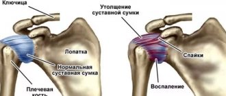
Causes of pain: from tumors to injuries
As in most other cases of painful sensations in various organs of the musculoskeletal system, only a specialist can answer the question of why the arm hurts from the shoulder to the elbow. Causes include various diseases and injuries. Discomfort may occur due to the following reasons:
- Organ damage due to trauma.
- Diseases of the shoulder joint of various origins.
- Abnormalities of the spinal column.
- Accumulation of anaerobic metabolism products in muscle tissue.
- Osteoporosis and its consequences.
- Neuralgia of the brachial and other nerves.
- Connective tissue diseases.
- Tumor diseases.
- Consequences of infections.
Injuries are often a consequence of athletes' passion for sports or professional activities. Also, many traumatic situations arise due to extreme entertainment.
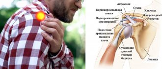
Shoulder myalgia symptoms
Pain in shoulder myalgia, in particular in the presence of myofascial pain syndrome, is often described by patients as aching, dull, tight, boring pain, which can intensify with load on this muscle, and in some patients it can manifest itself in sleep, when the impact occurs in a certain position to a trigger point in the muscle. A trigger point is a compaction of muscle fibers, which, upon palpation, is felt as a dense muscle cord or as a dense round formation and is accompanied by severe pain. Pain can manifest itself at a specific point (localization can be indicated with a finger), or it can be reflected; in this case, trigger points located in different muscles have certain zones where pain can spread.
Often, myalgia in the shoulder area is accompanied by a functional limitation of active movements in the limb, for example, it is painful for the patient to move his arm to the side beyond a certain degree.
The muscles most commonly presenting with localized or referred pain in the shoulder area.
Scalene muscle. Although the anterior scalene muscle belongs to the muscles of the neck, the pain from the trigger point located in it spreads to the upper limb. In this case, characteristic spread paths are noted: along the anterior and posterior surfaces of the shoulder girdle, the upper medial edge of the scapula, the outer surface of the shoulder and forearm, and onto the thumb and index fingers.
In the figure, the areas of most intense pain are marked in red. Spasm of these muscles can lead to a feeling of stiffness and limited movement in the cervical spine. The cause of tension in the flattering muscles can be, for example, carrying heavy bags in the hands, a prolonged cough, lung pathology that requires the inclusion of auxiliary respiratory muscles (chronic obstructive pulmonary disease, bronchial asthma).
A neurovascular bundle passes between the anterior and middle scalene muscles, and spasm of these muscles contributes to pinching of the bundle, which will manifest itself in the form of numbness, a tingling sensation, which is most pronounced in the hand, especially after physical work with this hand. This pathology is called scalenus syndrome or scalenus syndrome.
Pectoralis major and minor muscles. The localization of the trigger point in the clavicular part of the pectoralis major muscle and the pectoralis minor muscle are manifested by similar symptoms, namely: the reflected pain is localized along the anterior surface of the shoulder joint (the area of greatest pain intensity is indicated in red in the figure). Active movements that cause tension or stretching of these muscles, especially when lifting a weight forward with an outstretched arm, can increase pain; also, when sleeping in a prone position, trigger points can be affected, which leads to the occurrence or intensification of pain and frequent awakenings. There may be limited movement of the arm moving to the side.
When trigger points are localized in the left pectoralis major muscle, the pain can imitate angina attacks and a feeling of tightness in the chest.
With severe hypertonicity of this muscle, infringement of the lateral trunk of the brachial plexus and axillary artery often occurs, which is manifested by impaired sensitivity along the inner surface of the shoulder, forearm, ring finger and little finger.
One of the main causes of myofascial syndrome in these muscles is incorrect posture: a slouched back, in which the shoulders move forward, which leads to shortening of the pectoral muscles.
Infraspinatus muscle. With myofascial syndrome of the infraspinatus muscle, pain is localized in the anterior, lateral and posterior surfaces of the shoulder joint, and can descend along the anterior surface of the shoulder. The pain is often characterized as deep, feeling inside the joint (the places with the most intense pain are indicated in red in the figure). Patients note difficulties when servicing the posterior hemisphere of the body: it is difficult to wash the back, fasten the bra lock; attempts to comb your hair or brush your teeth cause sharp pain in the shoulder.
Coracobrachialis muscle. Referred pain in the presence of trigger points in this muscle is localized along the anterior surface of the shoulder joint and spreads down the outer surface of the shoulder, dorsum of the forearm and hand. The pain, as in the case of the infraspinatus muscle, will intensify when the arm is moved behind the back. Trigger points in this muscle, as a rule, appear secondary, i.e. in the presence of active pain points in the muscles adjacent to it, inflammation of the tendon of the head of the biceps, pathology of the shoulder joint, etc.
Arm pain: similarities and differences
Patients presenting with pain in the arm may describe completely different symptoms due to differences in the causes of the disease. But at the same time, there are often similarities in complaints. So, the muscles of the arm from the shoulder to the elbow hurt, combined with numbness due to nerve compression, and this, in turn, can be a consequence of both inflammation of the joints and dislocation. Referral of pain to the arm is observed with angina pectoris, colic, cholecystitis, and stomach ulcers.
So, based on the nature of the pain, they distinguish:
- acute, starting abruptly, which cannot be tolerated without medication - characteristic of injuries, damage to joints, nerves;
- having a baking character - together with shortness of breath and bluish discoloration of the skin, they are often a companion to myocardial infarction, in which case an ECG is required;
- dull, without strong expression, when the arm aches from shoulder to elbow - characteristic of a bruise, closed fracture, arthrosis, tear of muscles, ligaments and tendons.
Sensory disturbances can cause numbness in the hand, causing painful discomfort. Patients in this case also describe a symptom such as “crawling goosebumps.” Both signs are evidence of the onset of osteochondrosis, which must be treated promptly.
Diagnostics
This article does not present the entire spectrum of muscles and diseases of the structures of a given region that can manifest as myalgia. Diagnosis requires a careful individual approach to each patient with this problem. In most cases, it is possible to find the cause of pain based on the collected complaints, medical history and manual muscle testing. In some cases, instrumental diagnostic methods are required, such as radiography and MRI of the shoulder joint, to exclude its involvement in the pathological process. In addition, the pain process itself in the muscles surrounding the shoulder joint can cause inflammatory changes in the joint.
Early contact with a competent specialist will allow you to quickly identify the cause of pain and begin adequate treatment.
Do not engage in self-diagnosis; sometimes it can be difficult even for a specialist to determine the exact cause.
Diagnostic methods for pain in the ulnohumeral region of the arm
A patient who complains of discomfort and pain in the arm from shoulder to elbow is examined. The doctor details the complaints, examines the damage, visually assesses the color scheme and tone of the skin in the area where the pain is localized. By comparing the condition of the joints and studying motor activity, we can make the first assumptions about the presence or absence of damage. In cases where the arm is pulled from the shoulder to the elbow, a flexion resistance test helps.
But, as a rule, an examination cannot give an objective picture of the disease. Therefore, to make an accurate diagnosis, the following research methods are used:
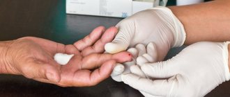
- general (clinical) blood test (CBC);
- blood test for biochemistry;
- X-ray examination;
- CT;
- MRI;
- densitometric study.
The CBC diagnoses inflammation in the body, and a biochemical study clarifies the presence of specific pathological abnormalities. Thus, osteoporosis is characterized by changes in calcium levels.
An x-ray is needed to determine the condition of the bones. Computed tomography and magnetic resonance imaging help to assess the condition of tissues and skeletal components. Densitometry reveals deviations from bone density standards, which is especially important for osteoporosis.
How is glenohumeral periarthrosis diagnosed?
Orthopedists and traumatologists who deal with various manifestations of joint diseases are well aware that glenohumeral periarthrosis is diagnosed mainly in women. The disease is localized mainly in the right shoulder and has a unilateral manifestation, however, during consultation, doctors, after examination and palpation, pay attention to other signs and also prescribe additional studies.
- A general clinical blood test shows an increase in ESR and a shift in the leukocyte formula.
- The X-ray image shows salt deposits, but there are no obvious changes: otherwise, instead of a diagnosis of periarthrosis, we would already be talking about arthrosis - a more serious degree of damage.
- MRI of the thoracic and cervical spine will help clarify the extent of joint damage.
If the patient cannot move his arm to the side by 60-120°, he most likely has periarthrosis
How to treat arm pain from shoulder to elbow
First of all, you need to remember the dangers of self-medication and not to make the disease worse. Only accurate diagnostics carried out in laboratory and outpatient conditions by specialists can identify methods of subsequent therapy. At the initial stage, medications will be prescribed to help relieve pain symptoms. Further, drug and physiotherapeutic treatment of pain in the shoulder and arm is carried out in a complex. Sometimes it is supplemented with herbal medicine.
The following types of medications are usually prescribed:
- Non-steroidal anti-inflammatory drugs - to relieve inflammation, bleeding and pain.
- Hormonal and anti-inflammatory - to support immune resistance.
- Painkillers.
- Chondroprotective - for the treatment and restoration of joint functions.
- Calcium-containing - to accelerate bone healing and relieve osteoporosis symptoms.
Vitamin D is also used to activate healing procedures. A number of drugs have a whole range of side effects. Therefore, when prescribing, specialists are guided by the dynamics of the body’s condition and expediency.
Musculoskeletal pain
Musculoskeletal pain (MSP) is extremely common and accounts for approximately one third of all acute and chronic pain syndromes. Their localization is very diverse, but the most favorite places are the lumbar and sacral spine, lower leg, shoulder girdle and neck. Moreover, in half of patients with MBS, pain occurs in several areas simultaneously [1]. SMS can occur at any age, both in the elderly and in young, able-bodied people, and are often found among students and schoolchildren. Patients suffering from pain experience constant restrictions on motor activity, which sharply reduces their quality of life, and professional and everyday activities deteriorate. Thus, pain is not only a medical, but also a social problem.
SMS (myofascial) occurs due to dysfunction of skeletal (striated) muscle tissue associated with muscle overload and muscle spasm. The reasons underlying the occurrence of this muscle spasm can be very diverse. Muscles respond with tension to any pathological impulse, being a nonspecific indicator of the pathological process, which is why myofascial pain is so common. This type of pain can be associated not only with pathology of vertebral segments or extravertebral structures (joints, ligaments, etc.), but also with pathology of internal organs. The latter should cause particular concern. Almost any somatic pathology can be accompanied by myofascial pain syndrome, since pain impulses from the affected organ lead to protective tension in the muscles surrounding it. Therefore, identifying the causes of myofascial pain always requires a detailed analysis: complaints, medical history and illnesses of the patient, data on general somatic, neurological, orthopedic and mental status. Only by excluding severe somatic pathology, such as tumors and metastases, abscesses, infectious and other diseases, can we talk about “benign” SMB.
The prerequisite for the development of such SMS is functional disorders in the musculoskeletal system and disruption of compensation for natural age-related aging processes. Risk factors for the development of BMS include [1]:
- age;
- heavy physical labor (especially long-term static loads, heavy lifting, body turns and vibration);
- psychosocial aspects (monotonous work, dissatisfaction with working conditions);
- anxiety and depressive disorders;
- obesity;
- smoking;
- drug addiction;
- severe scoliosis;
- history of headaches.
MPS is characterized by both acute and chronic pain syndromes. The initial pain that occurs is usually acute, sharp, intense, and it goes away when the irritant is eliminated and the damaged tissue or organ is restored. This pain performs a protective function, warns the body of danger and ensures the activation of systems aimed at eliminating the damaging factor. However, pain does not always occur in response to damage: often it has already been eliminated, but pain remains, no longer being a protective, but a damaging factor in the body. A component of such “pathological pain” is necessarily present in patients with chronic pain syndromes. Chronic pain often becomes an independent disease, being the only symptom that bothers the patient for a long time, and often its cause cannot be determined. Chronic pain affects an average of 15–20% of the population, and most often this pain is associated with musculoskeletal problems [2].
In addition to dividing pain according to the time factor into acute and chronic, the identification of local, radiating and referred pain and the determination of the mechanisms of its development (nociceptive, neurogenic, dysfunctional) are of great importance for differential diagnosis and determination of therapeutic tactics.
Local pain is always felt in or near the affected part of the body. It is usually associated with a pathological process affecting pain receptors (nociceptors) in the skin, muscles, tendons, ligaments, joints and bones. This is nociceptive (somatogenic, somatic) pain. The main mechanisms of such pain are inflammation and muscle spasm. It is often constant, but can change its intensity with movement and change in body position, can be sharp or aching, dull, and often diffuse in nature. Nociceptive pain disappears when the damaged organ or tissue is restored and responds well to therapy with narcotic analgesics.
Referred pain spreads within the dermatomes associated with the innervation of damaged spinal structures or internal organs. Often referred pain is caused by pathology of internal organs (Zakharyin-Ged phenomenon), for example, in diseases of the pancreas, aortic aneurysm, coronary heart disease, pathology of the gastrointestinal tract (GIT), kidneys, and gynecological diseases. The intensity of pain resulting from damage to internal organs usually does not change with movements in the spine. There is still no comprehensive explanation of the mechanisms of referred pain. It is assumed that it is formed due to the convergence (direct or indirect) of somatic and visceral afferent axons on the same groups of neurons of the central nervous system (CNS), at the level of the dorsal horn of the spinal cord, in the thalamus or sensitive zone of the cortex.
Myofascial pain syndrome is a variant of nociceptive local pain, as well as the most common cause of referred pain. The source of pain is considered to be myofascial trigger points, which can form in muscles, fascia or tendons. The trigger point is a local area of very high sensitivity; upon palpation, it is felt as a compaction or cord. Pressure on an active trigger point provokes sharp local pain with a flinch (jumping symptom) and pain in a distant but strictly defined place (referred pain). Each such point has its own zones of reflected pain. Referred pain is usually dull, aching, deep, and may be accompanied by paresthesia, limitation of movements and forced positioning in the lower back, arm or neck [3, 4].
There are the following criteria for diagnosing myofascial pain syndrome [5]:
A. “Big” criteria (all 5 signs must be present):
- complaints of regional pain;
- palpable “tight” cord in the muscle;
- area of increased sensitivity within the “tight” cord;
- characteristic pattern of referred pain or paresthesia;
- limitation of range of motion.
B. “Small” criteria (one of three signs must be present):
- reproducibility of pain or paresthesia on palpation of the trigger point;
- local contraction of the affected muscle during palpation or injection into a trigger point;
- reduction of pain when stretching a muscle, or during a therapeutic blockade, or injection with a “dry” needle.
It is obvious that myofascial syndromes can form in any muscle and cause pain in various parts of the body. Favorite places for the formation of trigger points are the muscles of the head and neck, shoulder girdle and lower back, which leads to the development of headaches, lumbago in the shoulder blade and neck, pain radiating to the buttock, thigh, foot, etc., forced position, such as torticollis .
It is believed that the formation of a trigger point is caused by repeated microtrauma or acute trauma that disrupts the structure and function of muscle fibrils. Intense or prolonged physical activity, especially with untrained or “unwarmed” muscles, leads to increased tension and the formation of tears at the muscle attachment points, in the muscle fibers and in their connective tissue sheaths. The appearance of pain and tonic muscle contraction is promoted by reflex tension in spinal pathology (dystrophic processes in the spinal segments, radiculopathy, developmental anomalies) and diseases of internal organs, suboptimal motor stereotype (poor posture, postural strain), hypothermia. Often, spasmed muscles become a secondary source of pain, which, in turn, triggers a long-term vicious circle “pain - muscle spasm - pain” [1] and the formation of chronic pain syndrome.
Radiating pain occurs when a root or nerve is damaged and is characterized by greater intensity and distal spread to the area of the corresponding dermatome. This pain is neurogenic in nature (neuropathic pain), that is, it is associated with damage or dysfunction of the nervous system, and not pain receptors. The neurogenic type includes pain due to mono- and polyneuropathies, trigeminal neuralgia, brain injury, etc. Such pain is usually accompanied by sensory disturbances, motor and autonomic disorders (decreased blood flow, impaired sweating in the painful area), and often cause emotional disturbances. Characteristically, pain occurs in response to mild stimuli that under normal conditions do not cause pain (allodynia). Neurogenic pain is unresponsive to morphine and other opiates in usual analgesic doses, indicating a difference in the mechanisms of neurogenic and opioid-sensitive nociceptive pain [6]. Neurogenic pain that occurs with radiculopathy is almost always accompanied by tension in the corresponding muscles and myofascial pain syndrome.
A separate group of pain syndromes consists of dysfunctional pain. They are based on changes in the functional state of the parts of the central nervous system involved in pain control. The main influence on their occurrence is exerted by emotional, social and psychological factors. The main difference from nociceptive and neuropathic pain is the inability to identify the cause or organic disease that explains the pain. Examples of such pain include fibromyalgia, tension headaches, and psychogenic pain in somatoform disorders [7]. Dysfunctional pain is usually present in the structure of any chronic pain syndrome and requires separate specific therapy.
Of course, most pain is of a mixed nature, and determining the presence of one or another component is necessary for the correct selection of therapy.
SMS therapy
Therapy for MPS is complex; both pharmacological and non-pharmacological methods are important in it [8].
Among the latter, interventions that help relax spasmodic muscles are especially recommended: post-isometric relaxation, massage, manual therapy, acupuncture, transcutaneous electrical stimulation, physical therapy [9]. The good effect of rehabilitation procedures aimed at muscle relaxation is further evidence that the basis for the formation of MPS is a dysfunction of the muscle. However, during the period of acute pain, it is better to avoid active manipulations and maintain rest and bed rest, and begin physical procedures in the subacute period [10].
The most common pharmacological therapies are the use of muscle relaxants, non-steroidal anti-inflammatory drugs (NSAIDs) and analgesics. The idea of prescribing muscle relaxants is obvious - by relieving muscle tension, we eliminate the cause of pain and break the vicious circle of “pain - muscle spasm - pain”. Baclofen, tolperisone, tetrazepam, tizanidine are used, each of the drugs has its own characteristics, for example, tizanidine has a fairly pronounced central analgesic effect, tolperisone has analgesic properties and has a vasodilator effect, which increases blood flow to the stenotic muscle. To eliminate pain, especially in the acute period, a variety of analgesics (NSAIDs, metamizole sodium, paracetamol, etc.) are widely prescribed; their effect can be enhanced by adding small doses of anticonvulsants, such as carbamazepine.
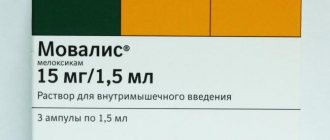
Nociceptive pain responds well to NSAID therapy. Due to the combination of analgesic and anti-inflammatory properties, they are successfully used to relieve acute pain in MPS. However, long-term use can lead to a number of complications from the gastrointestinal tract, hematopoietic organs, and kidneys. Therefore, when choosing an analgesic, one should be guided not only by the effectiveness, but also by the safety of the drug. For example, when treating patients with peptic ulcers, diabetic or other nephropathy, NSAIDs should not be overprescribed. Recently, selective NSAIDs, cyclooxygenase (COX) 2 inhibitors, such as Amelotex (meloxicam), have increasingly come into practice. The main therapeutic effects of NSAIDs - analgesic, anti-inflammatory and antipyretic - are based on reducing the synthesis of prostaglandins from arachidonic acid through inhibition of the COX enzyme. COX exists in two forms: COX-1 is constantly present in all tissues, COX-2 is synthesized against the background of inflammation. Most NSAIDs and non-narcotic analgesics inhibit both types of COX. Blocking COX-1 is responsible for most adverse drug reactions. Three groups of selective COX-2 inhibitors are currently registered in the Russian Federation: sulfonanilide derivatives - nimesulide; representatives of coxibs - celecoxib, valdecoxib; oxicam derivatives - Amelotex (meloxicam).
Amelotex occupies an intermediate position between non-selective and highly selective NSAIDs. The drug selectively blocks COX-2 in the area of inflammation and blocks COX-1 to a much lesser extent, thus the synthesis of prostaglandin, which protects the gastrointestinal mucosa, remains intact. A distinctive feature of the action of Amelotex is its “chondroneutrality”; unlike other NSAIDs, it does not have a negative effect on cartilage, which is important to consider in the treatment of patients with osteoarthritis and osteochondrosis. The results of clinical trials showed that meloxicam has an optimal balance of effectiveness and safety [11]. It is characterized by a long action (half-life of about 20 hours), which makes it convenient for the treatment of chronic pain syndromes. Amelotex has three formulations: parenteral for the treatment of exacerbations of MPS, oral for longer-term use, and an over-the-counter gel for external use, which is especially relevant for the treatment of local pain syndromes [12].
With topical use of NSAID-containing drugs, the likelihood of developing adverse reactions is reduced. Local forms of NSAIDs are safer than tablets; in addition, their use makes it possible to reduce the dose of drugs taken orally and parenterally [13]. When applied topically, Amelotex gel reduces or eliminates pain in the area where the gel is applied, including in the joints at rest and during movement. Helps increase range of motion. Ointments and gels with other analgesics, pepper plaster, and mustard plasters are also used locally. Compresses with dimethyl sulfoxide, procaine, NSAIDs and hydrocortisone have a good effect.
Invasive methods of influence - blockades on trigger points - are highly effective in eliminating myofascial trigger points. Injections may contain an analgesic (novocaine blockade), NSAIDs, corticosteroids, botulinum toxin, or be drug-free - “dry needle” [9, 14]. After puncturing the trigger point, the main symptoms (local and referred pain, “jumping symptom”) disappear and the muscle cord relaxes. Ischemic compression of the trigger point with a finger (acupressure) has a similar effect - as the pain decreases, the pressure on the point is increased, the pressure time is individual in each case.
To enhance the analgesic effect, NSAIDs are recommended to be used in combination with B vitamins, since vitamins of this group can potentiate the effect of NSAIDs, and, in addition, they themselves have analgesic activity. B vitamins are involved in many metabolic processes in the body, nucleotide synthesis, folic acid metabolism, catecholamine synthesis, they normalize metabolic processes necessary for normal hematopoiesis and the development of epithelial cells. It is also known about the active neurotropic effect of B vitamins, which are necessary for the synthesis of the myelin sheath of nerves, the conduction of nerve impulses and the implementation of synaptic transmission, i.e. for the normal functioning of the central and peripheral nervous system. Thus, B vitamins are effective for both nociceptive and neuropathic pain.
Today on the Russian market there is a combined injection drug CompligamV. This is a high-dose injection solution of neurotropic vitamins: B1, B6 and B12, which includes lidocaine. Due to this combination, the drug immediately has both a neurotropic and pronounced analgesic effect. It should be noted that the presence of lidocaine makes injections of the drug CompligamB much less painful than injections of conventional B vitamins. CompligamB can be used in complex therapy of both chronic and acute MBS, combined with NSAIDs. The combination of Amelotex + CompligamB can enhance the analgesic effect, reduce the duration of exacerbation of the disease and the timing of taking NSAIDs [15]. The latter is very important because it makes it possible to reduce the likelihood of adverse drug reactions, especially in patients at high risk (patients with peptic ulcers, hematological diseases). This combination treatment is superior in efficacy and safety to non-selective NSAIDs, such as diclofenac, as demonstrated in clinical trials [16].
Since patients with MPS may have neuropathic and dysfunctional components as part of the pain syndrome, it must be remembered that these pains have a development mechanism different from nociceptive ones; their distinguishing feature is their poor response to therapy with non-narcotic analgesics and NSAIDs. In this case, the potentiation of the analgesic effect and the pronounced neuroprotective effect of CompligamB may be very useful. In the complex treatment of chronic neuropathic and dysfunctional pain, drugs acting on the central nervous system are widely used: tricyclic antidepressants and selective serotonin reuptake inhibitors, or serotonin and adrenaline; anticonvulsants; opioid analgesics; small doses of antipsychotics. It is possible to use NMDA receptor antagonists, such as amantadine, which also has a positive effect on symptoms such as allodynia and hyperalgesia. Localized pain may respond to therapy with topical preparations containing biologically active substances from capsicum.
An important point in the treatment of pain syndrome is not only its effective relief, but also the prevention of further development of the pathological process, taking measures aimed at restoring structural and functional damage. To reduce the severity and prevent further development of the disease, chondroprotectors and neurotropic vitamins are used, primarily B vitamins. The use of the drug CompligamV for preventive purposes as a neuroprotector is quite justified in patients prone to MBS.
Literature
- Podchufarova E.V. Musculoskeletal pain in the back // Breast cancer. 2005; 12:836–841.
- Vorobyova O. V. Chronic pain syndromes in the clinic of nervous diseases: issues of long-term analgesia // Handbook of a polyclinic doctor. 2006; 6.
- Alekseev V.V., Barinov A.N., Kukushkin M.L. et al. Pain. Guide for students and doctors. Ed. Yakhno N. N. M.: MEDpress-inform, 2010. P. 303.
- Pilipovich A. A., Danilov Al. B. Myofascial pain syndrome: from pathogenesis to treatment // Breast cancer. Pain syndrome. 2012; 29–32.
- Danilov A. B. Back pain. Selected lectures on neurology II / Ed. V. L. Golubeva. M., 2012. pp. 179–193.
- Danilov A. B. Neuropathic pain. Selected lectures on neurology / Ed. Golubeva V. L. M., 2006. pp. 208–223.
- Danilov A. B., Danilov Al. B. Manage the pain. Biopsychosocial approach. M.: AMM PRESS, 2012. P. 582.
- Pilipovich A. A., Danilov Al. B. Differential approach to pain therapy: the role of non-steroidal anti-inflammatory drugs // Breast Cancer. Pain syndrome. 2013; 18–21.
- Özkan F., Özkan N. Ç., Erkorkmaz Ü. Trigger point injection therapy in the management of myofascial temporomandibular pain // A?RI. 2011; 23: 119–125.
- Hong CZ Treatment of myofascial pain syndrome // Curr Pain Headache Rep. 2006; 10: 345–349.
- Podchufarova E.V. Chronic musculoskeletal back pain // Neurology. Consilium medicum application. 2010; 1:46–53.
- Amelotex. Instructions for use. www.sotex.ru.
- Godzenko A. A., Badokin V. V. Local therapy of myofascial pain syndrome // Breast Cancer. Rheumatology. 2007; 26: 1998–2003.
- Lavelle ED, Lavelle W., Smith HS Myofascial trigger points // Med Clin North Am. 2007; 91:229–239.
- Gutyansky O. G. The use of the drugs Amelotex and Compligam B in outpatient practice in patients with pain in the back // Breast Cancer. Neurology. 2010; 6:1–4.
- Loginova G.V. Clinical effectiveness of the use of the drugs Amelotex and CompligamV for vertebrogenic lumbar ischialgia // Difficult Patient. 2010; 3:35–38.
N. V. Latysheva1, Candidate of Medical Sciences A. A. Pilipovich, Candidate of Medical Sciences A. B. Danilov, Doctor of Medical Sciences, Professor
State Budgetary Educational Institution of Higher Professional Education First Moscow State Medical University named after I.M. Sechenov Ministry of Health of the Russian Federation, Moscow
1 Contact information
How to avoid pain in the humeroulnar area?
Prevention is the best alternative to treatment. This simple truth also applies to cases where the arm hurts above the elbow. Strong, but regular exercise will help strengthen joints and muscles. Atherosclerosis loves people with excess body weight, so you should eat right and move more. Of course, you need to avoid traumatic situations, not get involved in fights and not look for extreme sports where it is unnecessary. Regular medical examinations and timely diagnosis will help to avoid a situation where the arm hurts between the shoulder and elbow.
Treatment
The most effective way to solve these problems is through surgery. Modern endoscopic equipment makes it possible to perform such operations with minimal trauma, quickly, with a short hospital stay, good functional and cosmetic results; many of them are simply impossible to perform using classical surgery using incisions. Treatment with injections and tablets does not eliminate the problem, it simply masks it, and, especially after the use of hormonal injections, often makes it completely unsolvable.
*Image from website
*Image from website
You should contact your doctor if your shoulder hurts for several days.
Why do my hands hurt?
To answer this question, you need to undergo a competent and comprehensive diagnosis at the Alan Clinic Center for Neurology and Orthopedics, which, with its comprehensive approach to identifying joint problems, will help establish your diagnosis. Diagnostics includes the following research methods:
- consultation with an orthopedic doctor;
- special orthopedic tests;
- dynamic active and passive tests;
- local palpation examination of damaged joints;
- assessment of the condition of the musculoskeletal system, posture, gait, uniform distribution of load on joints, range of motion, stability and strength of the joint;
- making and explaining the diagnosis;
- selection of individual complex motivated treatment.
If necessary, the doctor may also prescribe:
- MRI;
- Ultrasound;
- X-ray;
- lab tests;
- diagnostic puncture of the joint.
Drug treatment of periarthrosis involves prescribing:
- non-steroidal anti-inflammatory drugs;
- chondroprotectors;
- anti-inflammatory and warming ointments;
- biostimulants.
If it was not possible to stop the progression of the disease and the next examination showed arthrosis, solving the problem with conservative methods will be more difficult, and sometimes impossible. In such cases, intra-articular injections of Noltrex will help restore the viscosity of the synovial fluid and restore the usual mobility of the shoulder joint - in courses every 1-2 years, depending on the severity of the disease.
Has the disease progressed and turned into arthrosis? Ask your doctor about the course of intra-articular injections "Noltrex"