The problem of osteoporosis existed in all periods of human development, but in the 20th-21st centuries the disease acquired the character of a “silent epidemic”. In modern medicine, osteoporosis is considered as a systemic disease, the main signs of which are structural changes in the microarchitecture of bone tissue and a decrease in bone mineral density (BMD). If a patient is diagnosed with osteoporosis, treatment is prescribed immediately. Without complex therapy, the disease is prone to steady progression and a decrease in BMD to critical levels, which is associated with a high risk of fractures.
Osteoporosis is not only a medical, but also a social problem. According to statistics, the disease ranks 4th in prevalence, second only to diabetes, cardiovascular diseases and cancer. Advanced osteoporosis is characterized by high mortality and disability.
Another important aspect is a significant decrease in the patient’s quality of life. In fact, doctors often assess the severity of the disease and the effectiveness of prescribed treatment methods only based on laboratory and instrumental data, forgetting about the patient’s well-being and complaints.
Causes of the appearance and development of the disease
Diffuse osteoporosis is a metabolic disease of bone tissue, characterized by a progressive decrease in its density. An important role in its development is played by the natural loss of mineral density, which increases with age. That is why the disease is mainly observed in the elderly. However, there is no equal sign between old age and osteoporosis.
There are a number of risk factors that contribute to the earlier onset of the disease and its rapid progression:
- presence of bad habits – smoking and alcohol abuse;
- impaired motor activity – sedentary lifestyle, lack of any physical activity;
- improper diet - monotonous, mostly refined food (sugar, white flour products), lack of all elements necessary for the body in the diet;
- deficiency of calcium, phosphorus and vitamin D - can be caused by a lack of these elements in the diet, insufficient sun exposure, or other reasons (endocrine diseases, absorption pathologies, etc.);
- underweight, overweight (obesity);
- Osteochondrosis of the spine and arthrosis of the hip joints increase the risk of osteoporosis in the corresponding part of the spine and femoral necks.
Risk factors also include being Caucasian, a history of frequent fractures and injuries, and female gender. The listed factors were selected on the basis of large-scale multicenter studies and are reflected in all clinical recommendations for the diagnosis and treatment of the disease.
Gender is one of the most significant risk factors that cannot be modified. The causes of the disease in women over 50 years of age are hormonal changes in the body caused by menopause.
The increased risk of osteoporosis in women is associated with significant changes in endocrine regulation throughout life. Normally, estrogens, which are actively synthesized during puberty, prevent resorption (destruction, resorption) of bone tissue. Estrogens maintain homeostasis (constancy) and the balance of metabolism in the bones during the reproductive period, but with the onset of menopause, their concentration drops sharply. Estrogen deficiency, especially estradiol deficiency, leads to a decrease in mineral density due to increased bone resorption.
Many researchers are of the opinion that the likelihood of developing osteoporosis is influenced not by the fact of menopause itself, but by the duration of estrogen deficiency: the longer it is, the higher the risk of the disease. For example, among two women of the same age, the likelihood of osteoporosis is higher in the one who entered menopause earlier.
According to various researchers , every second to fourth woman over 51 years of age is diagnosed with diffuse osteoporosis after examination . For comparison, in men in this age group the incidence of the disease is 3 times lower. At the same time, the most common localization of the pathological process is the spine, which is associated with a more rapid loss of the cancellous component of the bone compared to the compact substance. The structure of the vertebral bodies of the thoracolumbar region is characterized by the predominance of spongy substance, and with its sharp decrease, osteoporosis of the spine occurs.
In 15-20% of cases, osteoporosis is in no way associated with all of the listed factors. In such situations it is called secondary, i.e. arising from existing diseases or exposure to negative external factors.
Possible causes of the secondary form of the disease:
- endocrinopathies associated with changes in hormonal status (hyperthyroidism, hyperparathyroidism, hyperprolactinemia and others);
- diseases of the gastrointestinal tract;
- genetic and autoimmune diseases;
- diabetes;
- liver and kidney pathologies;
- blood diseases;
- anorexia nervosa;
- taking certain medications (glucocorticosteroids, aluminum preparations, statins).
Prolonged immobilization (lack of movement for medical reasons) can also be a trigger for the development of the disease. The need for constant bed rest arises after severe injuries, fractures and operations, ischemic and hemorrhagic strokes, with dementia and other severe neurological or mental illnesses.
What influences the success of therapy
The success of therapeutic measures for osteoporosis is influenced by the following factors:
- timely consultation with a doctor as soon as the first symptoms of the disease appear;
- compliance with all medical prescriptions;
- adherence to the principles of proper nutrition;
- regular sports activities;
- degree of development of the pathological process;
- age category of the patient;
- duration of medication use;
- a correctly designed treatment regimen.
Symptoms of osteoporosis
The main reason for the late diagnosis of osteoporosis is the blurred clinical picture and minor symptoms that do not cause significant discomfort to the patient. This leads to delayed seeking of medical help. Another reason for late detection of the disease is the human factor. Osteoporosis is more typical for patients in the older age group, who tend to attribute existing symptoms to their advanced age.
Unfortunately, about half of the cases of the disease are detected only at the stage of complications - when pathological fractures appear. It is at this stage that the symptoms of osteoporosis become pronounced and depend on the location of the fracture.
The first nonspecific signs of the development of osteoporosis are symptoms of latent or overt calcium deficiency. These include:
- decreased performance and increased fatigue;
- paresthesia (crawling “goosebumps”, tingling) and muscle twitching;
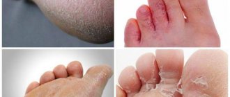
- excessive dry skin and brittle nails.
Warning should be caused by progressive changes in posture, the appearance of spinal curvatures, and dental problems (periodontal disease, progressive atrophy of the alveolar processes with loss of teeth).
Spinal osteoporosis has a variety of symptoms. The first visible signs of the disease are: changes in posture and a progressive decrease in height (2 cm or more in the last 12 months or 4 cm over a lifetime). If osteoporosis of the spine exists for a long time and occurs with serious complications (compression fractures of the vertebrae), then over time the deformation of the spinal column becomes pronounced. Thus, in the thoracic region a “widow’s hump” is formed - a kyphotic curvature, and in the lumbar region the physiological deflection is smoothed out.
Osteoporosis of the spine leads to persistent back pain that lasts from several months to several years. They are localized in the thoracic or lumbar region. The disease is characterized by increased pain while standing or moving , and lying on the bed they gradually subside and practically do not bother you. Osteoporosis of the spine is also sometimes accompanied by tension in the muscles of the back and shoulder girdle.
Signs
Osteoporosis is a dangerous disease, since its initial stages can be asymptomatic. However, doctors identify the following symptoms, the presence of which indicates possible osteoporosis:
- Increased fatigue and weakness for no apparent reason;
- Convulsive syndrome;
- Increased amount of dental plaque;
- Poor posture;
- Brittle nails and hair;
- Dry skin.
These symptoms are not specific, so patients seek medical help late.
Signs of osteoporosis in women remain unnoticed by either the patient or her relatives for quite a long time. Almost the only symptom of osteoporosis in women that attracts attention is pain. Patients complain of pain in the spine, in particular in the thoracic and lumbar regions. As a result, there is a limitation in the range of motion of these joints. The progression of the disease leads to changes in posture, and the patient’s height gradually decreases. But almost all patients associate these symptoms of osteoporosis with old age, heavy physical labor or lack of physical activity, so they do not turn to a specialist.
The reason for visiting a doctor is usually fractures. The most favorite place for a fracture in osteochondrosis is:
- Femoral neck;
- Vertebral bodies;
- Radius.
Almost half of cases of hip fracture lead to disability, and every fourth fracture is fatal.
Make an appointment
Degrees of osteoporosis
Osteoporosis of the spine and the severity of changes in it are assessed according to instrumental studies. After calculating many quantitative indicators, the doctor determines the degree of the disease.
Spinal osteoporosis has two classifications: according to the severity of deformation of the thoracolumbar vertebral bodies according to HK Genant and according to the degree of severity (K. Kruse, 1978).
Genant classification:
- stage 0 – there are no visible changes in the vertebrae;
- stage 1 – minor deformation due to a decrease in the height of the vertebra on one side (front, back, right or left) by 20-25%, but not more than 10-20% overall;
- stage 2 – moderate deformation: decrease in the height of one part of the vertebral body by 25-40% and a general decrease by 20-40%;
- stage 3 – significant deformation: reduction in the height of any area of the vertebral body by 40% or more.
General classification of the severity of vertebral osteoporosis according to Kruse:
- 0 degree – there are no changes in the vertebral bodies;
- 1st degree – borderline stage; osteoporosis of the spine is doubtful, but there are initial changes in the form of reduction of transverse trabeculae;
- 2nd degree – decreased bone density and radiologically determined transparency of the vertebral bodies, the presence of sharply striated vertical trabeculae;
- 3rd degree – pronounced resorption of bone tissue and the appearance of a wedge-shaped or biconcave deformity of the vertebra due to compression of the supporting platforms;
- 4th degree – terminal osteoporosis of the spine; sharp transparency of bone tissue, called “glass vertebrae”, wedge-shaped deformation of several vertebrae.
Non-steroidal anti-inflammatory drugs
Movalis
For osteoporosis, the medication is used in tablets and solution for injection. Injections are required only for the first 2-3 days during the exacerbation period, after which the patient is transferred to enteral treatment. The solution is injected deep into the muscle in an amount of 7.5-15 mg of the active ingredient. Injections are given strictly once a day.
The drug Movalis in the form of a solution for intramuscular administration
The tablets are taken at an individually selected time, strictly during meals, since the active substance can adversely affect the gastrointestinal tract. The dose is 7.5 mg also once a day. If necessary, the amount of Movalis is increased to 15 mg and reduced after achieving an adequate result.
Revmoxicam
The medication is also available in the form of injections and oral tablets. Treatment involves intramuscular administration of 7.5-15 mg of Revmoxicam in the morning. The drug is administered strictly once a day and no more than three days in a row. After an acute attack of osteoporosis is relieved, tablets are taken with meals at a dose of 7.5-15 mg. The tablets are taken daily in an individually selected course.
The drug Revmoxicam in tablets
Diclofenac
A classic anti-inflammatory, taken during an exacerbation of the disease. Take the medication in the form of tablets at a dose of 75 mg of Diclofenac. For severe pain, treatment can be prescribed with injections, which are given intramuscularly. The dosage is also 75 mg daily. The duration of treatment is always negotiated separately for each patient.
Anti-inflammatory drug Diclofenac
Attention! Nonsteroidal anti-inflammatory drugs are prescribed to patients with diffuse osteoporosis at any stage of development.
Porosity of bone tissue is the main sign of osteoporosis on x-ray
Types of osteoporosis
There are several classifications of the disease, but in clinical practice the most common is the division into 2 groups: primary and secondary. The primary form is caused by direct metabolic and structural changes in bone tissue. The secondary form is a consequence of existing chronic diseases of various systems and organs, genetic mutations and long-term use of certain groups of medications.
The primary type of disease has its own subtypes:
- postmenopausal – develops during menopause, most often in women over 50 with an asthenic or normosthenic body type;
- senile – observed in patients of the older age group (60-65 years and older);
- idiopathic – combines cases of the disease in young and middle-aged people.
The first two subtypes occur in 85% of all cases of the disease, with the majority of patients being older women. Such a high incidence of osteoporosis in them is associated with two factors: natural bone degeneration, characteristic of aging, and a sharp change in hormonal levels in women after 50 years.
Osteoporosis also has classifications based on prevalence, localization and morphological characteristics. Based on the extent of the decrease in bone density, diffuse and local forms are distinguished. Diffuse osteoporosis is characterized by a uniform decrease in BMD of all bone tissue. The local type of the disease is often accompanied by isolated changes in the epiphyseal-metaphyseal or adjacent areas of the initial sections of the bone diaphysis.
Due to the participation of the epiphyses in the formation of the joint and the close proximity of other designated areas of bone to the joint, in the non-medical environment the local type is also called osteoporosis of the joints. Diffuse osteoporosis is more common than local osteoporosis, and changes in bone tissue especially affect tubular bones and vertebrae.
Treatment of osteoporosis at ArthroMedCentre
ArthroMedCenter doctors prescribe medications for the treatment of osteoporosis. Physiotherapy is also effective. Additionally, the patient must change their lifestyle, improve their diet, and exercise regularly. Not only drug but also non-drug therapy is important. Non-drug therapy includes regular physical activity.
At the ArthroMedCenter in Moscow, doctors prescribe the following effective procedures for the treatment of osteoporosis:
- Hivamat therapy. A vibration massage is performed using an eclectic field. The procedure reduces pain, relieves swelling and improves blood circulation.
- SMT therapy. The procedure increases the activity of metabolic and recovery processes.
- Interference currents. The procedure reduces pain and promotes vasodilation. Blood circulation also improves and the regeneration process in damaged bone tissue starts.
Osteoporosis is a serious chronic disease. It is dangerous due to its long asymptomatic course. It is very important to undergo preventive examinations and examinations in order to promptly detect the onset of the disease and begin treatment immediately.
How is osteoporosis diagnosed?
After a conversation with the patient and a physical examination, the doctor prescribes a series of laboratory and instrumental studies . Diagnosis of osteoporosis begins with classical studies: general and biochemical blood tests, urinalysis, ECG. They are necessary for a quick assessment of the patient's general condition. In addition, a biochemical study allows you to determine the amount of calcium, phosphorus, and alkaline phosphatase, changes in which may indicate a disease.
A specific laboratory test for osteoporosis is a comprehensive determination of the level of osteocalcin, deoxypyridinoline (DPID) and c-terminal telopeptide of type I collagen (B-cross laps) in the blood. DPID, B-cross laps and pyridinoline (detected in urine) are a group of markers of bone tissue destruction. For a full diagnosis, a urine test is also prescribed to determine the daily excretion of calcium and phosphorus.
Mandatory instrumental studies to confirm the diagnosis are radiography of skeletal bones, X-ray morphometry and densitometry (full name - dual-energy X-ray absorptiometry). Traditional radiography of the thoracic and lumbar spine in 2 projections, pelvic bones including the femur and forearm can be used as a screening study to exclude or confirm the diagnosis.
The most complete information about the pathological processes occurring in bone tissue is provided by osteodensitometry with the determination of a number of indices. It is considered the “gold standard” for diagnosing osteoporosis. The main indicator indicating the presence of the disease is bone mineral density (BMD).
Densitometry is a more sensitive and accurate method than classical radiography. On radiographs, changes are usually visible only in the case of a pronounced loss of bone mass (35-40% of normal or more), and densitometry reveals a decrease in bone density even by 1-2%.
However, spinal osteoporosis is diagnosed by radiography in most patients. In this case, the most common reason for conducting research is persistent back pain that does not respond to standard therapy with non-steroidal anti-inflammatory drugs. The presence of compression fractures and wedge-shaped or biconcave deformities of the vertebrae on the radiograph, combined with the transparency of the bone tissue, are absolute criteria that the patient has severe osteoporosis of the spine.
The most complete picture of bone tissue mass is provided by quantitative computed tomography (QCT). If osteoporosis of the spine is suspected and the results of classical radiography are questionable, it can reliably confirm or exclude the diagnosis. During QCT, the ratio between cortical and cancellous substance in bones (for example, in the vertebrae) is determined and compared with the normal value for that bone. A significant disadvantage of the study is the high radiation exposure, so the method is not used as a screening method or for a one-time examination of the entire skeletal system.
In rare cases, with radiologically confirmed osteoporosis, a biopsy followed by histological examination is indicated. Most often, it is carried out for the purpose of differential diagnosis in young and middle-aged patients to exclude malignant processes and some systemic diseases.
Reasons for women
Osteoporosis is a disease that develops under the influence of many risk factors. There are primary and secondary osteoporosis. Primary osteoporosis develops with age. The reasons are considered to be:
- Complicated family history;
- Mature age;
- Short stature;
- Early menopause and late onset of menstruation;
- Instability of the menstrual cycle;
- Infertility;
- Breastfeeding for longer than one and a half years;
- History of bone fractures;
- Taking hormonal medications;
- Low physical activity;
- Pathology of the endocrine system (thyrotoxicosis, hyperparathyroidism, type 1 diabetes mellitus);
- Excessive drinking and smoking.
Secondary osteoporosis can be caused by the following diseases:
- Itsenko-Cushing's disease and syndrome;
- Thyrotoxicosis;
- Rheumatoid arthritis;
- Diabetes mellitus type 1;
- Conditions after gastric resection,
- Chronic renal failure;
- Chronic liver diseases.
There is a high risk of developing osteoporosis in people taking medications whose side effect is weight loss (immunosuppressants, glucocorticosteroids, anticonvulsants, gonadotropin-releasing hormone antagonists, aluminum-containing antacids).
Smoking, alcohol abuse, caffeine, excess or insufficient physical activity are factors that can cause osteoporosis. The likelihood of developing osteoporosis is higher in people who eat insufficient amounts of calcium, vitamin D, a lot of meat, or cannot tolerate dairy products. Generally recognized factors that increase the risk of developing osteoporosis are the presence of osteoporosis in parents, sisters and brothers, early menopause, and a sedentary lifestyle. Make an appointment
How to treat osteoporosis
Treatment for osteoporosis in men and women depends on the cause and symptoms. In secondary forms, the goal of therapy is to treat the underlying disease that caused osteoporosis.
The primary form of the disease requires long-term complex treatment. The patient must be interested in his own recovery and follow all recommendations daily. In case of an irresponsible approach to treatment, the result will be minimal.
Symptoms and treatment of the disease are largely determined by the degree and location of osteoporosis. The general treatment regimen consists of the following components:
- daily physical activity: walking, exercise therapy complexes, gymnastics (running and jumping are contraindicated!);
- correction of diet (inclusion of calcium-fortified foods) and lifestyle;
- drug therapy with antiresorptive agents, correctors of bone tissue metabolism, as well as bone anabolics containing calcium, magnesium and other components of bone tissue in digestible form, vitamin D;
- treatment aimed at unloading the affected areas of the spine and improving their blood supply, leading to the elimination of pain - osteopathy, physiotherapy, gentle manual massage.
If osteoporosis is detected in a woman after 50 years of age , treatment is recommended to begin with a visit to the gynecologist to decide on estrogen hormone replacement therapy (HRT). Usually it is prescribed to patients whose menopause has occurred independently at too young an age, or due to undergoing surgery.
Properly selected treatment for osteoporosis in women over 60 years of age should also take into account several factors: the duration of the menopausal pause and hormonal status, the presence or absence of calcium deficiency, the severity of pain and other neurological symptoms.
Drug therapy includes drugs to reduce bone resorption. They combine pharmacological treatment with physical therapy, osteopathic procedures, physiotherapy and massage. Treatment of osteoporosis in women over 60 years of age does not usually require estrogen replacement therapy.
Most symptoms and manifestations of osteoporosis go away with treatment, but the patient must protect himself and avoid injury. If there is an increased risk of pathological fractures, it is possible to wear special protectors for the hip or corsets for the spine.
How to restore bone health
The success of the therapeutic measures taken will directly depend on the patient’s compliance with all medical prescriptions. In addition to regularly taking medications, it is very important to adjust your lifestyle, establish proper nutrition, so that a sufficient amount of calcium and vitamin D3 enters the body with food.
The main goals of osteoporosis therapy are:
- reducing the risk of fractures;
- increasing bone mass density;
- improvement of markers of bone mass formation.
The patient is advised to control body weight and make all necessary efforts to build muscle tissue. You should also pay attention to saturating the body with calcium and vitamin D. These elements can be supplied with food, and the doctor often prescribes long-term use of medications that contain these minerals and vitamins.
Doctors' recommendations for the treatment of osteoporosis are determined by the stage of severity of the disease and the level of destruction of bone structures. Experts also take into account:
- age category of patients;
- general state;
- severity of symptoms;
- accompanying illnesses.
For elderly people with osteoporosis of the spinal column, therapy consists of taking medications - anabolic steroids, antiresorptive agents. The latter are designed to suppress the processes of destruction of bone structures. Anabolic steroids activate the formation of new bone tissue.
Medications are combined with drugs that contain a high concentration of calcium. If there are severe painful sensations, which often occur with compression fractures of the vertebrae, painkillers must be prescribed in a form convenient for the patient.
For patients diagnosed with a femoral neck fracture, surgical treatment is indicated. The main goal is to restore full function of the joint. Surgical intervention is necessary if there is a high degree of deformation of the chest due to compression injuries of the vertebrae. A certain diet plays a very important role in the treatment of osteoporosis.
Basic principles of nutrition:
- add to the menu dishes that contain a high calcium content. During the day, the human body should receive at least 1200 mg of calcium;
- minimize or completely abandon the consumption of sweets, baked goods, baked goods, and alcoholic beverages;
- include in the diet the daily consumption of fermented milk products, which ensures a regular intake of calcium in the body in soluble form;
- consume more foods that contain magnesium, phosphorus and potassium.
In addition to maintaining proper nutrition, it is important to pay attention to physical activity. In order to properly form a muscle corset that will support bone tissue, it is necessary to provide the body with regular physical activity.
If osteoporosis is detected on time and the patient follows all the doctor’s instructions, the disease will not progress further. The person will be able to lead a normal lifestyle.
Consequences and complications of osteoporosis
The main problem from which all other consequences flow is the increased risk of fractures. The insidiousness of the disease lies in the fact that the symptoms of osteoporosis may not appear for decades, and then one day a person can suffer a fracture and become disabled for the rest of his life. Moreover, they occur even with low-energy injuries or impacts. In advanced cases, even an awkward movement can lead to a fracture.
Diffuse osteoporosis is one of the main causes of disability in patients. The saddest statistics for pathological fractures of the femoral neck is that mortality in the first year after injury is 30-40%. Even with timely assistance, patients often become disabled and are not even able to care for themselves.
Osteoporosis of the spine with severe deformation of the vertebrae leads to constant pain that barely subside with rest. However, drug treatment alleviates the condition slightly. In the case of vertebral fractures with damage to the spinal cord, all functions for which this segment is responsible are disrupted, and the person becomes disabled.
How to detect the disease
The main danger of the disease is that in the initial stages of its development there are no manifestations that prompt a person to consult a doctor. Most often, the diagnosis of osteoporosis is made after a person goes to the hospital with a fracture. Such damage occurs even with minor injuries, bruises and falls. The most common fractures are the hip, spine, forearms and shoulder.
Other signs of osteoporosis include:
- decreased growth;
- reduction in body weight, since after eating a person becomes full faster;
- there are signs of esophagitis-reflux, which is accompanied by attacks of heartburn, painful sensations in the chest;
- the stomach begins to protrude forward;
- systematic pain occurs in the back and hip joints, difficulties in movement appear;
- the spine begins to sag so that the lower ribs and hip joints come into contact.
Most often, the disease develops when the body’s production of certain types of hormonal substances decreases. But scientists have found that today this diagnosis is given even to teenagers; it is not associated with hormonal imbalances. The reasons for the development of the pathological process are the deterioration of metabolic processes, a sedentary lifestyle, and lack of quality nutrition. In women, this disease is diagnosed quite often.
Prevention methods
Prevention of osteoporosis is a comprehensive program of measures aimed at reducing the risk of the disease:
- a complete diet;
- active lifestyle, regular physical activity;
- drug correction of deficiency of calcium and other components of bone tissue, vitamin D;
- Osteopathy sessions are special exercises aimed at improving posture and normalizing blood supply to the spine and joints.
Exercises to Prevent Osteoporosis
Spinal osteoporosis rarely develops in physically active individuals. Daily training strengthens muscles, bones and ligaments, increases load tolerance and improves flexibility. It is recommended to develop the optimal set of exercises together with a physical therapy trainer, taking into account individual characteristics.
To strengthen the back and joints of the lower extremities, exercises on a gymnastic mat are suitable: leg lifts, scissors, flexion and extension at the knee and ankle joints. From a standing position, you can perform 20 bends in different directions, while your legs should not bend at the knees or lift off the floor. Twisting a hoop or hula hoop is not only an excellent exercise for the thoracolumbar spine, but also a way to acquire a chiseled silhouette.
Physical activity
Swimming, gymnastics, static loads and walking with or without light weights on the legs are beneficial for the health of the spine and joints. It is not recommended to actively engage in heavy and traumatic sports, and in case of large body weight, running and jumping are prohibited - instead of preventing osteoporosis, problems with joints will arise. Walking in the fresh air is another option for physical activity available to everyone. It is recommended to spend 1-1.5 hours a day walking at an average pace, at a slow pace - 2 hours or more.
Products for the prevention of osteoporosis and general principles of nutrition
The main goal of correcting the diet is to make it as enriched as possible with micro- and macroelements necessary to maintain normal bone density.
Proper nutrition involves eating 5-6 times, three of which (breakfast, lunch, dinner) account for the bulk of calories. The last meal should be 3 hours before bedtime: you should go to bed with a slight feeling of hunger. “Unhealthy” foods are excluded from the diet: fatty, floury, sweet. The preferred heat treatment methods are steaming, stewing and boiling.
To prevent bone tissue destruction, the diet should contain foods enriched with calcium (including artificially): fermented milk products, cheeses of all types, sesame seeds, herbs, sardines and seafood. It is recommended to consume at least 500 ml of fermented milk products and 100 g of greens per day.
Preparations and vitamins for prevention
Even the most balanced diet is not always enough to meet the body's needs and cover the daily calcium requirement. Moreover, it is very difficult for an ordinary person to create the “correct” diet and calculate the amount of macro- and microelements received per day. To prevent and correct calcium deficiency, calcium supplements in the form of citrate and chelate can be used. The pharmacological market offers a lot of them - you can buy only calcium, or complex preparations containing most of the necessary elements. Most doctors recommend purchasing combination products that definitely contain vitamin D3, zinc and magnesium - they help prevent osteoporosis.
Preventive medical examinations
Visiting your primary care physician once a year is a must for maintaining health. A basic scope of diagnostic procedures (general examination, blood tests, urine tests, ECG) will allow you to notice the first changes and, if necessary, undergo an extended examination.
Senile osteoporosis often develops in older women, so there should be some caution. In older women, osteoporosis may have no symptoms, so it is detected by chance during an annual preventive examination. This statement also applies to men, however, the incidence rate among them is lower.
Postmenopausal osteoporosis is a common complication in menopausal women, so it is necessary to visit a gynecologist at least once a year. If necessary, you can take a sex hormonal panel, and in case of severe menopause with an abundance of symptoms, you can get a prescription for estrogen replacement therapy.
If you have chronic back pain, you should tell your doctor about it during a routine examination. After consulting a neurologist and determining the neurological status, the doctor will determine further tactics and prescribe additional laboratory and x-ray tests.
A visit to an osteopathic doctor once every six months will not only identify, but also eliminate both general and local overloads of the spine, improve posture, and normalize mobility and blood supply to the musculoskeletal system.
Physiotherapy
Physiotherapeutic procedures are effective in treating diseases of the musculoskeletal system.
Electrotherapy includes the following techniques:
- electrophoresis;
- phonophoresis;
- magnetic therapy;
- inhalation therapy;
- EHF;
- ultrasound.
The following treatment methods are also used:
- To improve blood circulation and get rid of swelling in the limbs, lymphatic drainage massage is prescribed. If there is pain, paresis, or weak muscle tone, the doctor prescribes electrical stimulation.
- Light treatment. It is an effective method as part of the complex treatment of diseases of the musculoskeletal system.
- Electromagnetic treatment. Helps in the treatment and prevention of diseases, reduces the severity of the inflammatory process, improves metabolic processes, and relieves post-traumatic consequences. The procedure is performed by exposure to alternating electrical impulses of a certain frequency.
- Treatment with water. It is represented by different types of therapeutic showers and baths. They have a positive effect on bone structures, joints and muscles. Hydromassage is effective because it improves blood flow.
- Massage. Thanks to therapeutic massage, you can get rid of clamps on nerve bundles, fight neuralgia, and remove stagnation of salts and toxins from the body. This procedure helps improve lymph flow and blood circulation. You must take at least one course every 6 months.
- Ozokerite. Helps reduce the severity of the inflammatory process, relieves pain and swelling, and removes toxins. After completing the course, the results last for several months.
- Acupuncture. Helps in rehabilitation after injury and surgery. Accelerates tissue regeneration and restoration of their integrity.
A mandatory step in the treatment of osteoporosis is a properly formulated diet. The body must receive the required amount of calcium and vitamin D. If it is not possible to meet the daily requirement of these elements, the doctor prescribes appropriate medications that contain these components. These multivitamins and calcium supplements will need to be taken over an extended period of time.
Therapeutic exercises should be prescribed by a doctor. In each specific case, an individual treatment regimen is selected. Therefore, it is not recommended to perform any exercises on your own. This can only be done after preliminary consultation with a doctor and work with a rehabilitation specialist.
General recommendations for a healthy lifestyle
Prevention of osteoporosis is primarily based on the principles of a healthy lifestyle. The rules for a healthy lifestyle are simple:
- rejection of bad habits;
- frequent split meals with a varied diet;
- compliance with the drinking regime - at least 1.5 liters of clean drinking water per day;
- maintaining a sleep schedule - healthy sleep cannot be less than 7-8 hours;
- maintaining psycho-emotional health - you should refrain from unnecessary worries and avoid stress;
- daily physical activity - a half-hour daily exercise program and an hour of walking are a mandatory minimum;
- a nightly 15-minute set of “stretching” exercises to eliminate constant muscle tension and maintain flexibility of the spine and joints.
Did you like the article? Add the site to your browser bookmarks
Sources
- Baranova I.A. Glucocorticoid-induced osteoporosis / I.A. Baranova // Practical pulmonology. - 2008. - No. 1. - P. 3-9.
- Baranova I. Caution: osteoporosis! / I. Baranova // Asthma and allergies. - 2004. - No. 4. - P. 18-19.
- Zulkarneev R.A. Prevention and treatment of osteoporosis / R.A. Zulkarneev, R.R. Zulkarneev // Kazan Medical Journal. - 2003. - T. 84. - No. 3. - P. 230-232.
- Lesnyak O.M. Diagnosis, treatment and prevention of osteoporosis in general medical practice. Clinical recommendations / O.M. Lesnyak, N.V. Toroptseva // Russian family doctor. - 2014. - P. 4-17.
- Postnikova S.L. Features of postmenopausal osteoporosis / S.L. Postnikova // General Medicine. - 2004. - No. 4. - P. 41-45.
- Rodionova I.V. Systemic osteoporosis and osteoporosis of the lower jaw / I.V. Rodionova [and others] // Nurse. - 2015. - No. 5. - P. 32-34.