Necrosis of cartilage tissue inside the knee joint is a complex pathology that leads to very serious consequences. The development of necrosis of the cavity of bone joints is called Koenig's disease and requires immediate treatment.
The etiology of the disease in question involves the gradual destruction and detachment of cartilage tissue from the bone of the joint, which creates difficulties in free movement, causing inflammation, limited mobility and severe pain. All this greatly reduces a person’s quality of life and requires qualified medical care.
Brief Definition
Before you understand what the prerequisites and symptoms of the pathology in question are, you should first understand, at least in general terms, what this disease is.
Koenig's disease is nothing more than osteochondritis dissecans of the knee. The disease has a fairly extensive clinical picture and belongs to the category of pathologies of a focal, idiopathic type.
As the pathological condition progresses, the patient's subchondral bone tissues are affected, which leads not only to a loss of cartilage stability, but also to its gradual destruction, leading to necrosis and pathological changes in the bone.
The impossibility of timely diagnosis, lack of treatment and neglect of prevention and therapy can provoke the premature development of osteoarthritis of various stages.
Any person on the planet is susceptible to Koenig's disease, regardless of age and gender. In the vast majority of diagnosed cases, the pathology develops in young adolescence. Despite the large risk group, it is worth noting that statistics show that diagnosis occurs in 21 cases out of 100,000.
Pathogenesis
Despite the fact that diagnosed cases are found in modern medical practice, the pathogenesis of osteochondritis dissecans remains incompletely understood.
Today, there are a large number of assumptions, each of which makes it possible to form a more or less complete picture. So, there are several theories. Let's try to look at a few of them.
Inflammatory processes
A large number of specialists around the world are inclined to believe that the pathogenesis of the disease in question lies precisely in inflammatory processes, which is confirmed by several histological samples. Thus, one of the authors described 24 such cases, while another noted 42 cases in 12 months.
It is worth noting that a fairly large number of histological analyzes were carried out, which, in the presence of symptoms, did not at all show the presence of inflammatory changes.
The theory about the origin of pathology from inflammation has no scientific or literary support.
Injuries
Two other American scientists noted 14 cases of pathology in which there was an assumption that the progression of tissue deterioration could be caused by injuries (both acute and chronic).
Considering this theory, one can trace a fairly obvious connection: pathological processes originate in the most vulnerable place of the joint, damage to which is extremely difficult to diagnose. Over time, bone tissue is destroyed, which causes necrosis and separation of the osteochondral fragment.
Systematic microtraumas
When talking about injuries, one should not ignore such a situation as microtrauma. Despite the apparent insignificance, repeated damage to tissue integrity (for example, minor blows to a limb) may well lead to very serious consequences.
Hereditary predisposition
A huge number of scientific works are devoted to the study of genetic factors.
Despite the large number of studies conducted, as well as the endlessly relevant discussions of medical workers around the world about the influence of the genetic factor, there is no exact confirmation of the existence of a connection between the formation of osteochondritis dissecans and heredity.
Ischemia (decreased vascular nutrition)
As you know, lack of blood circulation can lead to extremely negative, sometimes even irreversible, consequences. Thus, in connection with the results of numerous studies, the study of diagnosed cases and taking into account a number of other factors, a significant reduction in the number of vessels was noticed.
Considering this issue theoretically, we can come to the conclusion that circulatory disorders can be caused by various factors, for example, various types of trauma or abnormalities of the cardiovascular system.
What are the most likely causes of Koenig's disease?
Pathological damage to osteochondral tissues of this type is most often localized in the knee. Despite the systemic nature of the pathology and the relatively thorough study of its development, it is almost impossible to accurately determine the unambiguous causes of its occurrence. In this regard, it is generally accepted that Koenig's disease is a disease of a cryptogenic nature (with an unknown origin).
The most likely reasons include:
- hereditary factor;
- systematic traumatism of various segments of the musculoskeletal system;
- the presence of concomitant degenerative diseases of various types;
- diseases of the cardiovascular system;
- congenital anomalies of the skeleton, namely, osteochondral tissue;
- increased loads, specifics of work activity.
General symptomatic picture
Any unpleasant symptom that a person may notice in himself should be a signal to seek professional medical help. Especially when it comes to the musculoskeletal system.
The symptoms of the disease are quite extensive, which is due to the impossibility of establishing the true cause of its occurrence. Thus, the common symptoms of the disease include:
- discomfort in the knee area;
- pain of varying intensity, mainly aching in the knee;
- increased discomfort and discomfort during movement;
- attacks of pain without good reason;
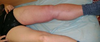
- local swelling, redness of tissues;
- difficulties in performing habitual movements, reduction in the amplitude of mobility of the lower limb, the appearance of a crunching sound;
- feeling of stiffness;
- joint inflammation of various etiologies;
- visually noticeable and palpable changes in gait, the appearance of lameness;
- lack of coordination.
Stages of Koenig's disease
The course of osteochondritis dissecans is predominantly gradual and measured. Having studied the features of the development of the disease, as well as getting acquainted with its symptomatic picture, we can distinguish 4 stages of damage:
Stage I - the person begins to experience minor discomfort and mild pain localized at the site of the lesion (mainly in the knee joints). At this stage, the cartilage tissue is not yet deformed, however, the process of damage to the bone marrow has already begun.
Stage II - more and more areas with necrosis (dead tissue) are formed, which causes an increase in the symptomatic picture. The progress of the disease becomes clearly visible during X-ray examination. Over time, the patient develops synovitis (inflammation of the synovial membrane) and the destruction of hyaline cartilage begins.
Stage III - a dead area of tissue separates from the surface of the bone, which provokes an increase in the symptomatic picture and causes limited mobility of the patient due to severe pain.
Stage IV - the affected tissues separate and form a body directly in the synovial bursa, which causes a serious inflammatory process and significantly reduces the patient’s quality of life.
What is the danger?
If the disease is not treated, a necrotic fragment remains in the joint. The dead tissue limits the mobility of the knee and increases inflammation. The joint is blocked and remains in an unnatural state all the time. These are excellent prerequisites for the development of deforming arthrosis with all the ensuing consequences and complications.
There are no special preventive measures against this disease. However, you can reduce the risks. To do this, it is important to lead a healthy lifestyle, give up bad habits in favor of daily morning exercises. It is also important to monitor the load on the lower limbs, and seek help at the first alarming symptoms.
Types of Koenig's disease
To date, there is no classification of osteochondritis dissecans. However, at the same time, it is customary to conditionally divide the diagnosed pathology into several types, depending on:
- patient's age (juvenile (8-12 years)/senile type (40-50 years));
- location of the pathology (knee);
Depending on the intensity of the symptoms, the collected medical history, as well as other features, an individual treatment plan is determined, which begins, first of all, with an accurate diagnosis.
List of sources
- Merkulov V.N., Yeltsin A.G., Mininkov D.S., Avakyan A.P. Treatment of osteochondritis dissecans of the femoral condyles in children and adolescents // Journal of the Russian State Medical University No. 4 (63) Special issue. Materials of the first joint scientific and practical forum of children's doctors in Orel 2008 (19-23/V) p. 136
- Shapiro, K.I. Frequency of injuries to large joints in adults / K.I. Shapiro // Diagnosis and treatment of injuries to large joints. – St. Petersburg, 1991 – pp. 3–5.
- Mironov, S.P. Classification and methods of treatment of cartilage defects / S.P. Mironov, N.P. Omelyanenko, E. Kon, A.K. Orletsky, I.N. Karpov, A.P. Kurpyakov // // Bulletin of Traumatology and Orthopedics named after. N.N. Priorova. – 2008. – No. 3. – P.81-85.
Set of diagnostic measures
If symptoms of joint pathologies appear, first of all you need to contact your general practitioner for a referral to specialized specialists or go directly to an orthopedic doctor or traumatologist.
In order to make the most accurate diagnosis and identify the most likely cause of the disease, an integrated approach is used, which involves a thorough examination of the patient using such examination methods as:
- radiography – makes it possible to generate up-to-date information about the condition, the degree of preservation of the tissue structure, as well as determine the exact location of necrotic processes (mainly at stages III-IV);
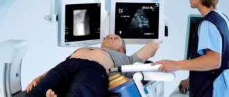
- CT (computed tomography) – creates optimal conditions for assessing the condition and area of damage to the soft tissues of the internal cavity of the joint;
- Ultrasound (ultrasound) or MRI (magnetic resonance therapy) are the most effective methods that allow us to identify the presence of a problem at the initial stages of its development, providing a detailed analysis of tissues;
- arthroscopy is a minimally invasive surgical intervention of a diagnostic nature, which allows one to study the condition of the cartilage using a micropuncture;
- laboratory test - a patient’s blood test allows you to form an idea of the level of his health, and also helps to identify the degree of rheumatoid factor.
Diagnostics - MRI and ultrasound for osteochondritis dissecans
In order to more accurately diagnose osteochondritis dissecans of the knee joint and select the correct treatment, experts recommend undergoing the following diagnostic measures:
- X-ray. The highest information content is possible only at stages 3-4 of the disease;
- Ultrasound and MRI of the knee joint. They are also highly informative and allow diagnosis to be made at the initial stages of the disease;
- CT. Allows you to detect the disease at 2-3 stages;
- Athroscopy. This method is famous for its increased reliability and reliability. Provides an opportunity to study the condition of the outer shell of the joints;
- Analysis of rheumatoid indicator. Allows you to determine the likelihood of rheumatic changes in the joint.
Treatment of Koenig's disease
Due to the peculiarities of the clinical picture of the disease, treatment of Koenig's disease is predominantly radical, which involves surgical excision of damaged tissue. However, the approach of specialists is comprehensive, which involves a combination of radical and conservative techniques.
Types of surgery
Despite the radical nature of the main treatment method, its use is the best solution to relieve the patient of the pressing problem.
Treatment of Koenig's disease surgically is carried out using arthrotomy - opening the joint cavity in order to eliminate necrotic fragments of the affected tissue. At the same time, the classical technique has a more gentle variation - surgical intervention using endoscopic methods with a minimum amount of damage (three small incisions/punctures).
Drug therapy
Prescribing medications in the treatment of Koenig's disease is one of the key stages and the second most important, because the success of the surgical intervention and the patient's rehabilitation period largely depend on the intake of medications.
Drug therapy involves the use of drugs such as:
- antibiotics;
- analgesics and antispasmodics;
- non-steroidal anti-inflammatory drugs (NSAIDs);
- vitamin and mineral complexes;
- chondroprotectors.
Due to the fact that as the pathology progresses, part of the cartilage tissue dies, special attention is paid to such a group of drugs as chondroprotectors. Thanks to the course of taking chondroprotectors, the regenerative process is accelerated, which ensures positive dynamics of the healing process and stimulates an improvement in the patient’s quality of life.
Artracam is considered to be one of the most popular and effective drugs in the chondroprotector group.
Modern methods of physiotherapy are used mainly in the initial stages of pathology, when it is necessary to activate metabolic processes by improving blood circulation in the affected joint, as well as during the rehabilitation period.
Among the widely used physiotherapeutic techniques in the treatment of Koening's disease, it is worth paying attention to such as electrophoresis, shock wave therapy, as well as magnetic therapy, etc.
Physiotherapeutic treatment is allowed only after a high-quality consultation with the attending physician, who takes into account not only the general condition of the patient, but also the dynamics of the development of the disease, the characteristics of its course and other factors.
What are the consequences of lack of treatment?
Regardless of why the patient did not begin timely treatment of a pathologically dangerous joint disease, its inevitable progress and active development will sooner or later lead to irreversible consequences.
Do you think that mild discomfort in the knee joint will “go away somehow on its own” or “just a coincidence”? No matter how it is. Advanced stages lead to the fact that the necrotic (dead) fragment is separated into the joint cavity, which leads to limitation of its mobility and intensive development of inflammatory symptoms. The blockade caused by the localization of such fragments provokes the appearance of deforming arthrosis.
Forecast
If you contact a specialist in a timely manner and have a mild form of the disease, the prognosis for patients is in most cases positive. At the same time, it is possible to achieve rapid restoration of health.
Unfortunately, in advanced stages of the pathological process one cannot count on a complete recovery of the patient.
Timely initiation of treatment measures and compliance with prevention recommendations make it possible to stop the development of the disease and maintain a person’s quality of life. In turn, if pronounced pathological processes are detected, the patient is placed on disability.