Publications in the media
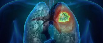
Atelectasis (collapse) of the lung is a loss of airiness in an area of the lung, occurring acutely or over a long period of time. In the affected collapsed area, a complex combination of airlessness, infectious processes, bronchiectasis, destruction and fibrosis is observed.
Etiology and pathogenesis • Obstruction of the bronchial lumen by plugs of viscous bronchial secretion, tumor, mediastinal cysts, endobronchial granuloma or foreign body • Increased surface tension in the alveoli due to cardiogenic or non-cardiogenic pulmonary edema, surfactant deficiency, infection • Pathology of the bronchial walls: edema, tumor, bronchomalacia, deformation • Compression of the respiratory tract and/or the lung itself, caused by external factors (myocardial hypertrophy, vascular abnormalities, aneurysm, tumor, lymphadenopathy) • Increased pressure in the pleural cavity (pneumothorax, effusion, empyema, hemothorax, chylothorax) • Restriction of chest mobility ( scoliosis, neuromuscular diseases, phrenic nerve palsy, anesthesia) • Acute massive pulmonary collapse as a postoperative complication (unrecognized and unsanitized obstruction of the main bronchus).
Genetic aspects are determined by the underlying disease (cystic fibrosis, bronchial asthma, congenital heart disease, etc.). Risk factors. Surgeries on the chest organs, COPD, tuberculosis, smokers, obese people and people with short and wide chests.
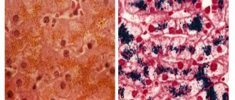
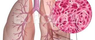
Pathomorphology • Capillary and tissue hypoxia causes fluid transudation. The alveoli are filled with bronchial secretions and cells, which prevents complete collapse of the atelectasis area. • The addition of infection causes fibrosis and bronchiectasis.
The clinical picture varies depending on the rate of development of bronchial occlusion, the extent of atelectasis and the presence of infection.
• Diffuse microatelectasis, small atelectasis, slowly developing atelectasis and middle lobe syndrome (chronic atelectasis of the middle lobe of the right lung due to compression by lymph nodes) may be asymptomatic.
• Extensive atelectasis due to acute occlusion is characterized by the following symptoms •• Pain on the affected side, sudden shortness of breath and cyanosis •• Cough •• Hypoxia with a significant decrease in paO2 with a tendency to its recovery during the first 24–48 hours due to weakening of blood flow in the atelectasis area • • Percussion: dullness of percussion sound over the area of atelectasis •• Auscultation ••• absence of breath sounds - with occlusion of the airways ••• bronchial breathing, if the airways are patent ••• moist rales with focal obstruction •• Decreased chest excursion •• Displacement apical impulse.
• Chronic atelectasis •• Shortness of breath •• Cough •• Percussion: dullness of percussion sound •• Auscultation: moist rales •• If infected: increased amount of sputum, rise in body temperature •• Recurrent bleeding from the affected area is possible.
Age characteristics • Early childhood: aspiration mechanism, pneumonia • Children: among the most common causes are mediastinal cysts, vascular anomalies • Elderly: among the most common causes are lung tumors, cicatricial stenosis, bronchiectasis.
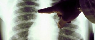
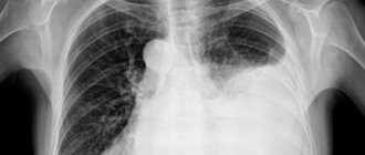
Special studies • Chest X-ray in two projections •• triangular-shaped intense homogeneous shadow with clear boundaries, with the apex directed to the root of the lung, with a decrease in the volume of the affected area of the lung •• With atelectasis of the lobe or lung - persistent displacement of the mediastinum to the affected side, dome the diaphragm on the affected side is raised, the intercostal spaces are narrowed •• diffuse microatelectasis - an earlier manifestation of oxygen intoxication and acute respiratory distress syndrome: a “ground glass” picture •• rounded atelectasis - rounded shading with a base on the pleura, directed towards the root of the lung (“comet-shaped” tail of blood vessels and airways). More often occurs in patients exposed to asbestos and resembles a tumor •• right-sided midlobe and lingular atelectasis merge with the borders of the heart on the same side (Arman-Delisle sign) • Bronchoscopy is indicated to assess the patency of the airways • EchoCG to assess the condition of the heart in cardiomegaly • CT or MRI of the chest.
Treatment
• The regimen depends on the patient's condition. Physical activity should be encouraged.
• Acute atelectasis (including acute postoperative massive collapse) •• The main cause of atelectasis should be eliminated, sanitation bronchoscopy should be performed, especially in cases of obstruction of the bronchial lumen with viscous sputum or vomit •• In case of foreign body aspiration - endoscopic removal.•• Adequate oxygenation, respiratory hydration mixtures •• In severe cases, mechanical ventilation with positive expiratory pressure or the creation of continuous positive pressure in the respiratory tract in persons with neuromuscular weakness •• Postural drainage (the head of the bed is lowered so that the trachea is below the affected area), breathing exercises, early postoperative mobilization of the patient •• Physiotherapeutic procedures, massage •• Broad-spectrum antibiotics are prescribed from the first day.
• Chronic atelectasis •• Postural drainage, breathing exercises (spirosimulator) •• Ventilation of the lungs with positive expiratory pressure or creation of constant positive pressure in the airways in persons with neuromuscular weakness •• Broad-spectrum antibiotics for purulent sputum •• Surgical resection of an atelectatic segment or lobe with recurrent infection and/or bleeding from the affected area •• If the obstruction is caused by a tumor, then the choice of treatment method is determined by the nature and extent of the tumor, and the general condition of the patient.
• Bronchodilators (salbutamol, fenoterol) - auxiliary value.
Complication : lung abscess (rare).
Prevention • Smoking cessation • Prevention of aspiration of foreign bodies and liquids, incl. vomit • In the postoperative period, the use of long-acting painkillers should be limited • Early postoperative mobilization of the patient • Breathing exercises.
ICD-10 • J98.1 Pulmonary collapse
Signs and symptoms
Signs and symptoms may be absent or include:
- cough, but not noticeable;
- chest pain (not common);
- difficulty breathing (fast and shallow);
- low oxygen saturation;
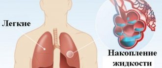
- pleural effusion (transudative type);
- cyanosis (late sign);
- increased heart rate.
It is a common misconception and pure speculation that atelectasis causes fever. A study of 100 postoperative patients followed by serial chest x-rays and temperature measurements showed that the incidence of fever decreased as the incidence of atelectasis increased. A recent review article summarizing the available published data on the association between atelectasis and postoperative fever concluded that there is no clinical evidence to support this suggestion.
Content
- 1 Signs and symptoms
- 2 Reasons
- 3 Diagnosis 3.1 Classification 3.1.1 Absorption (absorbing) atelectasis 3.1.1.1 Compression (relaxing) atelectasis
- 3.1.1.2 Cicatricial (grasping) atelectasis
- 3.1.2.1 Right middle lobe syndrome
Prevention
Smokers can reduce the risk of developing atelectasis postoperatively by quitting smoking, if possible, 6 to 8 weeks before surgery. After surgery, patients are encouraged to breathe deeply, cough regularly, and begin moving as early as possible. The use of devices that stimulate voluntary deep breathing (stimulus spirometry) and certain exercises, including changes in body position to enhance the removal of mucus and other secretions from the lungs, may help prevent atelectasis.
Prevention of atelectasis is also achieved by deep breathing. If possible, conditions that cause shallow breathing over a long period of time should be treated.
Recommendations
- ^ a b
Orenstein, David M. (2004).
Cystic Fibrosis: A Guide for the Patient and Family
. Lippincott Williams and Wilkins. paragraph 62. ISBN 9780781741521. - Mary Ellen's wedding; Gilis, Barbara A. (2005). Medical Terminology Systems: The Body Systems Approach: The Body Systems Approach
.
Philadelphia, PA: F. A. Davis Company. ISBN 0-8036-1289-3.[ page needed
] - "Atelectasis." Mayo Clinic
. Retrieved February 20, 2021. - Engoren, Milo (January 1995). "Lack of association between atelectasis and fever." Breast
.
107
(1):81–84. Doi:10.1378/chest.107.1.81. PMID 7813318. - Mavros, Michael N.; Velmahos, George S.; Falagas, Matthew E. (August 2011). "Atelectasis as a cause of postoperative fever." Breast
.
140
(2):418–424. Doi:10.1378/chest.11-0127. PMID 21527508. - Tarun Madappa. "Atelectasis." Medscape
. Retrieved 2018-02-02. Updated: November 28, 2021 - Air Vice-Marshal John Ernsting (2008). “The contribution of the RAF Institute of Aviation Medicine in 1945-1994. To ensure flight and aviation safety" (PDF). Journal of the Royal Air Force Historical Society
(43): 18–53. ISSN 1361-4231. - Lieutenant Colonel Rob "Mongo" Monberg. "Review of accelerated atelectasis: an old problem in a new environment" (PDF). IAMFSP.
- Woodring, John H. and James C. Reed. "Types and mechanisms of pulmonary atelectasis". Journal of Thoracic Imaging 11.2 (1996): 92-108.
- White, Gary K. (2002). Clinical Laboratory Core Competencies for Respiratory Care, 4th ed.
. Delmar Cengage Learning. p. 230. ISBN 978-0-7668-2532-1. - ^ a b
Robbins (2013).
Basic pathology
. Elsevier. item 460. ISBN 978-1-4377-1781-5. - Sheikh, Zeeshan; Weerakkody, Juranga. "Atelectasis of the lung." Radiopedia
. Retrieved February 20, 2021. - Kaplan Lecture Notes in Medical Pathology (2019). p.118
- Payne, K. R.; Jaques, P; Kerr, I. H. (1980). "Lung folding modeling peripheral lung neoplasm (Blesovsky syndrome)." Rib cage
.
35
(12):936–940. Doi:10.1136/thx.35.12.936. PMC 471419. PMID 7268670. - www.ncbi.nlm.nih.gov/books/NBK545316/.
First aid for pneumothorax
When pneumothorax occurs (closed, open or valvular), emergency care is required. If possible, assistance should be qualified and provided in a specialized hospital. In some cases, competent and timely provision of first aid can save a human life. In case of open pneumothorax or suspected lung collapse, a certain sequence must be followed.
Emergency care algorithm for spontaneous pneumothorax:
- Place the victim on an elevated surface to ensure the most favorable position for the respiratory system.
- Apply an occlusive dressing to the wound surface, which during emergency care can be any means that ensures the tightness of the affected part in the chest cavity. Available means include plastic film, adhesive tape, and rubberized fabric. The occlusive dressing is fixed with bandages or any fabric pre-treated with a disinfectant or iodine. Proper application of a bandage helps prevent the entry of an infectious agent onto the wound surface and prevent the development of a bacterial infection. The area around the wound must be treated with baby cream or Vaseline. In a hospital setting, the doctor will treat the peri-wound surface with a special ointment and apply a special hydroactive napkin.
- The patient must be anesthetized; the use of narcotic analgesics is allowed for severe pain.
- To remove air and drain the pleural cavity with drainage, the patient undergoes a pleural puncture in a hospital setting.
- Hormonal medications ( Dexamethasone ) are used to maintain normal blood pressure levels.
- If necessary, resuscitation measures are carried out.