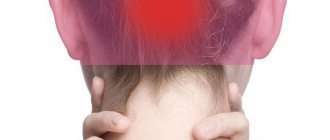
Causes of foot pain
Traumatic injuries
A foot bruise occurs when a heavy object falls, hits or trips. It manifests itself as moderate, gradually subsiding pain, swelling, and bruising. Support is preserved, sometimes limited. Symptoms disappear after 1-2 weeks. Foot fractures develop for the same reasons as bruises. They are characterized by explosive sharp pain, significant swelling, and severe dysfunction. The clinical picture is determined by the location of the fracture:
- Fracture of the talus.
Rarely seen. I am worried about pain at the base of the foot, under the ankle joint. Hemorrhages are detected in the area of the inner ankle. The ability to lean on the foot is lost. - Fractures of other tarsal bones.
Rarely diagnosed. Pain, swelling, and hemorrhages are found in the proximal portion of the dorsum of the foot. Turning the foot inward and outward is sharply painful. Support is impossible. - Metatarsal bone fractures.
Widespread. They manifest as pain in the middle and distal parts of the foot, and can be single or multiple. In the first case, the swelling is local, support on the foot can be partially preserved, in the second, due to significant swelling, the foot resembles a “pillow”, support is only possible on the heel.
Fractures of the bones of the foot in children are usually detected in the metatarsus area. With single fractures and no displacement, the pain may be moderate, but severe swelling and bruising indicate the presence of severe damage. Dislocations of the foot bones occur rarely and are accompanied by extremely intense, sudden pain that practically does not subside over time. The foot is swollen, grossly deformed, with extensive hemorrhages. The point of maximum pain corresponds to the location of the dislocation of the metatarsal or tarsal bones.
Marching foot (stress or fatigue fracture) is a special type of damage to the bones of the metatarsus, detected when the feet are intensely overloaded. Pain in the feet develops acutely 2-4 days after overexertion or gradually over 1-2 weeks. Patients complain of severe, sometimes unbearable pain in the middle part of the foot.
Chill develops with increased humidity and periodic cooling, manifested by burning, bursting pain, itching, sensitivity disorders, and the formation of areas of dense bluish-purple edema. With frostbite, the pain is initially slight, tingling, and appears against the background of coldness and pallor of the limb. Then the painful sensations become intense, burning, complemented by swelling, cyanosis, blistering, and in severe cases, necrosis.
Joint inflammation
All forms of foot arthritis are characterized by pain that intensifies when standing and walking, stiffness of movement, most pronounced in the morning, swelling, and hyperemia of the skin in the area of the affected joints. In rheumatoid arthritis, the joints are affected symmetrically on both sides; an acute onset with severe pain, fever, or a primarily chronic course is possible. The intensity of pain varies from weak, short-term to strong, permanent. Other manifestations correlate with the severity of pain.
In post-traumatic arthritis, one joint is affected, and the pain usually increases slowly over months or years. Nonspecific infectious arthritis develops against the background of bacterial, fungal, and viral infections and manifests itself with acute severe pain at rest and during movement, increasing swelling, local hyperthermia, and manifestations of general intoxication.
Gouty arthritis is characterized by damage to the first metatarsophalangeal joint. The attack begins suddenly, usually at night with very sharp acute pain, local swelling, redness of the skin, and increased body temperature. The pain persists for 3-10 days, then all symptoms completely disappear. Repeated attacks may develop over several months or years, with the intervals between attacks gradually becoming shorter. Tophi form on the skin.
With chondrocalcinosis (pseudogout), arthritis of a similar localization is sometimes observed, which can complicate diagnosis due to a similar clinical picture. Pseudogout is also accompanied by intense pain, swelling, and hyperemia of the joint; the difference from gout is the absence of general hyperthermia and tophi. An attack of chondrocalcinosis lasts about 1 week, on the first day the pain increases, then decreases.
Inflammation of bone structures
Simple periostitis of the foot is rare, usually the result of injury, manifested by moderate pain and slight swelling in the affected area. Periostitis ossificans develops after injuries, with arthritis, tumors, trophic ulcers, and some other diseases. It is characterized by moderate or minor pain in combination with increasing limitation of movements. With a long course, fusion of the tarsal bones is possible.
Osteomyelitis of the foot bones is provoked by open fractures, infected wounds, purulent diseases, and foot surgeries. It manifests itself as an increase in pain and a deterioration in general condition. The pain is tugging, bursting, boring, aggravated by any movement. The intensity of symptoms decreases after the formation of a fistula.
Foot pain
Specific infections
Gonorrheal arthritis in the area of the metatarsophalangeal joints is more often diagnosed in men and is manifested by intense pain during movement, fever, chills, local edema, and hyperemia. After the acute effects subside, the disease acquires a chronic relapsing course, reminiscent of the clinical picture of rheumatoid arthritis. Movement restrictions arise and deformities form.
With the development of osteoarticular tuberculosis, patients for some time are bothered by periodic mild pain in the foot, weakness, lethargy, and decreased ability to work. Then the intensity of the pain increases, the foot becomes swollen and hyperemic. Gait is disturbed. Gradually, the pain reaches an unbearable level, the general condition continues to deteriorate, fistulas form on the foot, and ankylosis of the joints forms.
Tendon diseases
Tenosynovitis most often affects the tendons of the dorsum of the foot. An acute nonspecific form of pathology is manifested by sudden moderate pain, limitation of movements, significant swelling, which can spread to the entire foot, ankle joint, and lower leg. With infectious tendovaginitis, the pain syndrome is more pronounced than with ordinary nonspecific. Fever and lymphadenitis are observed.
In acute crepitant tendovaginitis, along with pain during movements, there is a soft crunch, swelling is less than in other forms, and there is no fever. In the chronic course of aseptic tenosynovitis, a cord-like compaction forms along the tendon. Palpation is painful, movements are limited due to pain. The swelling is minor.
Osteochondropathies
Keller's disease I is detected in boys 3-7 years old and is accompanied by pain in the proximal part of the foot, closer to its inner edge. The pain intensifies when walking and feeling, forcing the child to rely not on the entire foot, but on its outer edge. Over time, the pain becomes constant and does not disappear even with rest. Lameness occurs. There is no swelling or hyperemia. All symptoms of osteochondropathy disappear within a year; without treatment, deformation of the foot is possible.
With Keller's disease II, which develops in girls 10-15 years old, pain is noted in the anterior parts of the foot, in the projection of the II-III metatarsal bones. At first, the pain is mild, appears during prolonged walking, then intensifies and becomes prolonged. Patients complain of increased pain when wearing shoes with thin soles or walking on uneven surfaces. Then the pain syndrome becomes permanent. Recovery occurs within 2-3 years, shortening of the affected metatarsal bone is possible.
Flat feet
Transverse flatfoot is manifested by aching, pulling, burning pain in the feet, and a feeling of heaviness in the legs. Symptoms intensify after prolonged walking or standing. The pain is localized in the forefoot along the inner edge. Hallux valgus is formed. Pain syndrome in the projection of the 1st metatarsophalangeal joint becomes longer and more intense, disturbing at night, combined with external deformation and limitation of movements.
In patients with longitudinal flatfoot, pain is localized mainly in the middle parts of the foot. Grade 1 is accompanied by mild pain after significant exercise. In grade 2, pain appears after light exertion, is felt at rest, and spreads to the ankle joint. At grade 3, the pain is constant and intense. There are difficulties when walking and using regular shoes. In the combined form of the disease, symptoms of longitudinal and transverse flat feet and diffuse pain are detected.
Other foot deformities
With the heel and cauda foot, the painful sensations are also diffuse, affecting the ankle joint and lower leg, complemented by a severe limitation of flexion of the foot to the plantar or dorsal side. With pes cavus, pain is localized in the proximal parts of the foot and is caused by compression of the tarsal bones by shoes. The pain syndrome is more pronounced in the case of a combination of cavus foot with transverse flatfoot.
Stiff big toe initially manifests as mild pain in the metatarsophalangeal joint after heavy exercise. Then the pain becomes prolonged and remains at rest. Due to the limitation of movements, the patient experiences overload of the outer parts of the foot, and pain appears in the zone of the V metatarsophalangeal joint. Subsequently, pain in the foot bothers you at night, there is no movement, and walking is difficult.
With hallux valgus, painful sensations are observed mainly in the inner parts of the foot and above the outer ankle. The pain syndrome intensifies with exertion, using uncomfortable shoes, and is combined with pain in the lower leg caused by constant muscle strain.
Congenital pathologies
Clubfoot is characterized by pain along the outer edge of the foot, which over time spreads throughout the foot, to the overlying parts of the limb. The foot is deformed, the sole is turned inwards. With congenital pachyonychia, extensive painful calluses and cracks form on the protruding areas of the feet. Changes in the feet are combined with onychodystrophy, the appearance of warty growths, peeling of the skin, and plantar hyperhidrosis.
Foot lesions in diabetes
Diabetic osteoarthropathy is a type of diabetic foot that primarily affects the metatarsal joints. Pain in the feet is initially mild and occurs against the background of existing swelling, hyperemia, and some deformation of the feet. Then the duration of the pain syndrome increases, pain appears at rest, and gross deformity forms.
The neuropathic form is characterized by the formation of zones of hyperkeratosis, painful ulcers, abrasions, and cracks on the plantar surface of the foot. In the ischemic form of diabetic foot, pain develops when walking, provokes intermittent claudication, and is combined with persistent swelling and weakened pulsation of the arteries. In severe cases, diabetic gangrene is detected, characterized by pain and swelling in the area between the affected and healthy tissues.
Vascular pathologies
Pain in the feet is observed with obliterating lesions of the arteries of the lower extremities. At first, they are observed only during walking and cause intermittent claudication. The pain occurs mainly in the sole area and spreads to the fingers and shins. Gradually the “pain-free” distance is reduced. In later stages, pain persists at rest. The limbs are pale, cold, the pulse is weak or undetectable. This symptomatology is typical for the following diseases:
- obliterating endarteritis;
- thromboangiitis obliterans;
- obliterating atherosclerosis.
In addition, pain in the feet is detected in trophoneurosis - erythromelalgia. The pain is very intense, burning, baking, starts with one finger, spreads to both feet. They arise suddenly, are provoked by compression or overheating of the limb, complemented by edema and pronounced hyperemia. They last from several minutes to several hours and interfere with walking.
Neurological causes
Morton's neuroma is characterized by burning local pain at the level of the heads of the metatarsal bones (usually in the 3rd interdigital space, less often in the 2nd), radiating to the fingers. Shots are possible. The pain is associated with wearing tight shoes and physical activity. A wave-like current is noted. As the pain progresses, it becomes constant, bothers you in any shoes, decreases, but does not disappear when you remove it, and is combined with numbness of the fingers.
Foot pain also occurs with tibial nerve neuropathy. The burning nature of the pain is typical, combined with sensory disturbances and muscle weakness. The localization of the pain syndrome is determined by the level of the lesion:
- Tarsal tunnel syndrome
: burning pain in the sole of the foot radiating down the back of the leg. - Medial plantar nerve
: along the inner side of the plantar radiating to the toes. - Common digital nerves
: starts from the arch of the foot, passes to the base of the II-IV fingers, then into the fingers themselves. - Calcanodynia
: pain occurs mainly in the heel area.
Neuropathy of the peroneal nerve is manifested by pain on the outside, and neuropathy of the sciatic nerve - on the posterolateral surface of the foot and lower leg. With femoral nerve neuropathy, pain is noted along the inner surface of the foot.
Other reasons
Among the tumor diseases in the foot area, there is chondroma, which is benign, manifested by short-term mild pain with unclear localization, intensifying with the growth of neoplasia and shoe pressure. Pain in the feet can also occur with depression and some mental disorders. Pain syndrome caused by mental disorders is characterized by inconsistency with the clinical picture of a particular disease, absence of anatomical changes, and sometimes a bizarre nature of sensations.
How to treat fasciitis correctly
The most effective and lasting treatment is always aimed at eliminating the cause of the disease. The main factors in the development of fasciitis are overload of the foot and decreased elasticity of the fascia. Therefore, actions aimed at unloading the arches and improving the quality of the ligaments lead to the most noticeable results. Most importantly, effective treatments for fasciitis are safe.
Plantar fasciitis is a disease that requires the active participation of the patient himself. Of course, it seems easier to give the injection and forget about the pain. But until the patient himself improves the functioning of his body, the symptoms will return and increase. Below are the most effective methods for finally getting rid of pain between the toe and heel:
- High-quality orthopedic insoles. It's better if they are individual. The elastic protrusions of the instep supports massage the aponeurosis, improve its blood circulation and elasticity. The right insoles support the arches well and relieve excess stress on the fascia.
- Physical exercise. A competent rehabilitator will describe a training regimen and “stretching” that will work most effectively for you. With regular exercise, it is possible to increase the elasticity of the fascia and muscles, which will significantly reduce discomfort.
- Night tires. Durable plates that are worn on the leg during sleep and improve the metabolism of the aponeurosis. During the night period, body tissues are most pliable, which gives the splint the opportunity to effectively increase their elasticity.
Medtechnika Orthosalon stores are professional establishments where you can undergo foot diagnostics and select the most suitable orthopedic products. A large selection of therapeutic and prophylactic insoles, massagers and splints allows you to individually approach any form of fasciitis. Experienced consultants will help you decide on the optimal models to improve the performance of your feet.
Diagnostics
The causes of foot pain are determined by orthopedic traumatologists. Data from an objective examination and additional studies are used to make a diagnosis. The following procedures are performed:
- Survey.
During a conversation with the patient, the circumstances, time of onset of pain and other symptoms are established. The nature of the pain syndrome and its relationship with various factors are determined. Find out your life history and family history. - Visual inspection.
The doctor assesses the appearance of the foot, identifies deformities, swelling, and violations of the integrity of the skin. Studies color, local temperature, pulsation of arteries, sensitivity, joint mobility, and other indicators. - X-ray of the foot.
Performed in 2 or 3 projections involving the entire foot or part of it. For flat feet, weight-bearing photographs are prescribed. X-rays show fractures, dislocations, areas of bone destruction and restructuring, and degenerative changes in joints. Assessing the relationships between the various structures of the foot plays a significant role. - Plantography and plantoscopy.
Methods for identifying flat feet and other foot deformities. They make it possible to establish the nature and severity of changes, features of the distribution of load on the feet, and the need to use orthopedic devices. - Ultrasound.
Ultrasound techniques (ultrasound, duplex scanning) are necessary for vascular pathology. Prescribed to study the state of the vascular network, vascular patency, blood flow speed, and other factors. - Electrophysiological methods.
Indicated for neurological disorders. The purpose of the examination is to clarify the level of nerve damage, the condition of the nervous and muscle tissue. - Lab tests.
They confirm the presence of inflammation, identify metabolic disorders and specific markers of rheumatological diseases. They make it possible to assess the general condition of the body in case of severe pathologies.
Taking into account the nature of the disease, consultations with other specialists are prescribed: endocrinologist, neurologist, vascular surgeon. According to indications, MRI is performed, and a biopsy of bones and soft tissues is performed to study their morphological structure.
Plaster cast on the foot
Joint diseases
The supporting apparatus of the feet contains a large number of small joints that can be susceptible to various diseases. Therefore, one of the most common reasons why the feet hurt after walking is joint diseases:
- arthritis - inflammation of articular cartilage of an autoimmune or infectious nature;
- polyarthritis - simultaneous inflammation of several small joints;
- osteoarthritis - destruction of the cartilage structure due to microtrauma and metabolic disorders;
- bursitis - inflammation of the joint capsules;
- gout - accumulation of uric acid crystals on the surfaces of cartilage;
- pseudogout is an arthritis caused by the accumulation of calcium compounds in the synovial fluid.
With osteoarthritis of the ankle, the joint of the foot hurts when walking, and after periods of rest the pain subsides. Arthritis, on the contrary, is characterized by pain that bothers you regardless of physical activity, during night sleep.
With bursitis, the foot hurts when walking on the bend or in the heel area, redness and bumps appear over the affected joints, and a local and general increase in temperature. Gout and pseudogout are characterized by severe pain, limited mobility, swelling, hyperemia of the skin and increased temperature over the affected area.
To effectively treat joints, it is necessary to determine the type of disease using complex diagnostics, which can be completed in our clinic.
Treatment
First aid
The limbs provide rest and an elevated position. For skeletal injuries, apply cold and immobilize with a splint or fixing bandage. In case of frostbite, an insulating bandage is applied; intensive warming of the feet is prohibited, as it can aggravate existing tissue damage. For non-traumatic foot pathologies, local anti-inflammatory and painkillers are used.
Conservative therapy
For fractures and dislocations, blockades, reduction or reduction, and application of a plaster cast are performed. The conservative treatment plan includes:
- Protective mode.
The specialist determines the mode of physical activity, the intensity of the load on the limb, and, according to indications, recommends the use of orthopedic devices and additional means of support (crutches, canes). - Drug therapy.
NSAIDs of general and local action are most often used. Infectious diseases require the prescription of antibacterial or antiviral drugs, vascular pathologies require medications to normalize blood circulation, diabetes mellitus require correction of antidiabetic therapy. - Non-drug methods.
Patients are referred for exercise therapy, massage, manual therapy, physiotherapeutic treatment (electrical stimulation, electrophoresis, UHF, laser treatment, etc.).
Prevention
It is important to be attentive to your health and pay attention to the slightest changes in the body. It is also recommended to follow simple rules:
- adjust your diet and lifestyle, add minimal exercise to your daily routine;
- if necessary, wear orthopedic insoles or shoes;
- choose the right shoe size, it should be comfortable;
- adjust your weight;
- walk barefoot more;
- perform self-massage.
Diseases of the ligamentous apparatus
The ligaments in the foot may hurt when walking due to injuries: sprains, bruises. If there was no obvious traumatic impact, but the tendon on the foot hurts when walking, then the causes of pain are regular microtraumas of the connective tissue resulting from excess body weight, too much physical activity, constant wearing of heels, flat feet, deterioration of metabolism due to aging or various diseases.
One of the most common diseases of the ligamentous apparatus of the feet, in which the sole of the foot hurts when walking, is plantar fasciitis - inflammation of the broad ligament that runs along the sole and connects the heel to the base of the toes. In the early stages of plantar fasciitis, the instep of the foot hurts when walking, and when the ligament in the area of the connection with the heel ossifies, a heel spur is formed, characterized by sharp pain in the heel area, intensifying at the time of loading.
Symptoms
With a variety of causes for heel pain, the symptoms of discomfort also vary. It is not for nothing that the doctor begins collecting anamnesis by clarifying the nature of the pain in order to understand in which direction to move. So, different diseases entail different sensations when they occur:
- throbbing pain in the form of alternating impulses;
- severe acute pain, constantly disturbing;
- periodic pain that occurs locally or everywhere;
- systematic morning and/or evening pain (often occurring immediately after overwork);
- redness;
- numbness;
- heat in the foot area;
- swelling and swelling;
- fever.
It is noteworthy that the initial stages of almost all diseases are asymptomatic.
Muscle inflammation
The muscles of the foot may hurt when walking after intense physical activity due to the accumulation of lactic acid in the muscle tissue. This pain does not require treatment and goes away within a few days.
Inflammatory processes in the soft tissues of the feet, caused by:
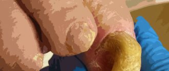
- corns;
- calluses;
- ingrown nails;
- infection of periungual tissues (panacirium).
They make walking difficult and cause suffering, but among all the causes of foot pain they are the most harmless. If you consult a doctor in a timely manner, these pathologies can be easily treated and do not lead to the development of complications.
It is important to correctly diagnose inflammation of the soft tissues of the feet, so as not to confuse them with symptoms of much more serious diseases, such as atherosclerosis of the lower extremities, diabetic foot.
Useful video from a doctor
Professor, neurologist, chiropractor and medical kinesiologist, MD. Vasilievna Lyudmila Fedorovna about pain in the foot:
Author of the article:
Kaplan Alexander Sergeevich |
Orthopedist Education: diploma in General Medicine received in 2009 at the Medical Academy named after. I. M. Sechenov. In 2012, she completed postgraduate studies in the specialty “Traumatology and Orthopedics” at the City Clinical Hospital named after. Botkin at the Department of Traumatology, Orthopedics and Disaster Surgery. Our authors
Vascular diseases
One of the most serious causes of pain in the legs is atherosclerosis of the lower extremities, in which the supply of oxygen and nutrients to the tissues of the feet is disrupted. The main causes of this disease are diabetes and smoking.
Diabetes mellitus leads to the development of the so-called diabetic foot, characterized by a whole complex of pathological changes. In the initial stages of the disease, symptoms such as decreased skin sensitivity, a feeling of numbness, nail deformation, swelling, itching, dryness, discoloration of the skin, and the ball of the foot may hurt when walking. In later stages, non-healing ulcers appear, which affect increasingly deeper layers of tissue down to the bones and lead to the development of gangrene, which requires immediate amputation of the legs above the affected tissue.