Definition of phalanx
Phalanx (plural: phalanges) refers to the bones found in the fingers, toes, paws, wings, hooves, and fins of animals.
These are long bones that are longer than they are wide. The phalanges are connected to each other at the interphalangeal joints, which can be used for flexion or extension. Some phalanges are fused with each other. There are 56 phalanges in the human body, with the thumbs and big toes having 2 bones each. The remaining fingers and toes have 3 bones with the fourth and fifth fingers having fused phalanges. This results in a phalangeal formula of 2 + 3 + 3 + 3 + 3 bones per limb. Depending on their position, these bones are attached to the bones of the palm (metacarpal bones) and foot (metatarsal bones) through ligaments at the metacarpophalangeal and metatarsophalangeal joints. Tendons connect the phalanges to the muscles of the palm and forearm, which direct the movement of the fingers. Similar attachments are seen for the phalanges in the toes.
The word phalanx comes from the Greek term for a military formation where soldiers stood in several rows and columns. Each digit has two or more bones, and each palm or foot consists of several digits. When these bone structures are visible (as in an x-ray or skeleton), it may appear as if the bones are arranged side by side, two or three rows deep, like an army formation.
What to do after forty
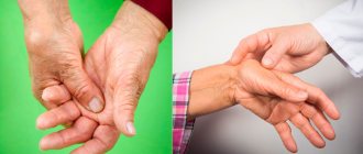
After forty, hormonal changes become the main cause of coxarthrosis, gonarthrosis and arthrosis of the hand. With the onset of menopause in women, the situation worsens, especially in the area of the fingers. The level of estrogen in the body decreases - it begins to actively lose moisture, including in the joint. Cartilage experiences a moisture deficiency, becomes brittle and vulnerable.
At this age, it is especially important to lead a healthy lifestyle - just being in the fresh air and giving up bad habits. The condition of joints and cartilage tissue depends on the style of nutrition, the supply of oxygen and vitamins.
Types of phalanx
The phalanges can be classified based on their position relative to the rest of the body. The bone closest to the metacarpals and metatarsals is the proximal phalanx, and the furthest one (usually located below the nail) is the distal phalanx. Some digits have more than two bones, and those located between the proximal and distal bones are called intermediate phalanges. While human fingers and toes have 2 or 3 phalangeal bones, there are primitive reptiles with 4, and some marine mammals may have up to 12 bones in number.
In horses, the proximal phalanx is called the pastern and looks like an hourglass. This is almost twice as long as the next intermediate phalanx. It is believed that the position of the hoof during running, which depends on the nature of the surface, influences the occurrence of fractures of these bones in racehorses. The proximal phalanges are also important when studying fossils to understand locomotion patterns, as they are skeletal elements in close connection with the substrate.
Injuries to the proximal phalanges include fractures and dislocations. They are usually treated with realignment under local anesthesia and splints to immobilize the bone and joint.
Type II: Intermediate phalanges
Intermediate phalanges are not present in all digits and are absent in the thumb and thumb in humans. These bones are intermediate in position, length and size. They are part of two interphalangeal joints interacting with the proximal and distal bones of the phalanx. In horses, the intermediate or middle bones of the phalanx are called crowns and form the upper border of the hoof. The shape and flexibility of this phalanx is important in animals such as bats, where the bone must flex and flex with air flow rather than restrict its movement.
Type III: distal phalanges
These bones, also known as terminal phalanges, play several roles. They support the fleshy underside of the fingertips in humans, which contain numerous nerve endings. Small, flat projections from these bones, called apical tufts, are present under the nails. A number of adaptations in the anatomy of the distal phalanx of the thumb are necessary for fine motor movement and for grasping objects. In large ungulates such as the giraffe, elongated, large distal phalanges are important for absorbing the stress of running. Similarly, arboreal mammals also have modified distal phalanges.
The distal phalanges of the toe bones taper outward, being widest at the base next to the proximal phalanx.
Recommendations
- Williams, Linda (22 June 2012). "A biphalangeal fifth toe is a common variation.'" news-medical.net
. Retrieved July 14, 2014. - Drake, Richard L.; Fogle, Wayne; Tibbits, Adam W. M. (2005). Gray's Anatomy for Students
. Illustrations by Richard Mitchell and Paul Richardson. Philadelphia: Elsevier/Churchill Livingston. ISBN 978-0-8089-2306-0. - ^ a b c
Gray, Henry (1918).
Anatomy of the human body
. ISBN 0-8121-0644-X. - ^ a b
"Apical bundles of phalanges". Center for Academic Research and Training in Anthropogenesis. Retrieved January 28, 2021. The fascicles support the fleshy palmar pad (also known as the distal pulp) on the palmar (volar) surface of the finger, as well as the nail on the dorsal surface. - ^ a b c d
Almesia, Sergio;
Moya-Sola, El Salvador; Alba, David M.; Streit, David S. (July 22, 2010). "The Early Origin of Human-Like Precision Grasping: A Comparative Study of Political Distal Phalanges in Fossil Hominins." PLoS ONE
.
5
(7):e11727. Doi:10.1371/journal.pone.0011727. PMC 2908684. PMID 20661444. - Shrewsbury and Johnson 1975, paragraph 784 harvnb error: no target: CITEREFShrewsburyJohnson1975 (help)
- Cooper et al., "Review and experimental assessment of the embryonic development and evolutionary history of fin and hyperphalanx development in dolphins (Cetacea: Mammalia)", ResearchGate
, DOI: 10.1002/dvg.23076. October 2021 - ^ a b
Mittra, ES;
Smith, V. C.; Lemelin, P; Jungers, W. L. (December 2007). "Comparative morphometry of the primate apical fasciculus". American Journal of Physical Anthropology
.
134
(4):449–59. Doi:10.1002/ajpa.20687. PMID 17657781. - Hamrick, M. W. (June 1998). "Functional and adaptive significance of primate pads and claws: evidence from New World anthropoids." American Journal of Physical Anthropology
.
106
(2):113–27. Doi:10.1002 / (SICI) 1096-8644 (199806) 106: 2 3.0.CO; 2-R. PMID 9637179. - ^ a b
Gough-Palmer, Antony L.;
McLachlan, Jody; Routh, Andrew (March 2008). "Paws to Think About: Comparative Radiological Anatomy of the Mammalian Forelimbs". Radiographics
.
28
(2):501–510. doi:10.1148/rg.282075061. PMID 18349453.
Functions of the phalanx
These bones form the structure of the fingers and toes and are critical to much of hominid evolutionary activity. view, Tool making, precision grasping, grasping and handling of equipment arose from the anatomical features of the phalanges. The contralateral thumb is due to the evolution of the tendon and ligament structure surrounding the bones. A number of changes in the anatomy of the distal phalanx of the thumb are necessary for fine motor movement.
In other animals, phalanges are adapted for rapid movement (horses and giraffes), for locomotion (arboreal mammals), for flight (wings in bats, birds), for swimming (fins in aquatic species) and for hunting (claws) and paws in carnivores).
Diagnostics
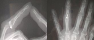
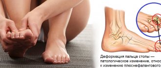
The appearance of signs of exostosis requires contacting an orthopedist-traumatologist. At the appointment, the doctor carefully examines the finger, palpates the growth and finds out the nature of the symptoms. If a dense bone formation is detected, an x-ray is indicated.
With its help, you can not only diagnose exostosis of the toe, but also evaluate its location and size. The images also provide data on the degree of deformation of the distal phalanges and allow you to plan the most effective course of treatment. In rare cases, CT and MRI are additionally prescribed.
Vestigial phalanges
The evolution of mammals gave a phalangeal formula of 14 bones per limb. However, in many taxa one or more digits have lost their direct function. Dogs, cats, cattle and many other ungulates have a big toe called a dewclaw. This phalanx is not usually used during movement.
In some monkeys, such as colobus monkeys, the thumb is vestigial. Many anteaters, armadillos and sloths have vestigial or missing phalanges. The giant armadillo and the nine-barred armadillo have vestigial fifth toes. In arboreal three-toed sloths there are no two numbers with accompanying phalanges, since they are not important for the animal’s lifestyle. The giant anteater has a fifth finger with only 2 phalanges. In lesser anteaters and silky anteaters, the fifth digit has lost all its phalanges.
- arboreal – Referring to organisms (especially animals) that live in trees.
- condyle - A spherical extension at the end of some bones that forms an interface with another bone.
- digit – Derived from the Latin word “digitus,” it refers to structures found on the extremities of limbs, such as fingers or toes.
- vestigial - Parts of the body that have lost function during evolution, although they are found as essential body parts in other, related species.
Types of finger fracture
Because of
- Traumatic fractures are damage to the finger bone due to trauma.
- Pathological fracture - a fracture of a finger in the area of pathological restructuring (affected by any disease - osteoporosis, tumor, osteomyelitis, etc.) Osteoporosis is the most common cause of a pathological fracture.
The nature
- Closed fractures (without breaking the skin)
— Incomplete
— Complete
- Open fractures (with skin damage)
— Primary open
— Secondary open
Based on the presence of offset:
- Fractures without displacement of fragments
- Displaced fractures.
Treatment tactics
In the early stages, you will be offered conservative treatment that will reduce pain and increase finger mobility. Some methods of physiotherapy for arthrosis will improve blood circulation and restore metabolism, activate tissue regeneration and reduce pain. The following techniques are also practiced:
- osteopathy and manual therapy - to strengthen muscles, improve mobility, restore periarticular blood flow;
- reflexology – helps relieve inflammation and swelling, improves the functioning of damaged interphalangeal joints;
- hirudotherapy – can replace several medications at once;
- physical therapy – relaxes muscles and strengthens muscles.
Intra-articular injections of the Noltrex synovial fluid prosthesis help stop the progression of arthrosis. The principle of action is simple: the gel gets inside the joints and takes on the functions of the missing lubricant, pushes apart the thin cartilaginous surfaces and nourishes them. The friction of the cartilages stops, they are no longer destroyed by mechanical stress, and the person does not experience pain.
If an orthopedist recommends intra-articular injections, then all is not lost
Hands need careful care, and it’s not just a matter of choosing the right cream or cosmetic procedures. We also need to take care of the interphalangeal joints, because they determine how confident and accurate our fine motor skills will be. Regularly stretch your fingers, do exercises, protect your hands from injuries and hypothermia - then the risk of developing arthrosis will be much lower.