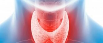
Technique for performing a blood test for thyroid hormones
The result will largely depend on how correctly the blood test for thyroid hormones is taken.
As a result, the correct diagnosis will be made and the correct treatment will be prescribed. With proper puncture, the risk of complications is minimized. For example, if the method of blood sampling is disturbed, end-to-end damage to the vessel is possible with further formation of a hematoma. And if antiseptic requirements are not observed, an inflammatory process may begin.
That is why blood sampling must be carried out by a specialist under appropriate conditions, using a disposable syringe or a special vacuum system.
Many clinical settings still use a needle to directly transfer material into a test tube. This technique is not only inconvenient, but also unsafe, as it is associated with an increased risk of blood contact with the environment.
Taking material with a disposable syringe is also considered a relatively outdated method. The obvious disadvantages of this procedure are the need for additional test tubes and test systems, as well as frequent cases of hemolysis during manipulation.
Modern laboratories have long been using new vacuum devices for collecting venous blood. The analysis apparatus consists of a test tube, inside of which there is a vacuum and a special chemical reagent, as well as a thin needle and a holding adapter. Such devices are durable. They completely eliminate the possibility of confusion in tests, contact of material with the environment and the hands of a healthcare worker, and also do not require the use of additional instruments. Blood collection using this method is painless, safe and fast.
Hormonal influence on the menstrual cycle
Depending on the content of LH, FSH, and estrogens in the blood, the menstrual cycle in women is divided into three phases, one replacing the other:
- Follicular (menstrual) - average duration 2 weeks (7-22 days), the end of the ovarian cycle. It begins on the first day of menstruation, when the functional layer of the endometrium is shed, coming out with menstrual blood and glandular secretions. During this phase, the dominant follicle matures, which has the largest number of FSH receptors and produces estradiol more than other follicles. The menstrual phase ends with a sharp release of LH from the pituitary gland, which gives rise to the next phase - the ovulatory phase.
- Ovulatory (proliferative) phase - average duration is approximately 3 days. The dominant follicle finally matures and becomes a Graafian vesicle, capable of admitting the egg. The ratio of FSH and LH changes. The phase is characterized by a wave-like release of LH, which stimulates active substances (prostaglandins and enzymes) that promote rupture of the walls of the Graafian vesicle and the release of the egg, that is, ovulation. This period is characterized by a decrease in estradiol and the development (not in all cases) of ovulatory syndrome. Many women feel ovulation by the appearance of pain in the lower abdomen and lower back. Ovulation occurs after the peak release of luteinizing hormone within 16-48 hours. Follicular fluid (5-10 ml) comes out with the egg.
- The luteal (secretory) phase - the average duration is 2 weeks, this is the most stable period of the cycle - the corpus luteum phase. After ovulation, the vesicle transforms into the corpus luteum, which secretes progesterone (called the pregnancy hormone), androgens (male steroids), and estradiol. Under the influence of these hormones, the endometrium thickens, secretes secretions, and prepares for the attachment of the oocyte.
At the end of the luteal phase, at the peak of the release of sex hormones, the production of FSH and LH decreases. If conception does not occur, the corpus luteum stops synthesizing estrogens and progesterone, after which it is destroyed. The negative feedback is interrupted, which contributes to the growth of LH and FSH and the beginning of a new cycle.
Development of the endometrium under the influence of hormones
What tests are done for thyroid hormones?
- TSH (more fully known as thyroid-stimulating hormone, or thyropine) is a substance produced by the pituitary gland. It activates the formation and production of hormones in the thyroid gland (such as T3 and T4). When the functioning of the pituitary gland is not impaired, the TSH level decreases against the background of increased thyroid function, and increases against the background of weakened thyroid function.
- Free T3 (another name is free triiodothyronine) is a substance synthesized by the thyroid gland that stimulates metabolic processes and activates oxygen uptake in tissues.
- Free T4 (we are talking about free thyroxine) is a hormonal substance produced by the thyroid gland and activates the processes of protein synthesis.
- AT-TG (meaning the presence of antibodies to thyroglobulin) - the level of these antibodies makes it possible to detect autoimmune pathologies of the thyroid gland, such as Hashimoto's disease, diffuse toxic goiter, atrophic autoimmune thyroiditis.
- AT-TPO (presence of microsomal antibodies, or antibodies to thyroid peroxidase) - we are talking about the presence of antibodies to the enzyme substance of gland cells. This analysis is very important for diagnosing autoimmune pathologies.
TSI/thyroid stimulating immunoglobulins
Thyroid-stimulating immunoglobulin-TSI is an antibody that stimulates the thyroid gland to enlarge and release excess thyroid hormone, causing hyperthyroidism. This test is sometimes called TSH receptor stimulating antibody.
Reference range: less than or equal to 1.3
TSI levels are elevated in 75% to 90% of patients with Graves' disease. The higher the level, the more active Graves' disease is considered to be. Some people with Hashimoto's disease also have these antibodies, which can cause repeated, transient episodes of hyperthyroidism.
The TSI test is usually performed to screen for Graves' disease. This is usually done in pregnant women with Dietsch's disease during the last three months of pregnancy to assess the risk of hyperthyroidism or Dietsch's disease in the baby.
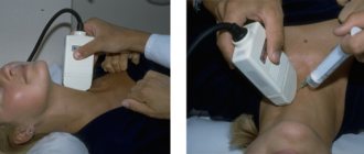
How is a thyroid hormone test taken?
The material is collected as follows:
- The health worker prepares instruments, laboratory directions (labels, enters information about the patient, makes notes in the journal and/or electronic system).
- The patient sits down on a chair. The health worker fixes his arm, first turning his palm up and extending the elbow joint as much as possible. For convenience, a special cushion is placed under the elbow area.
- Apply a tourniquet to the area of the middle third of the shoulder (pulse impulses on the wrist should be palpable).
- The specialist treats the skin in the area of the elbow with alcohol, asks the patient to make several movements, clenching and unclenching his fist (this will allow the vein to be filled to the maximum with blood), after which the patient fixes the fist in a clenched state.
- The healthcare worker punctures the vein (an acute angle must be maintained) and draws material into a test tube or special system, while simultaneously loosening the tourniquet. At this time, the patient loosens his fist.
- The specialist applies a cotton pad moistened with alcohol to the puncture site and removes the needle from the vessel. If a vacuum system was used, the tube with blood is first disconnected.
- The patient should sit for a while with his arm bent at the elbow joint to avoid bleeding. Usually 5-6 minutes are enough for this.
The health worker places the signed test tubes in a special container and then sends them to the laboratory.
Analysis of thyroid hormones for a child
Fluctuations in thyroid hormone levels in childhood are quite common. Statistics say that one baby out of five thousand babies born is diagnosed with a congenital thyroid pathology.
With a deficiency of thyroid hormones, children may lag behind in development, so doctors often prescribe special diagnostics to promptly identify the problem.
The normal TSH level in a child is always higher than in an adult. This hormonal substance is produced by the pituitary gland and serves as a kind of stimulator of the synthesis of T3 and T4. Accordingly, immediately after birth its level is higher than in adolescence.
At different age periods, normal TSH levels for children differ significantly:
- the first three days after the birth of a baby - from 1.3 to 16 mm/l;
- during the first four weeks of life – from 0.9 to 7.7 mm/l;
- after seven years and older - from 0.6 to 5.5 mm/l.
T4 and T3 levels remain stable throughout the entire period from newborn to adulthood (2.6-5.7 pmol/l and 9-22 pmol/l, respectively).
At the first signs of hypothyroidism, a decrease in T4 and T3 levels occurs with normal TSH values.
Secondary hypothyroidism occurs when the pituitary gland is damaged: all types of metabolism in the body are disrupted, the child becomes uncommunicative, apathetic, and lags behind in development - not only mentally, but also physically. The use of hormonal drugs at an early stage of pathology allows you to stabilize metabolic processes and stimulate the development of the baby.
Gitel E.P., Melnichenko G.A. MMA im. THEM. Sechenov, Moscow
In recent years, the interest of practitioners of various specialties in the pathology of the thyroid gland has increased, both due to its prevalence and the frequency of patients seeking medical help, and because this pathology has a significant impact on health, performance and quality of life. According to the Canadian Thyroid Research Foundation, 200 million people worldwide suffer from diseases accompanied by impaired thyroid function alone. Although there is no single classification of thyroid diseases accepted in all countries, nevertheless, in all classifications, indicators of the functional activity of the gland, parameters indicating its size and structural (morphological) characteristics are taken as the basis for assignment to a particular nosology. Even 25 years ago, the functional activity of the gland was judged mainly by the absorption of the isotope I-131 or technetium-99. In a significant proportion of people living outside iodine endemic areas, there is a correspondence between the level of radionuclide accumulation and hormone secretion, however, in an iodine endemic area, accumulation can be high not only in euthyroidism, but also in hypothyroidism. Regardless of the region of residence, the accumulation of the isotope may be paradoxically low in diffuse toxic goiter, if iodine-containing drugs were previously used, or in extremely intense biosynthesis of thyroid hormones. With the creation of radioimmune methods in the early seventies, it became possible to directly judge the level of thyroid hormones in the blood. However, until the mid-eighties, it was possible to determine only the total, i.e. simultaneously free and transport protein-associated functions of T3 and T4 (total T3 and total T4). These methods, while ensuring unconditional progress in the diagnosis and treatment of thyroid pathology, were not without serious drawbacks. Although the share of free fractions accounts for only 0.03% of total T4 and 0.3% of total T3, however, it is the fractions not bound to protein that provide the biological and metabolic activity of hormones.
Very often, the level of total, total hormones is increased or decreased in the absence of any pathology, for example, an increase in total (total T3 and T4) is typical for pregnancy and the newborn period, for taking estrogen-containing drugs, heroin, for some liver diseases, etc. . On the contrary, a decrease in the level of total T3 and T4 without any dysfunction of the gland is observed in diseases that occur with protein loss - systemic diseases, nephrotic syndrome, Cushing's syndrome, when taking androgens, anabolic steroids, danazol, glucocorticoids in large doses. In old age, in 20% of people the concentration of thyroxine-binding proteins decreases, which leads to a decrease in total T4, with actual euthyroidism. As an additional study, which makes it possible to bring the results of determining total T4 closer to the determination of free hormone, the calculation of the so-called free thyroxine index is used, but in this case there is a need to conduct a laboratory study of thyroxine-binding globulin. At the same time, neither the free T4 index nor a similar indicator for T3 (free T3 index) are indicators completely similar to the definition of free hormones themselves, since thyroxine-binding globulin is not the only protein that binds thyroid hormones. Table 1 summarizes the factors that influence the level of thyroxine-binding globulin and reduce the informativeness of determining total T3 and T4.
| Table 1. Factors affecting the binding of T3 and T4 by thyroxine-binding globulin. | |
| Patient age over 60 years | |
| Neonatal period | Hyperandrogenism |
| Estrogens and hyperestrogenic conditions | Therapy with large doses of glucocorticoids |
| Tamoxifen | Acromegaly |
| Oral contraceptives | Nephrotic syndrome |
| Perphenazine | Major collagenoses |
| Porphyria | Genetic factors |
| Infectious and chronic active hepatitis | |
| Genetic factors | |
| HIV infection |
Considering the above and relying on the experience accumulated by laboratories and clinicians over the time that has passed since the introduction into widespread practice since the early nineties of methods for determining free T3 and T4, there is no doubt about the greater diagnostic significance of determining free forms of thyroid hormones compared to total forms. However, the development of methods for the direct determination of free T4 and free T3 required considerable effort and time from researchers. More than twenty years have passed since the description of RIA methods for determining total T3 and T4 until the creation of reliable and accurate methods for the direct determination of free T3 and T4, comparable in characteristics to the gold standard - the determination of these parameters after the separation of free and bound fractions of hormones in serum by the method of equilibrium dialysis . The greatest development in the use of immunochemical methods for determining free forms of thyroid hormones was associated with the development and implementation of automatic chemiluminescent analyzers into laboratory practice since the early 90s. It would seem that the most reliable and accurate markers of thyroid function should be the results of determining the free fractions of T4 and T3. Their level does not depend on protein content. Most drugs that affect total T4 and total T3 do not interfere with free T3 and free T4 measurements. However, given that the majority of T3 is formed from T4, and the concentrations of free T4 are 3 orders of magnitude higher than those of free T3, we can say that the main marker for assessing the hormonal activity of the thyroid gland is free T4. In addition, the determination of free T3 as a mandatory test, without providing significant diagnostic information in most cases, reduces the economic indicators of the laboratory and increases its workload, and without clear indications, for example, if the conversion of T4 to T3 is impaired, there is no need to perform it.
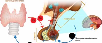
It is known that the production of thyroid hormones is regulated through a feedback mechanism that operates in the hypothalamus-pituitary-thyroid system. Physiologically active are free thyroid hormones, the level of which is regulated by thyroid-stimulating hormone (TSH) according to a feedback mechanism. There is an inverse logarithmic relationship between the concentrations of free T4 and TSH in the blood. Thus, a twofold increase in the level of free T4 corresponds to a 100-fold decrease in the concentration of TSH. This ratio makes the determination of TSH extremely important for assessing the functional state of the pituitary gland - thyroid gland. The disadvantage of radioimmunological methods for determining TSH in the first decades of their use (the so-called first-generation systems) was their low sensitivity, which made it possible to reliably differentiate only an increase in TSH levels from normal values, leaving the TSH concentration zone below 0.4 IU/l closed for diagnostic use, important for the differential diagnosis of latent thyrotoxicosis from the norm, leaving this prerogative to clinicians. At the end of the 80s, the use of non-isotopic variants of the label (fluorescent and chemiluminescent) and monoclonal antibodies made it possible to reduce the sensitivity threshold of methods for determining TSH to 0.1 IU/l (the so-called second generation). Further development is associated with the development of third-generation systems for use in automatic chemiluminescent analyzers with a sensitivity of 0.01 IU/L. In some systems, for example, in the third generation TSH system of the IMMULITE DPC analyzer (Diagnostic Product Corporation, USA), the analytical sensitivity reaches 0.002 IU/l. In this case, functional sensitivity, defined as the minimum concentration of the analyte measured with a coefficient of variation of less than 20%, is 0.01 IU/l. This sensitivity allows, in most cases, to differentiate the initial stages of thyroid dysfunction and practically eliminate the test with thyrotropin-releasing hormone. It is very important to use third-generation TSH methods to assess the effectiveness of suppressing pituitary TSH secretion in nodular lesions and thyroid cancer after surgical or conservative treatment, achieving an optimal TSH level depending on the malignancy or benignity of the lesion. So, in what situations does a clinician need to measure TSH and thyroid hormone levels? First of all, this is necessary in the presence of clinical signs of a particular thyroid pathology to confirm the diagnosis, secondly, to monitor the treatment of thyroid diseases and, finally, in real clinical practice. The study of thyroid hormones is a mandatory part of the general clinical examination in almost all hospitalized patients. This is partly justified, since there are huge groups of patients in whom primary hypothyroidism occurs under the “mask” of somatic, neurological, hematological or other pathology. One can only welcome the timely diagnosis of primary hypothyroidism in these cases, as well as the diagnosis of thyrotoxicosis in individuals whose only manifestation was paroxysms of atrial fibrillation. But the study of hormones “just in case” sooner or later confronts the doctor with the need to evaluate a common situation, which in the English-language literature is called “Low T3 syndrome” or “Euthyroid sick syndrome”. It must be said that there is no generally accepted Russian analogue for denoting this situation; the terms “Low T3 Syndrome” and “Euthyroid Patient Syndrome” are used.
Rice. 1. Algorithm for laboratory assessment of thyroid function (option A).
Rice. 2. Algorithm for laboratory assessment of thyroid function (option B).
Rice. 3 Algorithm for laboratory assessment of thyroid function (option C)
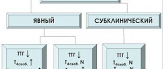
TSH is increased, free T4 is increased or normal, free T3 is decreased or normal.
- severe nonthyroidal pathology, including somatic and mental illnesses
- taking amiodarone, large doses of propranolol, x-ray iodine contrast agents
- recovery period after serious illness
- uncompensated primary adrenal insufficiency
TSH is normal, total T3 and T4 are elevated.
- all conditions in which the level of thyroxine-binding globulin is elevated
- heroin and amphetamine
- tamoxifen
TSH is normal, total T4 is increased, total T3 is normal or decreased.
- familial disalbuminemic hyperthyroxinemia
- amiodarone and radiopaque iodine-containing agents
- amphetamine
TSH is decreased, free T4 is increased, free T3 is decreased.
- artificial thyrotoxicosis due to self-prescription of T4
TSH is normal, T4, T3 are decreased.
- large doses of salicylates.
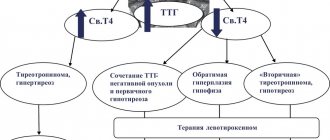
Rice. 4. Dynamics of TSH and free T4 during the treatment of hypothyroidism and hyperthyroidism.
In this case, as it has been believed since the 70s, the development of changes in the ratio of the level of free and reverse (biologically inactive) T3, as well as the levels of T4 and TSH, is based on adaptive reactions aimed at human survival by reducing the processes of catabolism in conditions of severe illness . In these cases, the erroneous diagnosis of a non-existent thyroid pathology and, moreover, its life-threatening treatment are completely unjustified. There are also genetically determined or acquired situations in which changes in the level of total T3 and T4 are associated with abnormalities in the production of hormone binding proteins. Modern algorithms for determining the functional activity of the thyroid gland require, first of all, two studies - determination of TSH by a highly sensitive method and free T4. Determination of free T3 is not a first-level test and is considered an additional test. Below are algorithms for examining patients depending on the clinical situation.
Option A (Fig. 1) provides for a situation where there is a clinically obvious disease, and the purpose of laboratory diagnosis is to confirm the diagnosis. Let us take this opportunity to recall that modern endocrinology requires mandatory confirmation of the diagnostic concept before starting treatment. Option B (Fig. 2) represents a situation where a laboratory test was carried out as a screening test in groups with a high probability of tyropathy. Since there are quite a lot of people involved in such screening, it is more reasonable to use at the initial stage the determination of only TSH (by the third generation method), which will exclude euthyroidism (TSH level is normal), and if an increased or decreased TSH level is obtained, additionally examine free T4, in order to identify both subclinical variants of common thyropathies and more rare clinical situations. Option C (Fig. 3) provides for rare, non-standard situations that arise when thyroid hormones and TSH are determined randomly. Another, and more common task of laboratories, along with the primary assessment of thyroid function, is dynamic monitoring of the treatment of patients with thyroid pathology.
Very important in assessing the adequacy of thyroid therapy is the choice of methods for this assessment and the frequency of its implementation. To assess the adequacy of treatment for primary hypothyroidism, it is recommended to examine the TSH . It is important to remember that the hormone level returns to normal no earlier than 2 months after replenishing the T4 deficiency. Thus, achieving the calculated dose of replacement therapy (approximately 1.6 mg/kg body weight) at one rate or another, depending on the clinical situation, should be accompanied by control TSH determinations after 2 months. An earlier study inevitably entails an erroneous conclusion about the inadequacy of therapy (Fig. 4). On the contrary, with thyrotoxicosis the level of thyroid hormones normalizes faster than the secretion of TSH is disinhibited (Fig. 4). Thus, the determination of free T4 or free T3 in isolated T3 toxicosis, starting from 3 months of treatment, serves as a more reliable indicator of euthyroidism and may indicate the desirability of additional prescription of T4 drugs according to the “blockade + replacement” scheme under the control of an additional determination of TSH. When assessing the values of free T4 in patients undergoing replacement therapy with thyroxine preparations, one should take into account the presence of a transient increase in free T4 by 10-15% within 3-4 hours after taking the last dose of the drug. When clinical and laboratory euthyroidism is achieved, it is advisable to carry out further laboratory monitoring (based on TSH levels) at intervals of 1 time every 6-12 months for life in case of hypothyroidism and 1 time every 1-3 months (depending on the clinic) during a one and a half to two-year treatment period diffuse toxic goiter. Obviously, in real clinical practice, along with assessing the functional activity of the thyroid gland, there is almost always a need to clarify the etiological factor of the disease, for example, to conduct a differential diagnosis between iodine deficiency goiter and autoimmune diseases of the thyroid gland or to clarify the nature of the nodular formation. Special tests are required to diagnose and monitor the treatment of thyroid cancer. Some modern automatic chemiluminescent analyzers have a wide range of tests in their menu to assess thyroid status. Table 3 presents the analytical characteristics of such tests used in the Immulite analyzer from DPC (USA). As can be seen from the table, the high sensitivity of these test systems and their operating range make it possible to carry out almost all the necessary studies of thyroid status.
| Table 3. Analytical characteristics of methods for assessing thyroid status used in the Immulite DPC analyzer | ||||
| Method | Units | Operating range | Sensitivity | Normal range |
| TSH supersensitive (III generation) | Chalk | up to 75 | 0,002 | 0,4-4,0 |
| TSH fast | Chalk | up to 75 | 0,01 | 0,4-4,0 |
| Free T4 | pmol/l | 2,75-77,22 | 0,43 | 10,3-24,1 |
| General T4 | nmol/l | 12,9-308,9 | 5,2 | 58,0-161,0 |
| Free T3 | pmol/l | 1,5-60,0 | 1,5 | 3,2-7,2 |
| General T3 | nmol/l | 0,61-9,22 | 0,54 | 1,3-2,7 |
| Antibodies to TPO | IU/ml | up to 2000 | 7 | 0-35 |
| Thyroxine binding globulin | nmol/l | 65-1480 | 20 | 240-721 |
| Thyroglobulin | ng/ml | up to 300 | 0,2 | 0-55 |
| Antibodies to thyroglobulin | IU/ml | up to 3000 | 20 | 0-40 |
One of the main advantages of using automated analyzers is the ability, in accordance with the diagnostic algorithm, to carry out subsequent tests step by step, depending on the results already performed from specific serum samples. In a similar way, additional studies necessary to clarify the diagnosis and adjust treatment can be performed. In any case, the time required for a complete step-by-step examination of the patient does not exceed 24 hours. This procedure allows for a complete examination of thyroid patients as quickly as possible without prescribing the simultaneous performance of the entire range of studies and significantly saves the material resources of laboratories. Often, in the practice of interaction between clinical laboratory service specialists and clinicians, situations arise when clinicians repeatedly refer a patient to assess thyroid function, since the results of determining TSH and free T4 are, in their opinion, “erroneous” and do not correspond to the clinical picture. Table 2 shows the most common reasons for this kind of discrepancy, taking into account which will allow clinicians to avoid some of these situations, and KDL doctors to expand their understanding of the pathphysiology of thyroid diseases.
| Table 2. The most common reasons for the discrepancy between the results of TSH and free T4 determination and the clinical picture. |
| Excessive thyroid hormone therapy (TSH ↓, fT4 →) |
| Recent adjustment of thyroid hormone therapy (TSH ↑, fT4 →) |
| Taking medications containing T3 (TSH ↓, fT4 →) |
| Patients on insufficient thyroid hormone therapy who have no complaints (TSH ↑, fT4 →) |
| Extrathyroidal pathology |
| Drugs affecting thyroid status (glucocorticoids, dopamine, etc.) |
| Total resistance to thyroid hormones (TSH ↑, fT4 ↑) |
| TSH-secreting tumors (TSH ↑, fT4 ↑) |
Thus, the use of automatic chemiluminescent analyzers makes it possible to introduce into laboratory and clinical practice algorithms for diagnosing the functional state of the thyroid gland, differentiated depending on clinical situations. A prerequisite for the effectiveness and accuracy of diagnostic programs is the presence in the menu of such analyzers of highly sensitive methods for determining third-generation TSH and free T4. The simultaneous possibility of using the widest range of analytically reliable methods for assessing thyroid status using the analyzer makes it possible, in combination with the use of instrumental methods, to verify the diagnosis and effectively monitor the treatment process of patients with thyroid pathology.
Literature
- Vetschev V.S., Melnichenko E.A., Kuznetsov N.S. and others // Diseases of the thyroid gland. M. 1996.
- Goncharov N.P. Problems of endocrinology, 1995, 3, pp. 31-35
- Starkova N.T. // Guide to clinical endocrinology, St. Petersburg, “Peter”, 1996.
- Shilin D.E. // In the collection “Diagnostic aspects of the use of Roche test systems. M., 1997, p.2
Thyroid hormone tests for men
Men most often have to take hormone tests if a married couple is unable to conceive a child. It is important to know not only the levels of sex hormones, but also the content of thyroid hormones.
In addition to problems in the reproductive sphere, blood sampling for analysis may be recommended in the following cases:
- in the presence of nodes, neoplasms in the thyroid gland;
- when losing weight or, conversely, sudden weight gain;
- with a sharp increase in appetite;
- with persistent sore throat, weakness, irritability;
- for arrhythmia not associated with heart disease.
Normal blood hormone levels in men are the same as in adult women:
- TSH – from 0.4 mU/liter to 4.0 mU/liter;
- Total T3 – from 1.2 nmol/liter to 2.2 nmol/liter;
- Free T3 – from 2.6 lmol/liter to up to 5.7 lmol/liter;
- Total T4 – from 54 nmol/liter to 156 nmol/liter;
- Free T4 – from 9.0 lmol/liter to 22.0 lmol/liter;
- AT-TPO – from 0 to 5.6 U/ml;
- AT-TG – from 0 to 18 U/ml.
RT3/reverse T3/reverse triiodothyronine
Reverse T3 is an inactive form of T3 produced in large quantities during stress.
Reference range: typically 10-24 ng/dL
This test is rarely prescribed by doctors, as they do not see the point in this measurement.
A group of doctors dedicated to optimal hormone balance believe that increased RT3 imbalance is the main sign of an underactive or dysfunctional thyroid gland. They believe that in the opposite direction, T3 should be reduced to the lower end of the normal range.
Analysis of thyroid hormones in pregnant women
Thyroid dysfunction in an expectant mother can negatively affect the course of pregnancy and labor. If a woman has hypothyroidism, then she is considered to be at risk for miscarriage. There is also a high probability that the born child will also have problems with the thyroid gland. And, as you know, hypothyroidism in a baby can cause problems in his general condition, weak immunity, and slower mental and physical development.
As a rule, a pregnant woman is routinely prescribed an analysis of T3 and T4 levels. The fact is that the TSH level during pregnancy is most often within the normal range (due to the increased content of somatotropic hormone, which stimulates the production of TSH).
For thyroid disease, tests are repeated every month. Additionally, 1-2 times during pregnancy, an ultrasound examination of the thyroid gland, an ECG, and an analysis of antibodies to TG and TPO are performed.
Normal indicators of the thyroid gland during pregnancy are as follows:
- TSH – from 0.4 to 4.0 µIU/ml;
- Total T3 – from 1.3 to 2.7 nmol/liter;
- Free T3 – from 2.3 to 6.3 pmol/liter;
- Total T4 – from 100 to 209 nmol/liter in the first trimester, from 117 to 236 nmol/liter in the second and third trimesters;
- Free T4 – from 10.3 to 24.5 pmol/liter in the first trimester, from 8.2 to 24.7 pmol/liter in the second and third trimesters.
It should be immediately noted that the reference values of various hormones may differ slightly in different laboratories. The fact is that when working with biomaterial, a large number of reagents are used, which determine the variants of the norm.
How to prepare for the procedure
Before examining the thyroid gland, it is necessary to remember that the biomaterial is collected on an empty stomach. To do this, it is important to refrain from consuming any food and drinks (except water) 8-10 hours before the procedure. Two days before the test, you should avoid vigorous physical activity, excessive fatigue, smoking and drinking alcohol. When the collection of biomaterial is repeated, in order to assess the effectiveness of the previously received treatment, it is necessary to stop all medications the day before donating blood. If you are constantly taking aspirin, tranquilizers or hormonal drugs, you should inform your doctor about this.
Antibodies
Many patients want to clarify: why does the analysis of thyroid hormones contain information not only about the hormones themselves, but also about some unknown antibodies? Why does a doctor need information about the so-called AT-TPO and AT-TG?
The fact is that an increase in the concentration of the antibodies presented indicates the presence of certain autoimmune processes in the gland. Such an analysis is not performed without indications: it is prescribed if the fact of autoimmune pathology has already been proven.
For the patient, changes in antibody levels are, in principle, unlikely to be informative. After all, the increase in the level of AT-TPO and AT-TG is not considered individually, but in combination with other indicative changes. Thus, an increase in their content against the background of a normal TSH value does not indicate the presence of pathology.
Gynecologist appointment
Published: 01/02/2017
Gynecology is a field of medicine that studies the sexual and reproductive health of the fair sex, and also helps women during pregnancy and childbirth. An appointment with a gynecologist is an obligatory part of every woman’s life.
Hormone tests after thyroid removal
After surgery to completely remove the thyroid gland (this intervention is called a thyroidectomy), the production of thyroid-stimulating hormones completely stops. As a result, the pituitary gland begins to work at an accelerated pace, trying to replenish adequate hormonal levels. At this stage, it is very important to start taking thyroxine so that complications such as thyroid coma do not develop. The first symptoms of this condition may be:
- apathy, constant drowsiness, clouding of consciousness;
- decreased body temperature;
- attacks of cold sweating;
- bradycardia, muscle atony;
- memory impairment;
- problems with kidney function;
- decreased intestinal motility.
The stated symptoms do not appear immediately, but gradually. Therefore, taking thyroxine after surgery is mandatory. Periodically, the patient must take a blood test to check the TSH level.
A low TSH level after removal of the thyroid gland may indicate taking excessively high doses of thyroxine, or a functional failure of the pituitary-hypothalamus system in the brain.
A high TSH level after thyroidectomy indicates excessive production of TSH - for example, in disorders of endocrine function, during treatment with certain drugs (antiemetics, antiepileptics, prednisolone, cardiac glycosides, morphine-containing drugs, oral contraceptives).
Tg/tiroglobulins
Thyroglobulin (TG) is a protein produced by the thyroid gland. Its presence in the blood is a sign that the patient still has a thyroid artery, whether the entire gland or remnants remain after surgery or radioactive ablation (RAI).
Thyroglobulin is primarily tested in patients with thyroid cancer to determine whether cancer tissue produces thyroglobulin before treatment, to determine whether treatment is being given, and to help detect recurrence after treatment. Because the most well-known types of thyroid cancer, namely papillary and follicular thyroglobulin, as well as elevated thyroglobulin levels can be a sign of cancer recurrence.
Reference range: if there is no thyroid gland, it should be less than 0.1 ng/ml. If there is still iron present, it should be less than or equal to 33 ng/ml.
Low levels of thyroglobulin are normal for people without thyroid disease. In patients with thyroid cancer, this means that thyroglobulin levels can be monitored later to detect recurrence.
Thyroglobulin levels after thyroid surgery or radioactive iodine (RAI) therapy are 0 or very low. If levels begin to rise after treatment for thyroid cancer, it may be a sign that the cancer has returned.
Conditions that cause inflammation of the thyroid gland, such as goiter, thyroiditis, or hyperthyroidism, can also cause thyroglobulin levels to increase. However, during treatment, these conditions are usually not imposed during the test.
How long does it take to test for thyroid hormones?
The period during which thyroid hormone test results can be obtained may vary. First of all, it depends on the capabilities of the laboratory itself. For example, in a public clinic, the procedure can take several days - for example, when using outdated equipment with first or second generation analyzers. And in a paid network of laboratories, the result can be obtained within a day: they usually use the latest analyzers that provide quick and accurate results. On average, it is believed that the study from the moment of blood collection to the delivery of results can last from 1-2 to 6-7 days. It is better to inquire about the exact period in the specific laboratory in which the diagnostics will be carried out.
How to take it correctly
Blood for analysis is taken from a vein. Depending on the symptoms, the doctor identifies a group of hormones that need to be tested. It is advisable to completely limit physical and psycho-emotional stress for 12 hours, not to drink alcohol, medications or food containing iodine.
Particular attention is required in preparation for donating blood samples for representatives of the fair half - it should be carried out during a certain period of the menstrual cycle, which is designated by the doctor. The donation procedure itself, as a rule, is scheduled for the morning hours, on an empty stomach.