See more neurological diseases starting with the letter “C”: Compression of the brain; Senile chorea; Sensitive ataxia; Serous meningitis; "Rigid person" syndrome; Alien hand syndrome; Restless legs syndrome; Bogorad syndrome; West syndrome; Gaye-Wernicke syndrome; Guillain-Barre syndrome; Piriformis syndrome; Carpal tunnel syndrome; Carotid sinus syndrome; Kleine-Levin syndrome; Klippel-Feil syndrome; Cauda equina syndrome; Crumpy syndrome; Lambert-Eaton syndrome; Landau-Kleffner syndrome.
For patients about piriformis syndrome
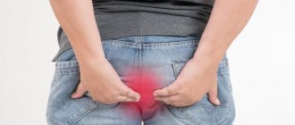
This is a combination of characteristic signs of compression of the sciatic nerve in the infrapiriform foramen. The main symptoms are pain in the buttocks and sacrum, peripheral paresis of the foot, sensitivity disorders. The main diagnostic methods are medical history, novocaine test, ultrasound, radiography, MRI and CT. Therapy includes medication, physiotherapy, kinesiotherapy and exercise therapy. Surgery is possible.
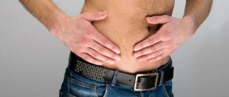
Diseases of the peripheral nervous system are one of the most common causes of disability in patients of working age. In the structure of these diseases, the predominant place is occupied by dorsopathies - degenerative-dystrophic diseases of the spinal column, the main clinical symptom of which is pain.
Pain in the lumbosacral region is the most common complaint. Piriformis muscle syndrome (PMS) occurs in at least 50% of patients with lumbosacral dorsopathies [1, 10]. At the same time, many issues of diagnosis and treatment of this syndrome have not been resolved and are at the stage of discussion and study. Differential diagnosis is especially difficult for lesions of various parts of the sciatic nerve [3, 4]. Pathological tension of the piriformis muscle due to compression of the L5 or S1 root, as well as with unsuccessful drug injections, leads to compression of the sciatic nerve (or its branches with a high origin) and accompanying vessels in the infrapiriformis space. SGM is often registered in gynecological practice.
Therefore, to make a correct diagnosis, the doctor must be guided by the following examination algorithm:
- Analyze the patient's medical history and complaints.
- Assess the clinical picture.
- Use additional diagnostic methods (x-ray, electromyography, etc.).
- Prescribe adequate therapy.
The assumption of the presence of SHM may arise if the patient has persistent pain along the sciatic nerve that does not decrease with drug treatment. It is much more difficult to determine the presence of this syndrome if there is only pain in the buttock area when walking or associated with certain positions (movements) of the pelvis.
With SHM, the following options for compression of the sciatic nerve are possible:
- compression of the sciatic nerve between the altered piriformis muscle and the sacrospinous ligament;
- compression of the sciatic nerve by the altered piriformis muscle as the nerve passes through the muscle itself (a variant of the development of the sciatic nerve).
The clinical picture of FMS consists of local symptoms and symptoms of compression of the sciatic nerve [14, 15].
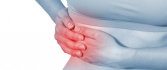
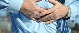
Local symptoms include aching, pulling, cerebral pain in the buttock, as well as in the sacroiliac and hip joints. It intensifies when walking, standing, adducting the hip, and also when squatting. The pain is relieved by lying down or sitting with your legs apart. With good relaxation of the gluteus maximus muscle, a dense and painful piriformis muscle is felt underneath it, and it is painful when stretched (Bonnet-Bobrovnikova symptom). With percussion in the area of the piriformis muscle, pain appears along the back of the leg (Vilenkin's symptom).
The clinical picture of compression of the sciatic nerve has a number of features associated with the topographic-anatomical relationship of its tibial and peroneal branches with surrounding structures [6, 7]. Pain during compression of the sciatic nerve is dull, cerebral in nature with a pronounced vegetative coloring (sensations of chilliness, burning, stiffness) with irradiation throughout the leg or along the zone of innervation of the tibial and peroneal nerves. Provoking factors for pain are heat, changes in weather, and stressful situations. Sometimes the Achilles reflex and superficial sensitivity are reduced. There is no increase in pain when coughing or sneezing. The gluteal muscles do not atrophy. The zone of hypoesthesia does not extend above the knee joint. With the predominant involvement of the fibers from which the tibial nerve is formed, the pain is localized in the posterior group of muscles of the lower leg and appears when walking and the Lassègue test. Palpation reveals tenderness in the soleus and gastrocnemius muscles. In some patients, compression of the inferior gluteal artery and the vessels of the sciatic nerve itself is accompanied by a sharp transient spasm of the vessels of the leg, which leads to intermittent claudication. The patient is forced to stop, sit down or lie down when walking. The skin of the leg turns pale.
The following clinical manifestations are possible depending on the level of compression of the sciatic nerve [16]:
- A very high level of damage (in the pelvis or above the gluteal fold) is characterized by paralysis of the foot and fingers, loss of the Achilles and plantar reflexes, anesthesia (hypoesthesia) of almost the entire leg and foot, loss of function of the biceps femoris and semitendinosus, semimembranosus muscles, hypoesthesia (anesthesia) in posterior outer surface of the thigh, inability to rotate the thigh outward, the presence of positive symptoms of tension, vasomotor and trophic disorders (hyper- or hypotrichosis, hypoor hyperhidrosis, changes in nail growth, formation of trophic ulcers in the heel area and the outer edge of the foot).
- A lesion at the level of the infrapiriformis foramen consists of two groups of symptoms - symptoms of damage to the piriformis muscle itself and symptoms of the sciatic nerve. The first group of symptoms includes pain on palpation of the upper inner part of the greater trochanter of the femur (the place of attachment of the piriformis muscle to the capsule of this joint), pain on palpation in the lower part of the sacroiliac joint, Bonnet's sign, pain on palpation of the buttock at the point of exit of the sciatic nerve from -under the piriformis muscle. The second group of symptoms includes symptoms of compression of the sciatic nerve and blood vessels. Painful sensations due to compression of the sciatic nerve are characterized by a feeling of constant heaviness in the leg, a dull, cerebral pain. At the same time, increased pain when coughing and sneezing, atrophy of the gluteal muscles, and a zone of hypoesthesia above the knee joint are absent.
- The lesion at the level of the hip (below the pelvic outlet) and up to the level of division into the peroneal and tibial nerves is characterized by impaired flexion of the leg at the knee joint and a specific gait; lack of active movements in the foot and toes, atrophy of paralyzed muscles, which develops after 2–3 weeks and often masks the pastiness of the leg; hypoesthesia (anesthesia) on the posterior outer surface of the leg, dorsum of the foot, sole and toes; violation of joint-muscular sensitivity in the ankle joint and interphalangeal joints of the toes; lack of vibration sensitivity on the outer ankle; pain along the sciatic nerve at the Balle and Gar points; positive Lassegue's sign; disappearance of the Achilles and plantar reflexes.
- The syndrome of incomplete damage to the sciatic nerve is characterized by the presence of pain of a causalgic nature (it is “burning” in nature, intensifies when lowering the leg, and is provoked by a light touch); severe vasomotor and trophic disorders (for the first 2–3 weeks, the skin temperature on the affected leg is 3–5 °C higher than on the healthy leg, then the lower leg and foot become cold and cyanotic). Often hyper- or anhidrosis, hypotrichosis, hyperkeratosis, changes in the shape, color and growth rate of nails are found on the plantar surface. Sometimes trophic ulcers occur on the heel, the outer edge of the foot, and the dorsum of the toes. X-rays reveal osteoporosis and decalcification of the foot bones.
In neurological practice, the following tests are recommended to recognize SHM [7, 9]:
- Detection of pain upon palpation of the upper internal region of the greater trochanter of the femur (place of attachment of the piriformis muscle).
- Detection of pain upon palpation of the lower part of the sacroiliac joint - projection of the attachment site of the piriformis muscle.
- Identification of Bobrovnikova–Bonnet and Bonnet symptoms. Passive adduction of the hip with simultaneous internal rotation is accompanied by increased pain; upon palpation, a dense and painful piriformis muscle is felt.
- Examination of the sacrospinous ligament. This test simultaneously diagnoses the condition of the sacrospinous and iliosacral ligaments.
- Tapping the buttock (on the sore side). This causes pain along the back of the thigh.
- Identification of Grossman's symptom. When struck with a hammer or folded fingers on the lower lumbar or upper sacral spinous processes, the gluteal muscles contract.
- Infiltration of the piriformis muscle with novocaine with assessment of the resulting positive changes. A significant reduction or disappearance of pain along the sciatic nerve is a dynamic test showing that the pain is caused by the compressive effect of the spasmodic muscle. Reflex tension in the muscle and neurotrophic processes in it, as a rule, are caused by irritation not of the fifth lumbar, but of the first sacral root. Since painful tension of the piriformis muscle is most often associated with irritation of the first sacral root, it is advisable to alternately carry out novocaine blockade of this root and novocainization of the piriformis muscle.
If diagnostics are difficult, electroneuromyographic studies are used to determine the damage to the peripheral neuron corresponding to a given nerve and a decrease in the speed of impulse transmission along the nerve distal to the place of its compression. Consultation with a gynecologist and urologist is necessary, since very often patients have a pathology that causes the occurrence of this syndrome (tumors, hematomas, etc.). It is recommended to conduct a computed tomography or magnetic resonance imaging scan of the muscles and pelvic organs to identify the causes of compression. Most often, the differential diagnosis of BMS must be made with discogenic compressive radiculitis L5–S2 (see table).
. Differential diagnostic criteria for compression of the sciatic nerve by the piriformis muscle and discogenic compressive radiculitis L5–S2.
Diagnosing BMS is a difficult task for a doctor, which must be solved taking into account all etiological and clinical factors.
Treatment of SHM requires from the doctor not only knowledge of the etiological and pathogenetic features of the disease, but also great patience. Patients are usually depressed or anxious, and their main complaint is pain that is difficult to treat. The doctor-patient contact is broken or very difficult to achieve. But at the same time, the doctor is obliged to explain to the patient the cause of his condition, encourage joint action to eliminate the disease, and increase confidence in the prescribed therapy. Treatment tactics depend on the severity and rate of progression of the disease. Pathogenetic therapy should be aimed at eliminating the pathological process and its long-term consequences [8]. In other cases, treatment should be symptomatic, the goal of which is to achieve long-term remission and improve the quality of life of patients. The main criterion for the optimal therapeutic effect on the patient is the combination of medicinal and non-medicinal methods. The basic principles of drug therapy for BMS are early initiation of treatment, pain relief, and a combination of pathogenetic and symptomatic therapy.
The arsenal of remedies offered to solve the problem of pain is quite wide, so the choice of drug should be reasoned. Non-steroidal anti-inflammatory drugs – NSAIDs – remain the first choice for pain relief [5, 11, 12]. The main mechanism of action of NSAIDs is inhibition of cyclooxygenase (COX-1, -2), a key enzyme in the arachidonic acid metabolic cascade, leading to the synthesis of prostaglandins, prostacyclins and thromboxanes. Due to the fact that COX metabolism plays a major role in the induction of pain at the site of inflammation and the transmission of nociceptive impulses to the spinal cord, NSAIDs are widely used in neurological practice [4, 10, 19].
All anti-inflammatory drugs have anti-inflammatory, analgesic and antipyretic effects, are able to inhibit the migration of neutrophils to the site of inflammation and platelet aggregation, and also actively bind to serum proteins. Differences in the action of NSAIDs are quantitative [5], but they determine the severity of the therapeutic effect, tolerability and side effects in patients. In this regard, for the treatment of severe pain syndromes, including for long-term use, drugs are needed that have anti-inflammatory and analgesic effects with minimal gastrotoxic reactions. At the same time, the problem of choosing a first-line drug becomes obvious. Traditionally, drugs with a predominantly anti-inflammatory effect are more actively used. However, for the treatment of BMS, adequate analgesia is of paramount importance [2], which allows us to recommend NSAIDs with a powerful analgesic effect, for example, ketorol as a first-line drug for the treatment of severe pain syndromes.
Ketorol (ketorolac) is a non-selective COX inhibitor. The mechanism of action is associated with non-selective inhibition of the activity of the enzymes COX-1 and COX-2, mainly in peripheral tissues, resulting in inhibition of the biosynthesis of prostaglandins - modulators of pain sensitivity, thermoregulation and inflammation. The drug does not affect opioid receptors, does not depress respiration, does not cause drug dependence, and does not have sedative or anxiolytic effects. In terms of the strength of the analgesic effect, the drug is comparable to morphine and is significantly superior to other NSAIDs [4]. In North America, the UK, some other European countries and Hong Kong, ketorolac is the only NSAID used for the treatment of pain as a rapid intravenous injection. It has been shown that when administered intramuscularly, the drug at a dose of 30 mg has an effect comparable to the effect of 10–12 mg of morphine or 50 mg of meperedine [17]. The advantage of ketorolac compared to narcotic analgesics is the absence of an effect on respiratory function, sedation and psychomotor effects.
In a multicenter study conducted by GD Innes et al. (1998), the analgesic effect of ketorolac for back pain was comparable to the effects of codeine with significantly fewer adverse side effects. The drug is rapidly absorbed and has high bioavailability (80–100%). After intramuscular administration and oral administration of the drug, the onset of the analgesic effect is observed after 30 minutes and 1 hour, respectively, the maximum effect is achieved after 1–2 hours, the duration of action is 6–10 hours.
Ketorol is prescribed intramuscularly at 1 ml (30 mg) or orally (10 mg) once or repeatedly (10 mg up to 4 times a day) depending on the severity of the pain syndrome. The maximum daily dose should not exceed 40 mg. The duration of the course of treatment should not exceed 5 days. When switching from parenteral administration of the drug to oral administration, the total daily dose of both dosage forms on the day of transfer for patients under the age of 65 years should not exceed 90 mg, for patients over the age of 65 years or with impaired renal function - 60 mg. In this case, the dose of the drug for oral administration on the day of transition should not exceed 30 mg. Taking into account the strong analgesic effect of the drug, it is used parenterally in the acute period of the disease, followed by a transition to selective NSAIDs, in particular nimesulide (Nise).
Nimesulide (Nise) reversibly inhibits the formation of prostaglandin E2 both at the site of inflammation and in the ascending pathways of the nociceptive system, including the pathways of pain impulses in the spinal cord. It has a slight effect on COX-1 and practically does not interfere with the formation of prostaglandin E2 from arachidonic acid under physiological conditions, thereby reducing the number of side effects of the drug [2, 18–20]. It has antioxidant properties and inhibits the formation of toxic oxygen breakdown products by reducing the activity of myeloperoxidase. Interacts with glucocorticoid receptors, activating them through phosphorylation, which also enhances the anti-inflammatory effect of the drug. Due to its high bioavailability, within 30 minutes after oral administration, the concentration of the drug in the blood reaches approximately 50% of the peak, and a clear analgesic effect is noted. After 1–3 hours, the peak concentration of the drug occurs and, accordingly, the maximum analgesic effect develops. The drug is superior in its tolerability to traditional NSAIDs, since it relatively rarely causes dyspepsia and other complications of the gastrointestinal tract.
It should be especially emphasized that the prophylactic use of NSAIDs in the absence of pain is inappropriate. Convincing evidence of a preventive effect has not been obtained, while the risk of complications increases significantly.
Another equally important component of the effect on pain is the use of muscle relaxants. When treating BMS, preference is given to tolperisone, since only this drug has a parenteral form. Tolperisone is a centrally acting muscle relaxant, has moderate adrenergic blocking and weak atropine-like effects, inhibits spinal polysynaptic nociceptive reflexes, due to which it has a direct analgesic effect. The muscle relaxant effect of the drug is realized at several levels: peripheral nerve, spinal cord, brain stem. The choice of tolperisone for outpatient practice is due to the good combination of effectiveness and safety of this drug. Unlike other drugs, it does not cause dizziness and does not affect the speed of reactions or the ability to drive. For severe pain syndromes, the drug is administered intramuscularly or intravenously slowly at 100 mg twice a day. In other cases, this drug is prescribed at a dose of 50–150 mg orally 3 times a day, depending on individual need and tolerability.
Pathogenetic therapy is aimed primarily at eliminating the consequences of nerve compression and subsequent ischemia, affecting the microvasculature, improving blood supply to the affected area, and relieving signs of neurogenic inflammation. One of the most important areas of treatment is the activation of neurotrophic processes and regeneration, especially in the case of external traumatic damage to the sciatic nerve or during compression-ischemic processes. Even in the case of reconstruction of anatomical continuity or decompression of the nerve trunk, regenerative sprouting proceeds very slowly and requires long-term conservative therapy. For this purpose, drugs of various groups are used, including anticholinesterase, antioxidant, vasoactive, and neuroprotective drugs.
The mechanism of action of anticholinesterase drugs is associated with the blockade of enzymes that destroy acetylcholine inside the synaptic cleft and thus increase the working concentration of the transmitter near the postsynaptic membrane. These enzymes include acetylcholinesterase and butyrylcholinesterase. The use of acetylcholinesterase inhibitors during BMS leads to stimulation of neuromuscular transmission, improvement of impulse transmission along peripheral nerves and autonomic fibers. As a result, the force of contraction of skeletal muscles and the contractility of smooth muscle muscles increases. Currently, there is a fairly large selection of drugs with anticholinesterase action. In clinical practice, neostigmine methyl sulfate and ipidacrine are most often used.
Ipidacrine is a reversible cholinesterase inhibitor, prevents the enzymatic hydrolysis of acetylcholine, stabilizes the transmitter in the synaptic cleft and enhances cholinergic impulse transmission. Blocks membrane potassium channels and promotes their depolarization. Stimulates synaptic transmission in neuromuscular endings, conduction of excitation in nerve fibers; enhances the effect of acetylcholine and other mediators (epinephrine, serotonin, histamine, oxytocin) on smooth muscles; restores neuromuscular transmission and conduction of excitation in the peripheral nervous system. The drug is prescribed intramuscularly at 15 mg once or twice a day for 10 days, followed by switching to oral administration at 20 mg three times a day for 1–3 months, depending on the rate of restoration of impaired functions.
Currently, clinical work has appeared on the use of drugs that improve the rheological properties of blood and endothelium-dependent reactions of the vessel wall in patients with compression neuropathies. In order to reduce the manifestations of oxidative stress, derivatives of thioctic acid and ginkgo biloba are successfully used. However, pathogenetically, the use of drugs with a polyvalent mechanism of action (ethylmethylhydroxypyridine succinate, Actovegin) is more justified.
Actovegin significantly improves the diffusion of oxygen in neuronal structures, which makes it possible to reduce the severity of secondary trophic disorders. Vasodilation and decreased peripheral resistance are secondary to activation of oxygen metabolism in the vascular walls. The drug is prescribed intravenously (drip) at 800 mg for 5–10 days, followed by switching to oral administration at 200 mg three times a day for 1–3 months.
Ethylmethylhydroxypyridine succinate belongs to the group of heteroaromatic antioxidants, antihypoxants with nootropic and anxiolytic properties. The mechanism of action of the drug is due to its antioxidant and membrane protective effects. It inhibits lipid peroxidation, increases the activity of superoxide oxidase, increases the lipid-protein ratio, reduces membrane viscosity, increases its fluidity, and stabilizes the level of endogenous antioxidants. Modulates the activity of membrane-bound enzymes (calcium-independent phosphodiesterase, adenylate cyclase, acetylcholinesterase), receptor complexes (benzodiazepine, GABA, acetylcholine), which enhances their ability to bind to ligands, helps preserve the structural and functional organization of biomembranes, neurotransmitter transport and improve synaptic transmission. The drug is prescribed at a dose of 300 mg intravenously or intramuscularly for 10 days, followed by switching to oral administration at 0.125 mg four times a day for 1–3 months.
Thus, the primary task is not only to relieve pain, but also to eliminate the cause of the disease. It should be noted that the range of drugs used for the treatment of peripheral diseases of the nervous system is currently growing, so in order to correctly select therapy, the doctor must not make a mistake in diagnosing piriformis syndrome, take into account all the pathogenetic mechanisms of its occurrence and, most importantly, the individual characteristics of the patients.
Information about the author: Marina Viktorovna Putilina – Doctor of Medical Sciences, Professor of the Department of Neurology of the Federal University of Internal Medicine of the Russian State Medical University. Email
Causes
The disease is provoked by pathological changes in the piriformis muscle:
- Damage.
- Inflammation.
- Fibrosis.
- Spasm.
- Increase in volume.
It can be caused by intramuscular injections, which can lead to intramuscular abscess and the formation of infiltrate. Main etiofactors:
- Injuries. This could be muscle overstretching, muscle fiber tearing, or fibrosis. In the latter case, the muscle shortens and thickens.
- Post-traumatic hematoma.
- Vertebrogenic pathology. These include spondyloarthrosis, osteochondrosis, intervertebral hernia in the lumbar region, spinal and vertebral tumors. Irritation of the fibers of the sacral plexus and spinal roots leads to a reflex spasm.
- Inflammatory processes, which include myositis, sacroiliitis, cystitis, prostatitis, endometriosis, prostate adenoma.
- Muscular overload that occurs during prolonged forced position of the pelvic-iliac segment. Increased load occurs when, with radicular syndrome, the patient tries to assume an antalgic position. Sports such as weightlifting and running can provoke the disease.
- Oncology of the sacral region and proximal femur. It causes anatomical changes in structures. Neoplasia can cause spasm.
- Asymmetry of the pelvis that occurs with shortening of the lower limb and scoliosis.
- Amputation of the hip puts the muscle into a permanent spastic state, which causes phantom pain.
Secondary pathology
Secondary syndrome is associated with the influence of external factors. These include the following:
- Traumatic injury to the buttocks. It can cause inflammatory damage to soft tissues, muscle spasms, and a combination of both. As a result, compression of the nerve occurs.
- Direct damage to the piriformis muscle, postoperative injuries, increased stress on the lower back. Under the influence of these factors, muscle spasms appear.
- Shortening of muscles. The syndrome is caused by changes in the biomechanics of the leg and lumbosacral region. This often causes compression and irritation of the sciatic nerve.
Secondary pathology is associated with microtraumas. They appear due to overuse of the piriformis muscle. These include long distance movement or direct impact.
Development mechanism
The piriformis muscle is attached with its narrow end to the greater trochanter of the femur, and its wide end to the sacrum. It is involved in external rotation and internal abduction of the hip. Passes through the greater sciatic foramen. The inferior gluteal, sciatic, pudendal and posterior cutaneous nerves, gluteal arteries and veins pass through it.
Due to persistent toxic contraction of the piriformis muscle, the size of the infrapiriformis foramen decreases. Because of this, compression of passing vessels and nerves begins, primarily the sciatic one. Compression of the vascular system impairs blood supply to the nerve trunk. This is an additional pathogenetic component of sciatica.
Walking for health. What should it be like?
The piriformis muscle is located next to other elements of the musculoskeletal system that are involved during walking. In order to strengthen muscle fibers and provide additional blood flow to the pelvic tissues, it is necessary to perform slow walks in the fresh air. The pace of walking should be moderate and unhurried.
A park with smooth alleys without holes, bumps and potholes is best suited so that the patient does not stumble and there is no deterioration in health. Rough terrain is not suitable in this case, since there is always a risk of tripping and damaging the affected limb. Do not suddenly accelerate, jump or run.
Clinical picture
In 70% of cases, the disease first affects the gluteal-sacral area. The pain is constant, nagging, aching. It intensifies when walking, squatting, or adducting the hip. To reduce discomfort, the patient is forced to spread his legs to the sides in a horizontal position or sitting. Over time, pain appears along the sciatic nerve - sciatica. There are lumbagoes running from the foot to the buttock. In the area where the pathology is localized, a decrease in pain sensitivity and burning occurs.
Hypotonia of the muscles of the foot and lower leg begins. With total compression of the nerve fibers, a “dangling foot” may appear. The patient begins to experience intermittent claudication. This is a consequence of vascular compression. It also causes a decrease in the temperature of the limb, pale skin and numbness of the fingers.
Contraindications
In some clinical situations, blockade is not recommended. Contraindications are:
- blood diseases with an increased tendency to bleed;
- the patient is taking blood thinning medications;
- active infectious process on the skin in the area of the planned injection;
- acute infectious and inflammatory disease, accompanied by an increase in body temperature and a deterioration in the general condition of a person;
- history of allergies to local anesthetics (contraindication for the use of novocaine or lidocaine);
- diabetes mellitus, adrenal disease (contraindication for the use of corticosteroids);
- myasthenia gravis;
- obvious tendency to hypotension.
Possible risks
The patient's ability to work is limited due to constant debilitating pain. Against this background, the following are possible:
- Emotional lability.
- Insomnia.
- Increased fatigue.
Peripheral paresis of the leg and foot causes muscle atrophy. With a long period of illness, the changes become irreversible. Persistent paresis leads to disability. Secondary spasm of the pelvic floor muscles is also possible. This causes discomfort when urinating and dyspareunia in women.
Diagnostic methods
It is important to differentiate the pathology from sciatic neuropathy and deep-seated piriformis muscle. For this purpose, clinical tests are carried out. The basic ones are:
Consultation with a neurologist with collection of anamnesis and determination of neurological status. There is pain on palpation of the sacroiliac joint and the ventromedial surface of the greater trochanter. You can provoke discomfort with a number of tests:
- Attempting to raise the knee on the healthy side.
- Passive internal rotation of the hip.
- Active internal rotation of the flexed hip.
- Bend your torso forward with straight legs.
Instrumental diagnostics include assessment of the condition of the leg muscles and conductivity of the sciatic nerve. Electronography is used for this. To establish pelvic asymmetry, injuries, and malignant neoplasms, radiography of the pelvic bones, ultrasound of the pelvic organs, MRI and CT of the spine are necessary. It’s easy to view the addresses of diagnostic centers in the capital and clarify the cost of prescribed procedures through the “Unified registration center for MRI/CT/ultrasound in Moscow.”
Additionally, you may need to consult an oncologist, vertebrologist, gynecologist, or urologist. When making a diagnosis, it is important to distinguish between piriformis syndrome and lumbosacral plexitis, radicular syndrome due to herniated disc, and toxic damage to the sciatic nerve. If the pathology is accompanied by intermittent claudication, it is necessary to exclude:
- Obliterating endarteritis of the lower extremities.
- Obliterating atherosclerosis.
How can you relieve pain during exacerbation?
Mechanical overload of the muscle should be avoided. It is possible to correct asymmetry caused by inequality in the length of the lower limbs by using a special heel on shoes. You can correct the asymmetry caused by a decrease in the size of one half of the pelvis by using a pad under the buttock. It is important to restore movement in the sacroiliac joint.
Choosing the right sleeping position is of great importance. When sleeping on the side, the patient should place a pillow between the knees to support the leg. Frequent changes of sitting position are necessary; you can use a rocking chair. Stops should be arranged during long driving periods - for short walks.
It is necessary to perform a set of exercises aimed at stretching the muscles, which include ischemic compression of trigger points. but it is very important to avoid compression of the nerves, so the exercises should be supervised by a specialist.
Therapy
Conservative therapy is a complex treatment. It includes pharmacological drugs and auxiliary treatments.
- Muscle relaxants are indicated to relieve spasms. A relaxing massage of the gluteal-sacral area can enhance the effect.
- The pain syndrome is relieved with non-steroidal anti-inflammatory drugs and analgesics. The best results are obtained by using drugs in the form of therapeutic blockades.
- Kinesitherapy will help restore the motor pattern. It includes myofascial release and post-isometric relaxation.
- For an anti-inflammatory effect, it is good to carry out physical procedures: hydrocortisone ultraphonophoresis, UHF therapy, magnetic therapy.
- Compresses with dimexide and acupuncture sessions are possible.
In most cases, such measures are quite effective. If the patient has pelvic asymmetry, inflammatory pathologies that were the root cause of BGM, they must be eliminated. When conservative methods are unsuccessful, surgery may be required. Dissection of the piriformis muscle and neurolysis are indicated for severe paresis of the foot.
Treatment
The piriformis muscle is located deep in the gluteal region. It is impossible to remove trigger points in it using classical massage techniques. The main treatment for problems caused by the piriformis muscle is manual therapy. In the presence of secondary disorders caused by pathology of the piriformis muscle, separate correction is required. Thus, neurovascular disorders in the affected limb, caused by compression of the vessels supplying the sciatic nerve, require the prescription of drugs that improve blood circulation. Depending on the severity of vascular disorders, drugs can be prescribed intravenously or in tablets. If the problems of the piriformis muscle are caused by a violation of its innervation, autogravitational traction of the spine is necessary.
As accompanying methods that accelerate the normalization of muscle tone and blood supply to the sciatic nerve, acupuncture, hirudotherapy, and some physiotherapeutic techniques are used. After pain relief, it is necessary to regularly perform special exercises for a long time to maintain normal muscle tone and form the correct pattern of movements.
Medical prognosis
With complex therapy, the prognosis is favorable. The effectiveness of surgical intervention is 85%, but the risk of relapse is quite high. Without correct therapy, persistent foot paresis occurs within a year.
Literature:
- Tukhbatullin M. G. Sharafutdinov B. M., Akhmedova G. M./Radiation diagnosis of neuropathies in piriformis syndrome//Practical Medicine - 2013.
- Romanenko I.V., Romanenko V.I., Romanenko Yu.I./Piriformis muscle syndrome//International Neurological Journal - 2014.
- Kanaev S.P./ Piriformis muscle syndrome. Complex clinical and instrumental research: new approaches to diagnosis: Abstract of the dissertation - 2005.
- Pravdyuk N. G., Shostak N. A.//Russian Medical Journal - 2014 - No. 28.// Myofascial syndrome (piriformis syndrome) - approaches to diagnosis, treatment.