The upper and lower jaw system has a very complex anatomical structure. The bone structure is characterized by a high dependence on effective blood supply and necessary nutrition for full growth and development. The full functioning of the skeletal system is directly related to the presence of all elements of the dentition. Due to the removal or long-term absence of molars, various pathological processes of the alveolar process of the upper and lower jaw develop.
In accordance with the anatomical features and existing pathologies, correction or augmentation is carried out, bone tissue is built up for successful prosthetics. Appropriate periodontal treatment makes it possible to securely fix the implant in the oral cavity without the risk of possible complications. Augmentation is often done using bone-replacing biomaterials of artificial or natural origin. An integrated approach to diagnosing pathology and a highly effective treatment method allows you to achieve the desired result.
What is the alveolar process?
Bone tissue, consisting of the basal layer, spongy tissue and cortical plate, plays an important role in reliable fixation of the dentofacial system. As a result of everyday physical activity, it undergoes morphological and histological changes. The alveolar process (AO) to the anatomical part that holds the elements of the dentition of the upper and lower jaw. It is formed from the moment the teeth erupt and atrophies after their loss.
The process consists of inner and outer cortical plates and cancellous bone tissue. It is permeated with small tubules through which blood vessels and nerves pass. The anatomical structure of the ridge has an unpaired symmetrical structure; the holes may differ in shape and size depending on the placement of the dental units. Alveoli are located in the center of the alveolar ridge; most often they have a cone-shaped shape. As a result of pathology or loss of elements of the dentition, a significant decrease in the volume of bone tissue occurs, which will require restoration of the alveolar process for subsequent implantation.
Mechanism of external respiration
The cells of the body are provided with oxygen and freed from carbon dioxide thanks to the blood passing through the capillary network of the alveoli. Oxygen and carbon dioxide, released from carbonate acid and its salts by the enzyme carbonic anhydrase, continuously move in opposite directions through the aerohematic barrier. It is found in red blood cells. The scale of diffusion can be judged based on the following figures: about 300 million alveoli forming lung tissue make up approximately 140 m2 of gas exchange surface and provide the process of external respiration. The above facts explain what the alveolus is and what role it plays in the metabolism of our body. In fact, it is the main element that ensures the breathing process.
Structure of the alveolar ridge
Taking into account the anatomical structure, the following parts are distinguished:
- Lateral. The outer wall located in close proximity to the cheeks and lips.
- Medial. The inner wall, which is directed towards the tongue and hard palate, has a compacted structure.
- Central. The location of the tooth sockets, the area has a large number of blood channels. This is where the molars and incisors are attached.
The alveoli and dental sockets are separated from each other by special bone partitions. In the alveolar part there are also interradicular septa. In the absence of functional loads on the ridge area, deformation of the alveolar process, changes in the anatomical structure and its reduction begin to be observed. The development of pathological processes in the upper and lower jaw often leads to a fracture of the alveolar process, which may require correction of this anatomical part.
Histological structure of the alveoli
Having examined the anatomy of lung tissue cells, let us now dwell on their species diversity. The alveoli contains two types of elements, called type I and type II cells. The first ones are flat in shape, capable of adsorbing particles of dust, smoke and dirt in the inhaled air. An important function in them is performed by pinocytosis vesicles filled with a protein substrate. They reduce the surface tension of the alveoli and prevent them from collapsing during exhalation. Another element of type I cells is the closing structures, which serve as a buffer and do not allow intercellular fluid to penetrate into the alveolar cavity filled with air. Groups of oval type II cells have foam-like cytoplasm. They are found in the alveolar walls and are capable of active mitosis, which determines the regeneration and growth of elements of lung tissue.
Functions of the alveolar process of the upper and lower jaw
- Fixation and maintenance of elements of the dental system.
- Active participation in chewing food.
- Makes it easier to bite into hard foods.
The state of AO is reflected in a person’s external data. The development of pathologies often leads not only to a deterioration in the performance of molars and incisors, but also affects the anatomy of the facial skeleton. Over the years, the oxygen supply to bone tissue is disrupted, which leads to various defects of the alveolar process. As a result, this can cause loss of teeth, the development of traumatic injuries, periodontal disease, periodontitis and other dental problems.
Idiopathic form of the disease
Idiopathic pulmonary alveolitis is less common than other types of the disease, and most often men face this problem. The causes of the development of the disease are not always known - many experts believe that the inflammatory process in this case is of autoimmune origin. Due to malfunctions of the immune system, antibodies to its own cells begin to be produced.
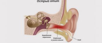
The main symptoms of the disease are cough and shortness of breath. The intensity of the symptoms constantly increases, so patients often consult a doctor after the onset of the fibrotic process. This form of alveolitis is considered the most dangerous, since in the vast majority of cases it ends in pneumosclerosis and respiratory failure.
Signs of pathologies of the alveolar ridge
- Predominant swelling of the mucous membrane in the area of the alveolar ridge.
- Pain when chewing food, swallowing saliva.
- Damage to gum tissue, bleeding.
- The appearance of multiple abrasions.
- Sharp expansion of interdental areas due to loss of incisors.
- Development of pathology of occlusion of elements of the dental system.
- The appearance of various speech defects, which can be expressed in a “lisp.”
In addition, hypertrophy of the alveolar process may develop, which is expressed in an increase in the volume of bone tissue due to histological changes. An external examination and x-ray may reveal a crack in the ridge or a complete separation of bone tissue from the fundamental cranial bone.
What diseases are characterized by the symptom of pulmonary dissemination?
Let's consider the most common diseases that can manifest as a disseminated pathological process in the lungs.
Tuberculosis
Mild tuberculosis is a fairly common, dangerous and serious disease. Its causative agent is bacteria - Koch bacilli, which are easily transmitted by contact and airborne droplets, can live for years in street dust and even in human lungs without causing any symptoms.
The disease manifests itself when the human immune system cannot independently restrain the active phase and growth of mycobacterium tuberculosis. Bacterial damage to the lungs in tuberculosis is usually visualized as multiple inflammatory foci - granulomas.
In the center of tuberculous granulomas there are foci of necrosis. Seals form around them - the pulmonary alveoli are filled with a liquid substrate, which contains the bacteria themselves, epithelial and plasma cells, dead lymphocytes, large Langhans cells, and macrophages.
In the initial stages, tuberculosis progresses almost asymptomatically; over time, the patient begins to notice weakness, deterioration in general health, cough and changes in breathing.
At the same time, something suspicious is rarely noted on auscultation. Sputum analysis does not show mycobacteria. Diagnosis of tuberculosis is possible based on the results of an X-ray examination and a tuberculin skin test.
Granulomatosis and diffusely located “ground glass” on CT are not a specific sign of tuberculosis. The former are also found in sarcoidosis, and the latter in pneumonia and other diseases.
To treat tuberculosis, the patient is prescribed a course of antibacterial therapy. It is important to prevent fibrosis (scarring of the lungs) as such changes can be irreversible.
Pneumoconiosis
Pneumoconiosis is a disseminated lung disease caused by inhalation of construction or industrial dust. Most often, pathological processes occur with a pronounced development of primary diffuse fibrosis, in which the pulmonary alveoli “stick together” as connective tissue grows in them.
Pneumoconiosis is also granulomatous in nature. CT scans clearly show multiple nodular densities of varying densities. Such compactions should be checked to determine whether the process is benign and whether there is an oncological threat.
Pneumoconiosis is a so-called “occupational” disease. Most often it affects:
- Electric welders;
- Builders and workers (the disease is caused by inhalation of dust from silicon dioxide, silicates, metals, carbons);
- Industrial workers (the disease is also caused by inhalation of dust from cotton, flax, grain, and some chemicals).
Silicosis is a common and severe lung disease, in which multiple diffuse fibrosis and nodular compactions are diagnosed. As a result, the patient's functional lung volume is significantly reduced. Occurs due to prolonged inhalation of dust with free silicon dioxide (contained in quartz sand and the surrounding air). Therefore, sandblasters, employees of relevant (abrasive blasting) enterprises, and residents of “sandy” regions are at risk.
Asbestosis is a chronic lung disease that also causes multiple pulmonary fibrosis. Occurs due to inhalation of asbestos dust or asbestos fibers. Employees of asbestos mining and asbestos processing enterprises are at risk.
Pneumonia and complications of pneumonia
Pneumonia is a group of inflammatory lung diseases. Against the background of this disease, the patient may experience more dangerous complications, which are also characterized by dissemination: acute respiratory syndrome (ARDS), interstitial lung disease, cobblestone symptom, honeycomb lung, pulmonary edema, fibrosis, etc.
In pneumonia with multiple diffusely located loci of damage, the vital capacity of the lungs is significantly reduced, since the inflammatory foci and infiltrates are large, the alveoli are filled with liquid exudate, not air.
Pneumonia with dissemination can be a consequence of pulmonary candidiasis, pneumoconiosis and other diseases. It is necessary to accurately establish the cause of pathological changes. Sometimes it is also necessary to exclude a malignant process (based on laboratory diagnostic results).
Sarcoidosis
Pulmonary sarcoidosis is an oncological disease. Its main features on CT are dissemination and mediastinal lymphadenopathy. Dissemination on scans is not as pronounced as with progressive tuberculosis, but there is a certain similarity. Diagnosis is complicated by pulmonary fibrosis. Along with disseminated damage to the respiratory organ, vasculitis, perivasculitis, and peribronchitis are present.
Multiple foci (from 2 mm to 1 cm) are often located along the bronchovascular bundles, interlobular fissures, costal pleura, and in the interlobular septa (perilymphatic type of dissemination). In sarcoidosis, granulomatosis is often (about 35% of cases) found not only in the lungs, but also in the bronchi. At the same time, their mucous membrane may not be changed - swelling, hyperemia, and areas of thickening of the epithelium indicate damage to the bronchi in sarcoidosis.
In the early stages, sarcoidosis develops asymptomatically; in later stages, the patient begins to be bothered by a nonproductive cough, breathing problems (for no obvious reason), discomfort and burning in the back, and heaviness in the chest.
Causes of alveolar process atrophy
- Injuries, mechanical damage to the area.
- Morphological changes associated with significant disruption of blood circulation.
- Formation of uneven edges of the alveoli after the removal of dental units.
- Osteomyelitis of the alveolar process, inflammatory processes of bone tissue.
- Neoplasms, cysts that lead to ridge degeneration.
- Loss of elements of the dental system.
Other causes of the development of pathological processes may include chronic inflammatory processes, fibrous osteitis, which is expressed in the thinning of the bone structure, or tumors of the alveolar process. Pathology may also be associated with hereditary factors and genetic predisposition. In all these cases, correction and immediate intervention by a dental surgeon are required, since the tissue itself does not recover.
Treatment of pathologies of the respiratory system in Kaliningrad
If you are bothered by a cough, shortness of breath, wheezing, or need a preventive examination, make an appointment with us for a consultation. The Pulmonology Center is staffed by competent, responsible and caring paid pulmonologists.
We offer:
- personal approach to each patient;
- rapid laboratory diagnosis of lung diseases;
- safe therapy at any stage of treatment: treatment of COPD, asthma;
- recovery programs, including rehabilitation after coronavirus in Kaliningrad.
Make an appointment at the Pulmonology Center by calling 8 (4012) 971-961.
Diagnosis of diseases of the alveolar process
In order to correctly select a therapeutic or surgical treatment method, an appropriate set of diagnostic procedures is carried out: blood tests, radiography. Additionally, MRI, CT of the upper jaw, and biochemistry may be prescribed. The last analysis is prescribed if there is a suspicion of metabolic disorders in the body. Densitometry and orthopantomogram are also prescribed as diagnostic procedures. Comprehensive diagnostics allows you to build the correct correction tactics.
Diagnosis of alveolitis
To determine how to treat pulmonary alveolitis , the doctor must accurately determine the form, type and stage of the disease. At the initial appointment, a physical examination is performed. In the acute form, breathing is heard with characteristic wheezing, resembling a crackling sound. The percussion sound is shortened.
Recommended instrumental and laboratory tests:
- chest x-ray to determine the source of inflammation and assess the reversibility of changes;
- CT to detect pathology in the early stages with mild symptoms;
- spirometry to assess vital signs of the respiratory system;
- ECG to check the functioning of the heart muscle;
- bronchoscopy for differentiation from malignant neoplasms;
- clinical blood tests to assess the intensity of the inflammatory process.
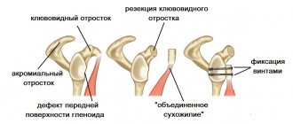
Treatment and restoration of the alveolar process
- Split-Control technology. The main purpose of this procedure is to expand the jaw bone to allow subsequent implantation. The procedure is performed as follows. The specialist saws the comb, places biomaterial, a bone tissue substitute, into the cavity, and applies sutures.
- Intercortical osteomia. It involves splitting the alveolar process to correct the bone structure. During the surgical intervention, the ridge is cut to form a movable fragment, which the dental surgeon then moves to another part where there is a lack of bone tissue. The moving part is fixed with special screws, and the cavity is filled with biomaterial.
The rehabilitation time after surgery and resection of the alveolar process can be several months. After this, you can begin to implant the implant. In each case, you must consult directly with your dentist. He will track the dynamics and determine the condition of the alveolar process in which there was a cleft or damage.
Pulmonary alveolitis: clinical recommendations
Symptoms of pulmonary alveolitis require treatment under medical supervision. The treatment regimen includes drug and non-drug therapy. First of all, patients need to eliminate the effect of the irritant if the disease is caused by exogenous factors. To improve the quality of life, the doctor will select a rehabilitation program based on physical exercise and oxygen therapy. After the course, the normal concentration of oxygen in the blood is restored, therefore shortness of breath decreases, performance and activity increase.
The prognosis for eliminating the symptoms of alveolitis will be favorable if the patient takes medications prescribed by a pulmonologist. For pathology the following are used:
- glucocorticosteroids to stop inflammation and relieve swelling;
- antifibrotic agents to protect tissues from irreversible changes;
- bronchodilators for better sputum removal and cough treatment;
- potassium supplements and diuretics to stabilize blood pressure and heart function.
In case of pronounced irreversible changes and ineffectiveness of conservative therapy, the patient undergoes organ transplantation. Indications for surgical intervention include a critical decrease in capacity, dyspnea, and oxygen deficiency. The prognosis for lung transplantation due to alveolitis is favorable. Survival rates reach 60%.
Benefits of bone grafting
- Reliable adhesion of the implant to the bone tissue.
- Minimal trauma. Minor swelling and hematoma.
- High efficiency. Allows you to start prosthetics in a short time.
Before performing correction of the alveolar process, it is necessary to make sure that there are contraindications, for example, the absence of an allergic reaction to biomaterial and drugs. Dental treatment is not carried out for cancer and autoimmune diseases, as well as poor blood clotting. In some cases, surgery may be delayed until the underlying conditions have resolved.
Pulmonary alveolitis: symptoms and treatment in adults
Treatment tactics directly depend not only on the cause of the disease, but also on the clinical picture. Patients are prescribed medications that prevent the progression of the pathology, as well as eliminate unpleasant symptoms.
Main signs of alveolitis :
- Cough. Typically dry, with minimal sputum production. There are no impurities of pus or blood in the mucus discharge. Wheezing may occur.
- Dyspnea. In the early stages of diffuse pulmonary alveolitis, the symptom is disturbing only after physical exertion, so many patients ignore it. Further, shortness of breath occurs during short walking and at rest.
- Pain behind the sternum. Discomfort increases with deep inspiration, which disrupts the respiratory rhythm and reduces the concentration of oxygen in the blood. The pain may radiate under the collarbone.
Symptoms also depend on the form of the pathology. Acute pulmonary alveolitis occurs with a significant deterioration in health. The patient complains of muscle pain, fever, and fever. With chronic alveolitis, weight loss, swelling, fatigue, and pale skin are observed.
Recommendations for rehabilitation
The rehabilitation period is aimed at restoring the functions of the affected area. After surgery it is recommended:
- Follow all doctor's recommendations.
- Avoid eating too cold or hot food.
- Follow the rules of oral hygiene.
- Avoid serious physical activity.
- Follow the recommendations for a gentle diet.
Additionally, oral baths using antiseptic solutions and the use of soft toothbrushes may be recommended to eliminate the risk of damage to the operated area. If you follow the recommendations, there are no risks of possible complications. Successful recovery depends on the skill level of the surgeon. Our AlfaDent clinic employs exclusively professionals who take advanced training courses and regularly improve their skills.
Symptoms of pulmonary dissemination
Like most lung diseases, disseminated lung disease does not have specific symptoms that can be used to accurately diagnose it. With dissemination, patients may experience standard respiratory symptoms:
- Dyspnea;
- Cough;
- Difficulty breathing (it becomes more frequent, it becomes difficult to hold it);
- Unpleasant sensations in the chest;
- Cough;
- Loss of appetite;
- Temperature increase;
- Weakness (due to impaired respiratory function and lack of oxygen);
- Decrease in blood oxygen saturation;
- Change in blood pressure.
Forecast
With timely and adequate treatment of acute exogenous allergic, as well as toxic fibrosing alveolitis, the prognosis is usually favorable. As the disease becomes chronic, the prognosis worsens.
Idiopathic fibrosing alveolitis is prone to gradual progression with the development of complications. Due to increasing irreversible changes in the alveolar-capillary system of the lungs, the risk of death is high. The five-year survival rate after surgical treatment reaches 50-60%.
Nutrition rules and some recommendations from doctors
Properly selected therapy helps to get rid of a disease such as alveolitis. Symptoms of mild forms of the disease disappear within a few weeks after the start of treatment. Nevertheless, patients are advised to quit smoking and get rid of other bad habits. You also need to eliminate contact with potentially hazardous substances.
Proper nutrition will also help speed up the healing process. Experts recommend adhering to the following rules:
- maintain a drinking regime (at least 2 liters of fluid per day);
- The diet should include dairy and fermented milk products, vegetables and fruits, freshly squeezed juices, honey, semolina, lean meats;
- dishes need to be steamed or boiled;
- Regular consumption of dried fruits, in particular dried apricots, raisins, prunes, etc., has a beneficial effect on the body’s condition.
Allergic alveolitis and its features
The cause of exogenous allergic alveolitis is contact with external irritants, but of natural origin. Allergens can include fungal and plant spores, pollen, particles of animal fur and fluff. Most often, harmful substances enter the body through inhalation. Farmers, furriers, and agricultural workers who constantly work with animals, rotted hay, and other irritants are susceptible to this disease.
A non-inhalation route for allergens to enter the body is also possible, but such cases are recorded extremely rarely.
The disease is accompanied by severe shortness of breath, coughing, attacks of which intensify during contact with potentially hazardous substances. In this case, an important part of therapy is identifying allergens and limiting contact with them. Without this, drug treatment will not have the necessary effect.
What does the treatment plan look like?
What should patients who are diagnosed with pulmonary alveolitis do? Treatment is tailored individually. Here, much depends on the form and stage of development of the disease. In any case, therapy should be aimed not only at eliminating the cause of inflammation, but also at inhibiting the fibrotic process.
- In case of allergic and toxic alveolitis, it is extremely important to protect the patient from contact with hazardous substances. Cytostatics and glucocorticoids are also used (sometimes they are administered directly into the respiratory system by inhalation).
- Patients are prescribed medications that dilute the mucus secreted by the epithelium and facilitate its passage.
- Prednisolone in small doses helps to cope with the inflammatory process.
- Autoimmune pulmonary alveolitis requires the use of immunosuppressants.
- Sometimes an oxygen concentrator is used - this helps to avoid hypoxia.
- In some cases, the doctor prescribes antibacterial medications, in particular penicillin.
Patients are also recommended special breathing exercises, which help preserve lung volume and cope with the fibrotic process.
Preventive actions
Unfortunately, today there is no specific prevention of such a disease as pulmonary alveolitis. However, it is worth taking some precautions. It is important to avoid contact with toxic substances and allergens. It is recommended to stop taking pneumotoxic drugs. If the use of these drugs is necessary, then it is important to constantly monitor the condition of the respiratory system.
As for patients who have already suffered from such lung diseases, they should be under the supervision of a rheumatologist and pulmonologist throughout their lives, regularly take tests and undergo medical examinations.
What complications can the disease lead to?
Under no circumstances should such a problem be ignored. The lack of timely therapy leads to the replacement of pulmonary structures with connective tissue. The lungs gradually lose their properties, the body does not receive enough oxygen. One of the dangerous complications is hypoxia of all organ systems. Serious metabolic disorders are also possible.
Gradually, the fibrotic process leads to the development of respiratory failure. The list of dangerous complications also includes pulmonary edema, which is the result of liquid blood components entering the cavity of the respiratory organs. Edema can develop at lightning speed - often this condition ends in the death of the patient. In other cases, the pathology develops gradually - the symptoms progress slowly, the patient’s condition worsens within 24 hours, which makes it possible to call a doctor in time.