Blood during urination in women is a symptom that appears due to injuries to the organs of the excretory system and various pathologies. It's called hematuria. It must be borne in mind that any change in urine color requires consultation with a doctor. Therefore, even if spotting appears only occasionally, do not neglect qualified medical care. This sign may indicate various diseases that occur in a latent form.
To get quality help, determine the causes of your illness and undergo treatment, contact a medical specialist in Belgorod. By following the recommendations of our doctors, you can improve your well-being and quality of life.
Urine formation
The excretory system is primarily necessary to filter the blood and remove metabolic products from the body.
The urine formed in the kidneys is the result of careful filtration of blood coming from the cardiovascular system: in the nephrons, harmful and ballast substances are released into the urine and the reverse transfer of useful substances (such as proteins and minerals) into the blood. As a result, a light yellow liquid with a specific chemical composition is formed. Substances contained in urine:
- various organic components, including glucose, uric acid, urea, ketone bodies, amino acids and creatinine;
- PMmineral substances in the form of ions: calcium, magnesium, chlorine and others;
- water is the main component of urine.
The organic components of urine are predominantly represented by nitrogenous compounds formed as a result of the breakdown of protein in the body. With an excess of nitrogenous compounds in the blood, we can sometimes talk about impaired renal function. The urine may also contain a moderate number of cells. Thus, normally no more than two red blood cells can be detected in urine (in the visible field).
Additional structures of the excretory system are involved in the removal of urine from the body. From the kidneys, fluid enters the ureters and is deposited in the bladder. During urination, fluid leaves the body through the urethra. The process of urination can also be affected by other organs that release their secretions through the urethra. These are the prostate gland, seminal vesicles, bulbourethral glands and testicles.
Causes of gross hematuria
The medical term for so-called gross hematuria is gross hematuria. The urine has a visible red or dark brown tint. This condition may indicate a dangerous pathological process in the body.
Main reasons
- Kidney stones or the formation of stones in the bladder. Stones can form in the excretory system due to poor diet, primary kidney pathologies and other conditions. A large stone can damage the mucous membranes of organs and form small foci of hemorrhage.
- Infection of kidney tissue. Pathogenic microorganisms can enter the kidney through the circulatory system and damage the nephrons. Symptoms of this disease usually include fever and back pain.
- Infection of the lower urinary tract, that is, damage to the bladder or urethra by microorganisms. The appearance of an unpleasant odor in urine is a common sign of pathology.
- Polycystic kidney disease. This is a dangerous disorder characterized by damage to kidney tissue.
- Blockage of the renal vein as a result of thrombosis (blood clots in a vessel).
- Various injuries. Physical damage to the urethra often occurs during prolonged catheterization.
- Pathologies of the prostate gland. Blood in the urine can appear with an enlarged prostate (adenoma) and inflammatory processes in the tissues of the organ.
- Oncological diseases. The growth of a malignant tumor in the tissues of the kidney, bladder, prostate or other genitourinary organs can be complicated by hematuria.
Careful visual examinations help quickly determine the cause of the disorder.
Emergency care for acute pyelonephritis and hematuria
Acute pyelonephritis is a nonspecific infectious inflammation of the pyelocaliceal system and renal parenchyma. ARF is caused by Escherichia coli, Klebsiella, Proteus, Pseudomonas.
Routes of entry of an infectious agent into the kidney:
- ascending - foci of chronic inflammation are located in the female genital organs, lower urinary tract and less commonly in the colon;
- hematogenous - the source is an acute or subacute inflammatory process outside the urinary tract (mastitis, boil or carbuncle, etc.).
Predisposing factors for the development of acute pyelonephritis are hemodynamic or urodynamic disorders in the kidney or upper urinary tract. Acute pyelonephritis is more often observed in women.
The infection, once in the kidney, receives favorable conditions for its development in zones of hypoxia, where the inflammatory process occurs. An infected thrombus or embolus in the terminal vessels of the renal cortex causes an infarction followed by suppuration. The occurrence of multiple small suppurating infarctions in the cortex is classified as apostematous nephritis. The development of an extensive infarction with subsequent suppuration leads to the formation of a renal carbuncle.
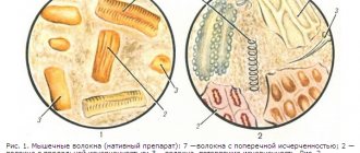
With regard to the pathogenesis of the disease, primary and secondary pyelonephritis are distinguished (Fig. 1). Secondary pyelonephritis is based on organic or functional changes in the kidneys and urinary tract.
Depending on the passage of urine through the upper urinary tract, i.e. from the kidney to the pelvis and further along the ureter, acute pyelonephritis is distinguished between non-obstructive (if it is preserved) and obstructive (if it is disturbed).
Manifestations of acute pyelonephritis:
- chills;
- high fever (38–39°C and above);
- pain in the lower back (side);
- often nausea and vomiting;
- general intoxication.
Often acute pyelonephritis is preceded by frequent, painful urination at the end (clinical picture of acute cystitis).
Non-obstructive acute pyelonephritis can begin with dysuria and on the same day or 1-2 days later lead to high body temperature, chills and pain in the affected kidney. Chills may be replaced by heavy sweat with a short-term decrease in body temperature; pain in the lumbar region in some cases appears during urination and precedes chills and fever (vesicoureteral reflux!), and after them does not recur (rupture of the fornix of one or more calyces and resorption of urine - fornical reflux!).
Obstructive acute pyelonephritis (occlusion of the ureter by a stone, products of chronic inflammation of the kidney, external compression - retroperitoneal fibrosis, cancer of the internal genital organs, enlarged lymph nodes, etc.) begins with gradually increasing or acute pain in the lower back on the affected side, followed by chills and increased body temperature.
Complications of obstructive acute pyelonephritis:
- development of a purulent process;
- severe impairment of renal function;
- bacteriotoxic shock;
- urosepsis;
- toxic hepatitis;
- paranephritis;
- pyonephrosis.
Diagnostics
When collecting anamnesis, pay attention to:
- for recent hypothermia;
- chronic pyelonephritis;
- urolithiasis;
- diseases of the female genitalia, prostate gland;
- previous operations on the kidneys and urinary tract, etc.
They check that the pulse rate corresponds to body temperature, detect pain in the hypochondrium during palpation of the abdomen and a positive symptom of tapping on the lower back (Pasternatsky’s symptom) from the affected kidney.
Laboratory diagnostics. In a general urine test, leukocyturia is often noted, which may be absent in obstructive acute pyelonephritis, since urine from the affected kidney does not enter the bladder.
In a general blood test, leukocytosis is noted, often with a shift in the blood count to the left (the number of band neutrophils is 20% or higher).
In a biochemical blood test, an increase in urea and creatinine levels is possible, often in elderly and weakened patients, with chronic renal failure or damage to the only functioning kidney.
When culturing urine (carried out before antibacterial therapy), the causative agent of the disease is isolated and its sensitivity to antibacterial drugs is determined.
To clarify the diagnosis and form of acute pyelonephritis, the following is carried out:
- Ultrasound;
- X-ray examinations;
- computed tomography;
- magnetic resonance imaging.
Acute pyelonephritis is differentiated from the following conditions:
- infectious diseases that occur with fever and chills and are not accompanied by pain localized in the lumbar region;
- surgical pathology of the abdominal organs, in which severe pain is often observed, and a pronounced increase in temperature and dysuric phenomena are rare.
Main directions of therapy
The emergency care algorithm for acute pyelonephritis is presented in Figure 2.
- Normalization of urine passage from the kidney:
- installation of a ureteral catheter or stent;
- installation of a catheter in the bladder if vesicoureteral reflux is suspected (lower back pain during urination);
- nephrostomia.
- Antibacterial therapy for acute pyelonephritis is in most cases empirical and depends on the severity of the disease:
- mild - oral drugs from the fluoroquinolone group;
- moderate and severe - parenteral aminoglycosides in combination with or without ampicillin, fluoroquinolones, third and fourth generation cephalosporins in combination with or without aminoglycosides.
- Surgical treatment after further examination is prescribed:
- if antibacterial therapy is ineffective within 3 days;
- severe course of the disease;
- purulent pyelonephritis
Clinical pharmacology of individual drugs
Gentamicin is effective against infections caused by gram-positive and gram-negative microorganisms, bacteria of the intestinal group. The drug is rapidly absorbed when administered intramuscularly, the therapeutic concentration in the blood is reached after 1 hour and persists for 8–12 hours. A single dose is 80–160 mg, a daily dose is 160–320 mg. Side effects: nephro- and ototoxicity. Contraindications for use: decreased kidney function and decreased hearing.
Fluoroquinolones (ciprofloxacin, norfloxacin, ofloxacin) are active against a large number of gram-positive and gram-negative bacteria and are available in forms for oral and parenteral administration. The drugs are well absorbed in the intestine and widely distributed in body fluids and tissues, secreted mainly by the kidneys. The half-life is 3–7 hours. The most widely used drugs for the treatment of acute pyelonephritis are ciprofloxacin (medociprin, siflox, ciprovin) - 500 mg 2 times a day, norfloxacin (nolicin, norbactin) - 400 mg 2 times a day and ofloxacin (zonocin, oflo , ofloxacin) - 200 mg 2 times a day. The use of fluoroquinolones is contraindicated in children under 14 years of age, pregnant women, as well as in case of individual intolerance.
Common therapy errors:
- conservative management of obstructive pyelonephritis;
- continuation of intensive antibacterial therapy without effect for more than 3 days without further examination;
- prescription of antibacterial drugs from the group of semisynthetic penicillins (ampicillin, oxacillin);
- the use of drugs from the group of uroseptics (nitroxoline, palin, negram, etc.).
Indications for hospitalization are most cases of acute pyelonephritis, especially if there is a suspicion of an obstructive nature of the lesion.
Analysis of clinical cases
Patient S., 18 years old. Complaints of pain in the left lumbar region, increased body temperature to 38°C, accompanied by chills, nausea, increased frequency and painful urination. History: 3 days ago there was hypothermia, after which urination became more frequent and painful, the temperature increased and pain appeared. I took nitroxoline without effect. On examination: pain on palpation in the left lateral parts of the abdomen, Pasternatsky’s sign is sharply positive on the left. Diagnosis: acute non-obstructive pyelonephritis on the left. The patient was hospitalized in the urological hospital. Treatment: antibacterial therapy with gentamicin (80 mg 2 times a day IM).
Patient K., 29 years old. Complaints of sharp pain in the right lumbar region, radiating to the perineum, increased body temperature to 39°C with tremendous chills, nausea, and repeated vomiting. Medical history: he has been suffering from urolithiasis for a long time; previously, small stones passed on their own several times. Two days ago, sharp pain appeared in the right lumbar region, she took Baralgin and No-shpa with a temporary effect. A day after the onset of pain, she noted a rise in temperature with chills, nausea and vomiting. On examination: the patient groans in pain. The tongue is dry, heart rate 90 beats/min. The abdomen is soft, sharply painful on palpation in the right lateral regions, Pasternatsky's symptom is sharply positive on the right. Diagnosis: right-sided renal colic. Acute obstructive pyelonephritis on the right. The patient was hospitalized in the urological hospital. Treatment: catheterization of the right ureter; if this is impossible, nephrostomy on the right. Intensive detoxification and antibacterial therapy.
Patient Sh., 67 years old. Complaints of chills, increased body temperature to 39.5°C, lower back pain on the right. History: 3 months ago, a radical cystoprostatectomy was performed for bladder cancer, plastic surgery of the bladder with an intestinal flap according to Studer. Preoperative examination revealed aplasia of the left kidney in the patient. A month after the operation, constant nagging pain appeared in the right lumbar region; he did not consult a doctor. Over the past week, the amount of urine excreted has decreased and swelling of the legs has appeared. Three days ago he noticed an increase in temperature against the background of increased pain in the right lumbar region, the temperature progressively increased, reached 39.5°C and was accompanied by chills, the volume of urine per day did not exceed 200 ml. On examination: the patient is pale, asthenic in build, and there is swelling of the lower extremities. Palpation reveals severe pain in the right lateral parts of the abdomen, Pasternatsky's symptom is sharply positive on the right. Diagnosis: acute obstructive pyelonephritis on the right. Stricture of the right ureteroneocystanastomosis. Oliguria. The patient was hospitalized in the urological hospital. Treatment: emergency nephrostomy on the right.
Hematuria is a pathological symptom characterized by blood in the urine.
The main causes of renal bleeding are presented in Table 1.
Essential hematuria (Table 2) combines a number of conditions in which the etiology and pathogenesis are unknown, and clinical, radiological and morphological studies do not allow us to accurately indicate the cause of bleeding.
Hematuria can be caused by taking non-narcotic analgesics, anticoagulants, cyclophosphamide, oral contraceptives, vincristine.
With kidney tumors, when the tumor grows in the pelvis or calyces, the integrity of the vascular wall is disrupted. If the tumor does not communicate with the pyelocaliceal system, the venous outflow from the area of the kidney with the tumor node is disrupted, the fornical veins dilate and rupture.
With prostate cancer, the tumor grows into the walls of the bladder or prostatic urethra, the veins of the bladder in its cervical section are compressed by the tumor, and venous stasis occurs. Free-floating villous tumors of the bladder, located near its neck, during urination are carried away by the flow of urine into the urethra, clog its lumen and, when pinched, swell, burst and begin to bleed.
With cystitis and prostatitis, the mucous neck of the bladder becomes inflamed, injured and bleeds easily when it comes into contact with other walls at the end of urination.
When renal hemo- and urodynamics are disturbed, the venous plexuses of the fornix, with impaired venous outflow or a significant increase in intrapelvic pressure, become overfilled with blood, the veins encircling the vaults of the calyces in a ring form expand and their integrity is disrupted, which leads to hematuria from the upper urinary tract.
With necrosis of the renal papillae, the blood supply to the papilla, and sometimes to the entire Malpighian pyramid, is disrupted, necrotic tissue is rejected, and bleeding occurs.
With benign prostatic hyperplasia, venous congestion occurs in the pelvic organs, and the integrity of the vessels is disrupted.
There are two types of hematuria:
- microscopic - the presence of red blood cells in the urine (more than three in the field of view) is determined only by microscopic examination of urine sediment;
- macroscopic - blood in the urine is determined visually; if only in the first portion of urine - initial (initial), in the last portion of urine - terminal (final), in all portions of urine - total.
Clinical picture
The appearance of red blood cells in the urine gives it a cloudy appearance and a pink, brown-red or reddish-black color, depending on the degree of hematuria. Hematuria can occur suddenly, be one-time or repeated. Its cessation is deceptive, since the cause of hematuria, as a rule, is not eliminated, and examination in case of repeated bleeding with a great delay often reveals an already advanced tumor.
With a kidney tumor, hematuria may be the first and for a long time the only symptom of the disease. In most cases, it is painless, but with profuse bleeding with the formation of clots in the renal pelvis and their passage through the ureters, dull, less often colicky pain occurs in the lumbar region. With papillary tumors of the bladder, hematuria is usually painless and profuse without the formation of clots. With villous tumors of the bladder located near its neck, the urine stream may be interrupted during urination.
With tuberculous kidney damage, total hematuria is observed in the very early stages of the process, which may be accompanied by persistent pyuria.
With benign prostatic hyperplasia, hematuria occurs for no apparent reason or during catheterization of the bladder due to a violation of the integrity of the loosened mucous membrane of the posterior urethra.
In acute cystitis and prostatitis, terminal hematuria is noted against the background of severe dysuria.
With urolithiasis, hematuria usually develops following an attack of renal colic; the urine stream may suddenly stop during urination.
Possible complications: with massive bleeding with the formation of shapeless clots, acute urinary retention is possible due to tamponade of the bladder with these clots. Rarely, shock may develop due to blood loss and pain.
Diagnostics
During the survey you need to find out:
- conditions for the occurrence of hematuria;
- degree, nature and duration of hematuria: - initial hematuria - a pathological process in the urethra (tumor, inflammation, foreign body, burn, etc.); – terminal hematuria - a pathological process in the neck of the bladder (acute cystitis, prostatitis, stones and bladder tumors); – total hematuria is a pathological process in the kidney, ureter or bladder (tumors, benign prostatic hyperplasia, renal tuberculosis, pyelonephritis, necrosis of the renal papillae, nephroptosis, etc.);
- the presence of blood clots in the urine, their shape: – worm-shaped – bleeding from the upper urinary tract and the formation of clots in the ureter; – shapeless clots – bleeding from the bladder;
- the presence of pain and its connection with changes in the color of urine: – pain in the lumbar region on the affected side – disruption of the passage of urine from the kidney due to blood clots, urolithiasis; – painless hematuria with subsequent development of renal colic (normal-colored urine) – tumor of the kidney or upper urinary tract;
- presence or absence of dysuric phenomena;
- whether there were injuries to the kidneys, bladder, urethra (in case of injury, blood is released from the urethra outside the act of urination).
Visual assessment of urine:
- scarlet blood - bleeding continues;
- brown urine - bleeding has stopped;
- brick shade - intense uraturia.
The color of urine may change when taking medications and foods:
- pink - when taking pyramidon;
- saffron yellow - nitroxoline;
- brown - rhubarb and hay;
- raspberry - purgena;
- red - phenolphthalein and beets;
- red-brown - madder dye.
With the syndrome of prolonged compression and crushing of tissues, myoglobin from the muscles enters the blood and penetrates into the urine, which gives it a red-brown color.
To determine the nature and source of bleeding in a hospital setting, the following is carried out:
- cystoscopy - in all cases of gross hematuria, with the exception of inflammatory diseases (acute urethritis, cystitis, prostatitis);
- digital rectal examination;
- Ultrasound of the bladder kidneys, prostate, transrectal ultrasound;
- excretory urography, retrograde reno- and urethrocystography, computed tomography, osteoscintigraphy according to indications in a hospital setting.
Laboratory diagnostics include:
- clinical analysis of blood, urine;
- blood chemistry;
- microscopic examination of urine sediment;
- urine culture for sterility;
- determination of the level of prostate-specific blood antigen.
With hemoglobinuria, the color of urine does not change even after standing for a long time, and with hematuria, red blood cells settle to the bottom of the vessel and the upper layers of urine become yellowish.
Hematuria in women should be differentiated from bleeding from the genitals. To do this, examine the average portion of urine during spontaneous urination or urine obtained from the bladder by catheterization.
Main directions of therapy
The emergency care algorithm for hematuria is presented in Figure 3.
| Figure 3. Emergency care algorithm for hematuria |
With the development of hypovolemia and arterial hypotension, restoration of bcc: crystalloid and colloid solutions intravenously.
If acute cystitis is suspected, after collecting urine for culture to ensure sterility, a broad-spectrum antibiotic is prescribed.
Therapy with hemostatic drugs should be carried out in a urological hospital, after diagnosis, under the control of blood clotting.
Clinical pharmacology of individual drugs
Etamsylate (dicinone) activates the formation of thromboplastin, the formation of blood coagulation factor III, and normalizes platelet adhesion. The drug does not affect the thrombotic time, does not have hypercoagulable properties, and does not contribute to the formation of blood clots. Etamsylate is administered in 2–4 ml (0.25–0.5 g) intravenously at once or drip, adding to conventional solutions for infusion.
The hemostatic effect develops:
- for intravenous administration - after 5–15 minutes; maximum effect - after 1–2 hours; the effect lasts 4–6 hours or more;
- when administered intramuscularly, the effect occurs somewhat more slowly;
- When taken orally, the maximum effect occurs after 3 hours.
Etamsylate should not be used for hemorrhages caused by anticoagulants.
Aminocaproic acid inhibits fibrinolysis by blocking plasminogen activators and partially inhibiting the effect of plasmin, and has a specific hemostatic effect in bleeding associated with increased fibrinolysis. To quickly achieve an effect, under the control of a coagulogram, a sterile 5% solution of the drug in saline is administered intravenously, up to 100 ml.
Protamine sulfate has an antihemorrhagic effect in bleeding caused by heparin during its overdose, after operations using extracorporeal circulation and the use of heparin. Apply 1 ml of a 1% solution intravenously in a slow stream over 2 minutes or drip; 1 mg of protamine sulfate neutralizes approximately 85 units of heparin. Contraindicated in hypotension, adrenal insufficiency, thrombocytopenia.
Common treatment errors: prescribing hemostatic drugs before identifying the source of hematuria.
Gross hematuria is an indication for emergency hospitalization in a urological hospital.
Analysis of clinical cases
Patient M., 30 years old. Complaints of cramping pain in the lumbar region on the left, aggravated by movement, after physical activity, blood in the urine, increased body temperature to 38.2°C. The disease began with dysuria in the form of frequent, painful urination, a day later pain appeared in the lumbar region, and blood in the urine. History: 7 years ago, a diagnosis was made of stage I nephroptosis on the left. The disease manifested itself as periodic attacks of pyelonephritis. On examination: the patient is restless. The kidney area is visually unchanged, painless on palpation, the effleurage symptom is weakly positive on the left, there is no pain along the ureters, the bladder is empty by percussion. Diagnosis: acute ascending left-sided pyelonephritis. Nephroptosis on the left. The patient was hospitalized in the urology hospital.
Patient J., 77 years old. Calling an emergency room due to intense blood in the urine and the release of blood clots during urination. History: no urological diseases. On examination: the patient is asthenic, the skin is pale. The kidney area is not visually changed; bimanual palpation of the right kidney reveals a mass formation in the lower segment; the effleurage symptom is negative on both sides. A digital rectal examination reveals that the prostate is enlarged 1.5–2 times, has a tight-elastic consistency, the median groove is smoothed, the rectal mucosa above the gland is mobile, palpation is painless. Diagnosis: tumor of the right kidney. The patient was hospitalized in the urology department.
Patient Ch., 24 years old. Complaints of constant bleeding through the urethra. History: About an hour ago I fell and hit my crotch on a pipe. On examination: bruising in the perineal area, discharge of scarlet unchanged blood from the urethra, outside the act of urination. A digital rectal examination reveals that the prostate gland is of normal size, has a tight-elastic consistency, the median groove is pronounced, the rectal mucosa above the gland is mobile, palpation is painless. Diagnosis: urethral injury. The patient is indicated for hospitalization in a hospital for examination and determination of treatment tactics.
Thus, the success of treatment of patients with pyelonephritis and hematuria is largely determined by the adoption of adequate measures in the provision of emergency medical care.
E. B. Mazo , Doctor of Medical Sciences, Professor, Corresponding Member of the Russian Academy of Medical Sciences, Russian State Medical University, Moscow
Table 1. Causes of renal bleeding
| Causes of hematuria | Pathological changes in the kidney, blood diseases, etc. |
| Congenital anomalies | Cystic diseases of the pyramids, papillary hypertrophy, nephroptosis, etc. |
| Mechanical | Trauma, stones, hydronephrosis |
| Hemodynamic | Disorders of the blood supply to the kidney (venous hypertension, infarction, thrombosis, phlebitis, aneurysms), nephroptosis |
| Hematological | Blood coagulation disorders, hemophilia, sickle cell anemia, etc. |
| Reflex | Vasoconstrictor disorders, shock |
| Allergic | Glomerulonephritis, arteritis, purpura |
| Toxic | Medicinal, infectious |
| Inflammatory | Glomerulonephritis (diffuse, focal), pyelonephritis |
| Tumor | Benign neoplasms, malignant neoplasms |
| Essential | (See Table 2) |
Table 2. Causes of essential renal hematuria
| Nature of the process | Immediate cause |
| Pathological processes | Pyelovenous reflux, calyceal venous channel, |
| in the papillary-fornical zone | venous varicose veins |
| kidneys (fornical bleeding) | |
| Diseases of the papillae and pelvis | Cystic processes in the papilla, “spongy kidney”, necrosis of the papillae, endometriosis, varicose veins |
| Pathological kidney mobility | Nephroptosis |
| Renal sinus diseases | Adhesions around the main renal vessels, sinus lipomatosis, aneurysms, varicose veins |
| Diseases of the renal interstitium | Microfibromatosis, interstitial hyperplasia with arterio- |
| Infectious and inflammatory | Pyelonephritis, necrotizing papillitis, hepatitis |
| processes | (hepatorenal syndrome) |
| Renal vascular damage | Infarction, venous thrombosis, compression of the main renal vein, varicose veins of the intrarenal veins, intraparenchymal aneurysms, angiomatous formations in Fornix |
| Blood diseases, dyscrasia | Sickle cell anemia, hemophilia, erythremia, |
| The harmful effects of drugs that disrupt the function of the blood coagulation system | Anticoagulant therapy, cytostatics |
Causes of microhematuria
Externally imperceptible penetration of blood components into the urine can occur during inflammatory and infectious diseases.
Possible reasons:
- Damage to the renal glomeruli due to autoimmune inflammation (glomerulonephritis) and other inflammatory kidney diseases.
- Thin basement membrane disease. This is benign microhematuria caused by a slight impairment of blood filtration.
- The formation of benign tumors (polyps) in the urethra or bladder.
- Infectious diseases with a moderate course, usually without pain.
It is not always possible to clearly separate the etiology of microhematuria and macrohematuria. Certain pathological processes can lead to the penetration of varying amounts of blood into the urine. This is especially true for oncology, infection and trauma.
Risk factors
Urologists are aware of certain forms of predisposition to hematuria. These are pathologies, anomalies and various signs that affect the state of the human excretory system.
Main risk factors
- Age. For many men, the presence of blood in the urine occurs at the age of 40-50 years as a result of an enlarged prostate gland.
- Recent infection. The inflammatory process in tissues may persist for several days. Thus, post-infectious glomerulonephritis is a common cause of microhematuria in children.
- Unfavorable family history. The presence of diseases of the excretory system in close relatives indicates an individual risk of developing hematuria.
- Taking medications that affect the kidneys. These are aspirin, non-steroidal anti-inflammatory drugs and antibiotics.
- Increased physical activity. Hematuria is especially common for people who prefer long-distance running.
- Inflammation of the genital organs in women. Such ailments imply an increased risk of infection spreading into the excretory system.
Taking risk factors into account allows a person to prevent diseases of the excretory system.
Diagnosis of pathologies manifested by blood during urination
The range of diagnostic procedures prescribed by a doctor depends on the specific clinical case. Therefore, initially he collects a detailed medical history and clarifies your complaints. After this you may be assigned:
- general urine analysis;
- urine analysis according to Nechiporenko and Zimnitsky;
- ultrasound examination of the pelvic organs and kidneys;
- X-ray examination of the urinary tract;
- computed tomography;
- cystoscopy;
- magnetic resonance imaging;
- general and biochemical blood tests;
- tests for sexually transmitted infections.
Additional symptoms
Change in urine color is rarely the only symptom of the disease. Often, hematuria occurs against the background of pathological signs associated with the functioning of the kidneys and other organs.
Other signs:
- increase in body temperature;
- pain in the back, lower abdomen, or perineum;
- the appearance of an unpleasant odor in urine;
- change in urine volume (diuresis);
- difficulty urinating or urinating too frequently;
- dizziness;
- nausea and vomiting;
- weakness and fatigue;
- painful urination.
If such symptoms appear, you should make an appointment with a doctor. In case of an acute infectious process, the patient may require urgent treatment.
Glomerulonephritis
This disease causes damage to the vascular glomerulus of the kidneys, where the process of filtering urine takes place. During inflammation, the walls of the nephron become permeable and some of the blood enters the glomerulus, and the red blood cells acquire an irregular shape. It is on this basis that glomerulonephritis is quite easily diagnosed based on a urine test. The color of the urine becomes the “color of meat slop.”
Diagnostics
To undergo the examination, you must make an appointment with a urologist. During the consultation, the doctor will ask the patient about complaints and collect anamnestic information to identify risk factors for diseases. An initial examination sometimes reveals specific symptoms. To clarify the diagnosis, the urologist will need the results of instrumental and laboratory tests.
Necessary research
- Additional general urine analysis. The doctor will need to make sure that other samples also contain abnormal numbers of red blood cells. In addition, it is important to study the chemical composition of the liquid.
- Cystography is a visual examination of the inner lining of the bladder. The specialist inserts a thin tube equipped with a camera into the urethra. Using this method, stones, signs of damage or other changes can be detected.
- Ultrasound examination of the genitourinary system. Using ultrasound, you can study the structure of organs in real time. Imaging of the kidneys, bladder, and prostate is usually done.
- Additional visual examination methods, such as urography or computed tomography. Obtaining high-precision images gives the specialist the opportunity to detect even minor areas of hemorrhage.
The tests prescribed depend on the patient’s symptoms and individual medical history. Sometimes the doctor does other tests, such as a blood test or radionuclide kidney scan.
Why does blood appear when women urinate?
Most often, the symptom indicates the presence of fungal, viral and parasitic pathogens. Against the background of inflammation of the genitourinary system, blood clots occur during urination in women, as well as hematuria.
The color of urine may change due to menstrual flow. In addition, in the presence of hard deposits in the kidneys, hematuria often appears. As the stone passes through the organs of the excretory tract, they are traumatized, which is why hematuria is observed.
With severe physical exertion, you may experience bleeding. In this case, there is no discomfort; the phenomenon is short-term in nature. A reddish coloration of urine can occur when consuming certain dried fruits, beets, blackberries, and medications.
Hematuria may occur against the background of some gynecological diseases. It is also provoked by trauma, as well as recent cystoscopy. During diagnosis, a specialist may accidentally injure the mucous membrane of the urinary tract.
In some cases, pathologies are not accompanied by specific symptoms. For example, pyelonephritis can occur without burning, increased temperature and decreased overall tone of the body. And it is blood during urination that will be that alarming “bell”, when it appears, you need to consult a competent doctor as soon as possible.
Treatment
Hematuria is a clinical sign and not a disease. Accordingly, the doctor needs to accurately determine the cause of the disorder and select treatment for the corresponding disease. The main task is to restore the functions of the excretory system and eliminate dangerous conditions such as bleeding, infection or the growth of a malignant tumor.
Features of treatment tactics
- Asymptomatic (isolated) hematuria usually does not require treatment.
- In case of infection, antibiotics are first prescribed. In case of chronic bacterial infection, it is recommended to test the sensitivity of microflora to antibiotics to select effective medications.
- Surgical intervention is most often prescribed for obstruction of the excretory tract by stones, tumors and anatomical defects of organs.
- In most cases, patients require a special diet.
Thus, hematuria is a common symptom of diseases of the excretory system. In rare cases, this symptom does not indicate the presence of pathology. During the consultation, the urologist will be able to explain why there is blood in the urine.