Disc herniation of the cervical spine is an advanced form of degeneration of the cervical intervertebral segments, which is complicated by displacement of the nucleus pulposus of the disc with its extension beyond the anatomical boundaries. The disease is characterized by pronounced neurological and reflex manifestations, including the cerebral type. This is explained by the fact that in this section the ridge connects to the head, and the spinal cord passes into the brain, and a dense neurovascular network is concentrated here. The pathological process is mainly a complication of long-term osteochondrosis, the symptoms of which were ignored by patients for a long time.
MRI of the cervical spine.
Even a hundred years ago, in the field of view of neurologists, pathology with such a serious localization fell into the category of isolated cases, and it was mainly the lot of older people. Today, cervical intervertebral hernia is one of the most common diagnoses among all possible problems with the spine. Now, unfortunately, it too often concerns the working population. The diagnosis of “cervical hernia of the spine” predominates in people 20-55 years old. The incidence rate in men and women is approximately the same: 52% of patients are male patients, 48% are female.
In general, in the structure of all diseases of the human body, cervical hernias occupy 5th place in the number of hospitalizations, 3rd place in the need for surgical treatment. According to data from authoritative sources, at least 20% of people with cervical hernias suffer from severe vertebroneurological disorders, which often lead to disability. In approximately 35%-40%, the pain syndrome becomes chronic, which is why patients live in constant stress and emotional tension.
The risk group includes people suffering from systemic connective tissue diseases, obesity and various metabolic disorders, diabetes mellitus, and orthopedic pathologies. Also, professional athletes, office workers, teachers, machine operators, seamstresses, draftsmen and other people whose cervical region lingers in an immobilized position for a long time or performs monotonous monotonous movements have a high predisposition to displacement of the disc fragment in the cervical region.
What is a herniated cervical spine?
What is the essence of the diagnosis? In local pathological changes in the intervertebral disc, resulting in prolapse of the nucleus pulposus, which occurred due to the impaired integrity of the fibrous ring of the disc. The process of herniation is preceded by advanced degenerative-dystrophic pathogenesis in a specific spinal motion segment. But let's look at how everything happens in reality to make it more clear. Let's gradually get to the main point, starting with a brief excursion into anatomy.
- There are only 5 intervertebral discs in the cervical spinal system, and 7 vertebrae. They are located between each successive pair of vertebral bodies, connecting them to each other. Between the 1st and 2nd vertebrae (atlas and axis) the disc is not provided by nature, it does not exist. This means that pathology can occur at levels such as C2-C3, C3-C4, C4-C5, C5-C6, C6-C7. Most often it develops in the C5-C6 and C6-C7 segments.
- Each disc element consists of a jelly-like substance (nucleus pulposus), which is enclosed in an annular fibrous rim (annulus fibrosus). The disc performs a shock-absorbing function, absorbing and absorbing shocks and shocks in the spine during human physical activity. It also, together with other structural components, provides the upper part of the spine with the necessary potential for mobility and flexibility.
- The main material composition of the disc is represented by water (80%) and collagen fibers (15%) immersed in a matrix of proteoglycans. Collagen provides elasticity to the disc and retains proteoglycans (hyaluronic acid). And proteoglycans are responsible for attraction and control of water balance, creating the necessary intradiscal pressure. A normal ratio of all designated structural substances is possible only with good metabolism at the vertebral levels and complete delivery of nutrients to them.
- The structure of the disc is also avascular, or the absence of its own blood supply. Therefore, its supply of nutrition occurs exclusively through the blood vessels of the adjacent vertebrae, which enters through the marginal endplate. At a certain spinal level from C2 to C7, circulatory and metabolic processes may be inhibited. For example, due to constant physical overload or a sedentary lifestyle, possibly due to obesity, autoimmune pathologies or after local trauma. As a result, osteochondrosis develops.
- Since in osteochondrosis the blood flow in the vertebral bodies is reduced and the permeability of the marginal plate is impaired (sclerosis), the transport of important metabolites into the disc, as we said, does not occur in the required quantities. This leads to a gradual decrease in the specific fluid content in fibrous and pulpous tissues, that is, the loss of their main component - water. Dehydration serves as a stimulus for thinning, flattening, and disintegration of the intervertebral lining.
- Then cracks appear on the inside of the degenerated ring, and the nucleus pulposus changes its normal position. The jelly-like substance sags and moves into the defective part of the ring. As a result, a disc deformation occurs in a certain direction, protruding beyond the boundaries of the vertebrae.
- At first, the cervical disc protrudes slightly, but until the ring ruptures, a hernia will not form. This early stage is called protrusion. As soon as a protrusion is diagnosed, this means that all the prerequisites and favorable conditions for the appearance of a hernia have already been created. If the processes of degeneration at the prehernia stage are not stopped in a timely manner, the fibrous ring continues to collapse. The core, in turn, is increasingly dislocated towards the periphery and exerts compression on the weak area of the ring.
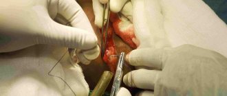
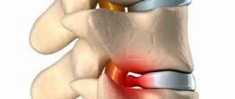
Hernia and coracoid osteophytes.
Ultimately, the ring-shaped structure ruptures with a fragment of jelly-like cartilage falling out through the formed hole. Open prolapse of part of the nucleus pulposus into the intervertebral space through a peripheral through breakthrough of the disc is a herniation of the cervical spine.
Anatomy of a normal cervical disc
Between each vertebrae there is a disc, which is a strong, elastic, shock-absorbing cushion. Each disc is composed of an annulus fibrosus, which encloses a gel-like substance called the nucleus pulposus. Nerve roots exit the spinal canal through small passages between the vertebrae and the disc. Pain and other symptoms are caused when a damaged disc presses on a nerve root or spinal cord. A cervical disc herniation occurs when the annulus fibrosus ruptures and the nucleus pulposus extends beyond it.
Types and stages of intervertebral hernias in the neck
We told you that discs can be pre-herniated and truly herniated. At the stage of protrusion in the cervical region, the cartilaginous layer is considered small up to 0.2 cm and corresponds to stage 1 of the pathology. Protrusions exceeding 0.2 cm are classified as medium and large sizes (2, 3, 4 degrees). So, let's give a more precise description of each stage.
- Stage 1 – the integrity of the disc is preserved, the size of the bulge is up to 2 mm inclusive.
- Stage 2 – moderate severity, the integrity of the fibrous ring is compromised, the protrusion is increased from 2 mm to 4 mm.
- Stage 3 – severe, the ring is torn, the disc displacement is strong, equal to 4-6 mm.
- Stage 4 (extrusion or sequestration) – a critically severe degree of deformation, which can end in sequestration at any time. The size of the hernia at this stage is > 6 mm, and can reach up to 8 mm or more.
Sequestration is the most dangerous form of the disease, when a sagging cartilage fragment is completely torn off from the disc with a piece of dead cartilage entering the spinal space. Such pathogenesis can, in a short period of time, cause serious irreversible damage to the nerve, including its death, which leads to paralysis.
In general, all disc prolapses at the level of the neck, which are greater than 4-6 mm in volume, are considered clinically extremely unfavorable, since they seriously impair cerebral circulation, depress and disrupt the unity of the two main parts of the central nervous system - the brain and spinal cord.
Unacceptably large sizes (> 4 mm) in this section are associated with the highest risks of partial/complete immobilization of the arms and legs, as well as paralysis of the torso from the affected area and below. In addition to paresis and paralysis, severe dysfunction of the genitourinary and reproductive organs develops with high frequency.
CT slice.
The species diversity of such disc lesions in the cervical region is similar to lumbar and thoracic hernial protrusions. The pathological focus is classified according to its location (direction) relative to the spinal canal and vertebral bodies. According to this criterion, the following types of hernias are distinguished:
- lateral, or lateral – located strictly on the sides of the vertebral bodies;
- anterior, or ventral - grow anteriorly, that is, oriented in the opposite direction from the spinal canal (less dangerous view);
- posterior, or median – the protruding component faces clearly towards the center of the spinal canal (the most dangerous form);
- posterolateral, or paramedian - the deformed element is tilted several degrees away from the median axis leading directly into the spinal canal.
The share of lateral and posterolateral localizations is approximately 85%, median and ventral – 15%. Unfortunately, the relatively favorable location of the neck hernia - anterior - is the least common (5%) among patients.
Differential diagnosis
A herniated disc in the cervical spine must be differentiated from the following conditions:
- Cervical stenosis
- Cervical zygapophyseal (facet) arthropathy
- Discitis
- Epidural, subdural, or intradural abscess
- Osteomalacia
- Parathyroid disease
- Polymyalgia rheumatica
- Ankylosing spondylitis
- Reiter's syndrome
- Enteropathic arthritis
- Diffuse idiopathic skeletal hyperostosis
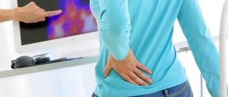
Symptoms and pain with a cervical hernia
A pathological focus within the cervical spine is usually accompanied by local pain in everyone, which can radiate to certain parts of the body innervated by the cervical-vertebral nerves. The pain, depending on how advanced the hernia is, can be unbearable or moderately tolerable. In addition, it can be either shooting, burning or aching. The symptomatic spectrum is quite diverse, patients mainly complain of:
- acute painful syndrome in the neck and collar area, aggravated by turning and tilting the head;
- stiffness of movement, crunching in the neck, tension in the neck muscles;
- frequent dizziness and intense headaches, mostly one-sided in the back of the head;
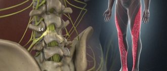
- pain and paresthesia in the form of crawling goosebumps, tingling, numbness, other unnatural signs in the hands (mostly in the wrists, fingers, forearm);
- weakness, paresis of one of the upper limbs, difficulty raising the arm up;
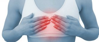
- pain in the shoulder, in the area of the shoulder blades, diaphragm, and occasionally in the leg;
- pain, numbness in one half of the face;
- discomfort and noise in the ears;
- hoarseness, feeling of soreness and foreign body in the throat;
- spots before the eyes, decreased visual acuity and blurred vision, other visual disorders;
- vestibular disorders, for example, unsteadiness, unsteadiness of gait;
- poor memory ability, absent-mindedness, increased fatigue;
- irritability, depressed mood, depression;
- light sleep or insomnia;
- surges in blood pressure, more often blood pressure tends to increase in severe pathogenesis with compression of the cervical artery that supplies the brain.
The pathology in most people provokes excruciating pain, which in dominant cases appears in the upper back, arms and head.
Results of treatment at Dr. Dlina Clinic
Our clinic has been open to patients for 20 years. During this time, effective methods of providing assistance have been developed. Long-term observations show that timely diagnosis and comprehensive treatment of diseases of the musculoskeletal system demonstrates positive dynamics:
- The condition of the spine and the entire body improves in 98% of cases. This result is clearly demonstrated by imaging techniques for examining joints and the spinal column.
- When using our techniques there are no contraindications or side effects - all techniques are completely safe and suitable for treating patients of various age categories.
- Long-term effect of therapy - proper treatment prevents recurrent attacks and acts as a preventive measure, strengthening the condition of the spine and muscle structure.
Dr. Dlin’s clinic helps patients cope with diseases of the spine and joints, get rid of headaches and discomfort. We guarantee that you will feel better after the first session.
Diagnosis of the cervical spine
When a patient comes to the clinic, the neurologist first collects and analyzes the patient’s medical history. This is followed by testing of the spinal cord using special non-hardware tactics for examining the spine. Non-hardware diagnostics are based on neurological tests, which involve checking the problem area of the back using motor and palpation techniques, thereby establishing:
- presence and degree of neurological deficit;
- dependence and nature of the pain syndrome when performing certain movements of the neck, limbs, etc.;
- the consistency of the reflex response and the volume of amplitude of movements of the cervical level in various physiological directions;
- impaired sensitivity in areas of the body associated with SHO (hands, facial area, etc.).
Based on complaints and neurological tests, a specialist can make a preliminary diagnosis, and during the initial examination. But treatment will not be prescribed until suspicions of a hernia are confirmed by the results of hardware imaging. The generally established diagnostic standard for the purpose of detecting pathology and obtaining reliable detailed information about the clinic of hernial protrusion is magnetic resonance imaging (MRI). Using volumetric layer-by-layer MRI images in different projections, as a rule, facts about:
- the development and degree of structural changes in the intervertebral discs at any stage;
- localization of the damaged fibrocartilaginous lining, and in the case of sequestration - the number and location of sequestration;
- the specifics of the growth of the bulge (direction vector);
- exact dimensions of the formation down to a millimeter;
- the presence and severity of pinching, compression of components of nerve nodes, arteries, spinal canal and spinal cord;
- intra- and paravertebral edema, inflammation, atrophy;
- condition of muscle tissue, ligaments, tendons, intervertebral joints;
- presence/absence of concomitant diseases on the examined part of the spinal column - stenosis, arthrosis, tumors, blood supply problems, etc.
X-ray does not fully reflect the clinical picture, since it visualizes only the outlines and position of the bone elements of the spine, the distance between the vertebrae, and osteophytes. X-rays do not show soft tissues, which include cervical discs, spinal cord, nerve plexuses, vascular and musculo-ligamentous structures. The slice-by-slice computed tomography method, although it involves the use of more advanced radioactive technologies than conventional x-rays, is also not informative enough in terms of the value of the obtained clinical signs in the diagnosis of this disease.
No conclusion is made about a cervical hernia based on X-ray or CT images alone; these are two auxiliary tactics, the advisability of which in each case is determined individually. Only MRI can thoroughly examine the problem area. Magnetic resonance imaging also has a significant advantage - it works on the phenomena of nuclear magnetic resonance and, unlike devices with ionizing radiation, does not have a negative effect on the human body.
Localization of the cause of pain
Not every disc herniation can cause pain. Some people are diagnosed with herniated discs after magnetic resonance imaging for other conditions.
In most cases, symptoms force the patient to seek medical help. A visit to the doctor is usually accompanied by a routine and neurological examination, an x-ray, computed tomography and/or magnetic resonance imaging is performed to verify the extent and localization of the disease.
The truth about treatment: analysis of the effectiveness of all tactics
Do medications help?
In the acute period, doctors prescribe medications for pain, inflammation and swelling for local and internal use to relieve painful manifestations of the disease. The basis of such drug therapy is NSAIDs:
- Indomethacin;
- Ibuprofen;
- Meloxicam;
- Diclofenac.
In addition to non-steroidal anti-inflammatory drugs for pain, specialists prescribe medications from a series of analgesics:
- Analgin;
- Spasmalgon;
- Ketorol.
If the clinical picture is aggravated by muscle hypertonicity, muscle relaxants are recommended to relax the spasmodic muscle structures. Among the most used muscle relaxants are Mydocalm and Siralud.
With constant debilitating pain, which NSAIDs and analgesics cannot cope with, a transition to treatment with strong hormonal or anesthetic drugs is carried out. These include glucocorticosteroids, lidocaine and novocaine. Using powerful hormone-containing and anesthetic agents, only as prescribed by a doctor in specialized conditions, the patient is given a course of spinal blockades. In the very near future, a person with such a critically unsafe diagnosis needs to undergo surgery.
All of the listed drugs act purely symptomatically: there will be no effect in terms of reducing the hernia component. In fact, they only temporarily relieve pain by blocking the transmission of nerve-pain impulses in the problem area. They also have a moderate anti-inflammatory effect. All intervertebral deformations and degenerations still do not disappear.
Note that limiting yourself only to drug therapy, even if it helps to get rid of pain, is the height of recklessness. With this approach, a hernia of the cervical spine will soon remind itself, and next time in a more vivid manifestation. But the worst thing is that, living only on painkillers, intervertebral pathogenesis will actively progress, increasing the risk of disability more and more every day.
Achieving stable remission through a drug regimen in complex combination with other conservative methods, as observations show, is not always achievable. If the initially prescribed complex treatment for 3 months, maximum 6 months, does not lead to tangible and lasting improvements, the patient is recommended to undergo surgery.
Long-term internal use of pain medications or their injection method is fraught with the development of side effects - stomach ulcers, liver and kidney diseases, hematopoietic and immune systems. External agents - gels, creams, ointments - have less negative reactions. But in terms of the strength of the analgesic effect they provide, they are significantly inferior to oral medications and injections.
It is impossible not to say a few words about popular chondroprotectors, the action of which is aimed at improving metabolism and activating nutrition in osteochondral structures. Remember, their benefits have been clinically confirmed exclusively for osteochondrosis that has not developed into a cervical hernia. It is acceptable to use such drugs for small protusions as a preventive measure for further degeneration of the cervical intervertebral disc. But! When hernia has already occurred, chondroprotectors are ineffective or “don’t work” at all. When a global restructuring of the disk contents into an irreversible (!) non-viable state has already occurred, they do not provide any therapeutic or prophylactic benefit.
Reviews of classes in our center
Feedback from a patient of the Innovative Medical Center
Patient's review Innovative Medical Center
Feedback on recovery after knee surgery
26.01.2021
I would like to express my gratitude to all the staff of the medical center for the treatment provided. I came to the center for treatment of heel spurs. The initial appointment was with Dr. Bogdanov V.Yu., who, after an examination, prescribed a course of UHT and hyvomat. I would like to express my special gratitude to the physiotherapist Tatyana Ivanovna Shmakova for her professionalism and excellent human qualities. After several procedures, the quality of life improved significantly. The pain gradually goes away. I hope it goes away forever. Thank you very much again. The medical center is very well located, not far from the metro, and has its own parking lot. The center is very cozy and equipped with modern equipment. If the need arises, I will definitely contact this institution.
read in full Olga
04.12.2020
I came to the center with back pain, the spasm would not go away. After several sessions of kinesiotherapy and massage, the spasm went away and my back felt better.
read in full Elena
All reviews
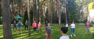
The effect of gymnastic exercises
Of course, it will not be possible to get rid of the disease through special gymnastic exercises for the neck. Exercise, even the best, is not able to “retract” the fallen pulpous element back or destroy it without a trace, or “patch” the gap in the fibrous ring. This can only be accomplished during surgical treatment.
However, exercise therapy has been proven to improve well-being and, importantly, prevent the development of atrophy of the muscles of the cervical and cervicothoracic complex and upper extremities. Let us immediately emphasize that the productivity of LH action is significantly reduced in the penultimate and final stages of the hernia. Still, unique physical training makes the greatest contribution to the restoration of musculoskeletal potential after surgical repair of a hernia. In general, the effect of physical therapy both during conservative therapy and at the time of postoperative rehabilitation is due to:
- stimulation of blood circulation;
- activation of metabolic processes;
- increasing the production of nutrients necessary to maintain the functions of the spinal structure;
- strengthening and increasing endurance of the neck muscles;
- correct, gentle development of motor and support functions of the neck;
- increasing the distance between the vertebrae, which helps prevent or reduce radicular/vascular compression.
But in order to really benefit from gymnastics, you need to get a training program from a specialist that is specially designed just for you, taking into account all the characteristics of the clinical picture on MRI. Plus, indicators of age and body weight, level of physical fitness, and associated health problems are taken into account.
It is advisable to take the first course in a physical therapy room under the supervision of a professional instructor in order to thoroughly understand and hone the technique of each individual exercise. When performing training, it is important to follow 5 rules:
- Regularity of exercise therapy.
- Gradual increase in load.
- Smooth and accurate movements of the cervical spine.
- Complete abolition of physical exercise if the slightest pain or changes in sensitivity appear in any part of the body.
- Based on point No. 5, urgently contact a treating specialist so that he can conduct a high-quality examination and make adjustments to the therapeutic exercise plan.
Taking and applying any physical activity on your own from the Internet, at random, or on the advice of other patients is highly not recommended! In the acute phase, exercise therapy is contraindicated! If you neglect these rules, the likelihood of achieving disastrous results is too high. Consequences of an illiterate campaign - exacerbation of symptoms of hernia SHO
, increased frequency of relapses, progression of disc displacement, even greater reduction of the spinal lumen, compression and death of nerve endings, damage to the spinal cord/brain, compression and narrowing of the cervical arteries, separation of the hernial sequestrum. As a result, the deadline for the operation will not move away, but will noticeably get closer.
Non-surgical treatment methods at Dr. Dlin’s Clinic
Dr. Dlin’s Clinic uses the most modern and effective techniques that help eliminate pain and restore the patient’s previous mobility:
- Di-Tazin therapy is an original three-component technique that combines manual therapy, photodynamic laser therapy and electrophoresis. The complex effect allows you to quickly eliminate pain, activate blood circulation, normalize metabolic processes, stop inflammation, and prevent the destruction of joint tissue.
- Manual therapy is a proven method of manual intervention that helps cope with many diseases of the musculoskeletal system, relieves pain, and restores the ability to move without restrictions.
- Restorative massage is a manual technique that improves blood supply and nutrition of cells, relieves muscle tension and increases the body's stability during periods of stress and tension.
- Osteopathy – massage actions are aimed not only at the spine and muscles, but also at internal organs for comprehensive healing and strengthening of the body.
- Therapeutic and physical training complex is a proven method of treatment and prevention of joint diseases, which reduces pain, improves blood supply to all organs, and correctly distributes muscle load.
- Shock wave therapy is a restorative procedure that uses low-frequency acoustic waves, accelerates the healing process of damaged tissue, and reduces back pain due to hernia and other diseases of the spine.
- Medicinal electrophoresis - the effect on the body of a constant electric current in combination with active medicinal substances, has analgesic, anti-inflammatory, regenerating, relaxing properties.
- Physiotherapy is targeted stimulation of the body using various methods (exposure to air, water flows, magnetic fields, laser beams, ultrasound). Normalizes muscle tone and metabolic processes, improves oxygen supply to tissues and organs.
In total, Dr. Dlin’s Clinic uses over 20 non-surgical techniques to treat the spine and joints. The staff is staffed with highly qualified doctors: vertebrologists, neurologists, osteopaths, chiropractors. The medical staff is constantly trained in the best clinics in the Middle East, Europe, and America.
Impact of massage
The leading function of massage is to create a trophic effect. This means that massage techniques make it possible to increase blood circulation in the area of interest, ensure better lymph outflow and the supply of necessary metabolites and oxygen to tissue cells. Another goal of massage is to prevent atrophic phenomena in muscle structures and reduce intradiscal compression. However, we note that in no way can special techniques for massaging the cervical-collar area eliminate the hernia and reduce its volume.
This type of treatment is recommended strictly according to indications, since it is not equally useful for all types of hernias. It is prescribed especially with caution for medium and large protrusions, nerve pinching in the projections of the neck. Such manipulations in a neglected, complicated clinic can give a completely opposite effect, since it is easy to turn a large hernial mass in an unfavorable direction using this method of influence. Therefore, before prescribing a massage, the doctor must weigh the pros and cons.
At the beginning of the development of a hernia, massage can serve as an excellent measure to prevent the progression of degenerative-dystrophic pathogenesis in a still slightly changed disc. But we emphasize that massage techniques, if the doctor gives the go-ahead for them, should be carried out by a good massage therapist from the neurology department. And, finally, this is an auxiliary treatment tactic, which makes sense to use purely in conjunction with other basic treatment programs.
HOW DOES A HERNIA DEVELOP?
It all starts with poor posture. Intervertebral discs are strong rings of connective tissue that protect the delicate gel-like core. Good posture, physiological curves and a straight back are the conditions created by wise nature for shock absorption and softening of stress while walking.
Poor posture leads to other problems. The vertebrae are displaced and compress the blood vessels. The connective tissue of the disc ceases to receive enough nutrition, becomes thinner, and the core gradually shifts under the increased load of the displaced vertebrae. Microcracks on the ring separate until the core breaks the wall completely and comes out.
Cervical physiotherapy
Physiotherapeutic procedures also do not treat a hernia, but fight its symptom complex according to approximately the same principle as massage and exercise therapy. The goal of standard physiotherapy sessions (magnetic therapy, electrophoresis, laser and ultrasound therapy) is to reduce swelling around the affected intervertebral disc, promote blood flow to the weak area, and relieve muscle strain.
If this goal is fully achieved, the patient feels much better after a course of physical therapy, noting an increase in the range of motion in the neck, a persistent disappearance or reduction in pain and other previously disturbing signs. But, according to clinical studies, the effect of physiotherapy is noted by only 50% of patients with this diagnosis. In some cases (up to 40%, as a rule, with stage 3-4 pathology), on the contrary, after treatment with physiotherapy, a deterioration in well-being is observed.
Laboratory research
Laboratory tests are necessary to exclude other diseases (rheumatoid arthritis, ankylosing spondylitis, Reiter's syndrome and polymyalgia rheumatica). These studies may include the following tests:
Rheumatoid factor (increased in rheumatoid arthritis)
HLA-B27 (positive in ankylosing spondylitis)
Erythrocyte sedimentation rate (increased in polymyalgia rheumatica)
In addition, blood tests can rule out infectious processes such as discitis, epidural abscess or vertebral osteomyelitis:
Changes in the number of leukocytes (increased, with a shift to the left - with bacterial infection)
Blood culture (positive if infection is present)
Red blood cell sedimentation rate (increased during infection)
Complications and consequences of the disease
The cervical region is the narrowest part of the spinal system, in which the largest arteriovascular highway passes. The department includes the largest arteries - the left, right vertebral and basilar, each of which forms up to 6 groups of vascular branches.
In the back of the neck there is also an important cervical nerve bundle (node), which consists of the hypoglossal, lesser occipital, greater auditory, transverse, and supraclavicular nerves.
Thus, even slight deformations of the disc, displacement of the vertebrae in an overly narrow area, densely penetrated by nerves and blood vessels, can cause a real catastrophe:
- clamping of arteries and nerve roots, constant severe pain;
- blocking the circulation of blood and cerebrospinal fluid between the spinal cord and brain with severe cerebro-vertebral signs;
- neurotic nature of auditory and visual disturbances, frequent loss of consciousness, coordination disorders;
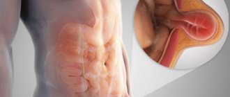
- serious dysfunctions of the gastrointestinal tract (nausea, vomiting, fecal incontinence), urinary system (uncontrolled urination) and genital organs (impotence, frigidity, infertility);
- muscle weakness of the arms, including complete or partial paralysis;
- depression of respiratory functions due to severe damage to the spinal substance and nerve nodes, up to the cessation of breathing;
- insufficient blood supply to the brain, which can lead to cerebral ischemia and stroke.
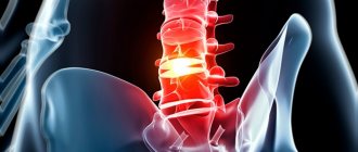
MRI
MRI of the cervical spine is the best method for diagnosing disc herniation. MRI scans can be performed with or without contrast. MRI can detect which disc is damaged and whether there is root compression.
MRI also makes it possible to clearly visualize changes in the structure of bone tissue or tumors, abscesses, and changes in the spinal cord (stenosis). In addition, MRI is absolutely harmless and can be prescribed repeatedly (to monitor dynamics).
What complications can this disease cause?
Complications always occur from complete disregard for treatment or unscrupulous implementation of the treating doctor’s recommendations. In such cases, the disease rapidly progresses and can lead to the development of ischemic stroke, vertebrobasilar insufficiency, radiculitis or complete paralysis.
As it progresses rapidly, the intensity of the pain syndrome constantly increases, performance and normal lifestyle are disrupted, and this can lead a person to disability.
What's included in the program:
Doctor appointments, diagnostics, treatment and rehabilitation
| Name of service | Number of services |
| Repeated appointment (examination, consultation) with a neurologist | 1 |
| Laser therapy (HILT therapy (1 field) | 10 |
| Electrophoresis with sinusoidal modulated currents (SMT-phoresis) 1 field | 10 |
| Medical massage of the lower limb - 1.5 units. | 5 |
| Reflexology (1 session) | 10 |
| Primary examination (consultation) with a physiotherapist | 1 |
| MRI of the lumbosacral region (KUNTSEVO) after treatment | 1 |
| Program Cost | RUB 61,580 |
| 25% discount | RUB 49,335 |
FORECAST AND PREVENTION
If you consult a doctor at an early stage, when the hernia is not sequestered, the prognosis for treatment of a cervical hernia of the spine is positive. An individually selected program, consisting of several treatment methods, can significantly reduce the size of the hernia within 4-6 weeks until it is completely eliminated. This quickly relieves the condition. Upon completion of the course, individual recommendations for maintaining health are developed for the patient. It is advisable to continue to see a doctor, conduct a follow-up examination every 3-6 months, and receive maintenance therapy.