Description
Extensive laboratory testing of thyroid function.
The thyroid gland is one of the most important organs of the human endocrine system. The main function of the thyroid gland is to produce thyroid hormones. They regulate most metabolic processes in the body.
Why is it important to have your thyroid examined?
The thyroid gland affects the entire body as a whole, and even the most insignificant, at first glance, deviations from the norm have an impact on the body’s metabolism, cardiac, nervous and reproductive systems. The earlier a thyroid pathology is detected, the easier it is to treat.
Why is it better to undergo a comprehensive examination?
The thyroid gland synthesizes 2 main hormones: T3 and T4, the formation of which is regulated by TSH (synthesized in the pituitary gland), it is important to see the whole picture. In addition, TG and TPO are involved in the formation of hormones in the thyroid gland, to which, in some forms of pathology, antibodies are formed in the thyroid gland, therefore, to assess the function of the thyroid gland, a comprehensive examination is necessary. The value of laboratory tests increases with simultaneous ultrasound examination (US).
Symptoms of thyroid dysfunction (in children and adults):
- Sudden weight changes.
- Unstable menstrual cycle in women and teenage girls.
- Change in appearance: problems with skin, hair, nails.
- Disorders of the gastrointestinal tract and cardiovascular systems
- Memory deterioration, slow thinking and speech.
- Increased sweating, hand tremors and increased body temperature.
- Weakness, irritability, tearfulness.
- Decreased immunity, tendency to colds.
Who should have a thyroid examination?
Everyone without exception: women and men, children.
For the purpose of prevention and if one or more of the above symptoms appear, it is recommended to undergo a comprehensive examination of the thyroid gland: take tests, undergo an ultrasound and, based on the results of the examination, consult a doctor.
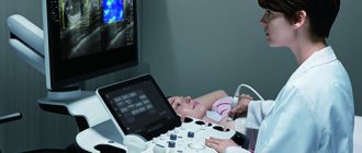
How to prepare for an ultrasound
The method is accessible and simple, does not require special preparatory operations. You can eat absolutely any food product. But there are a number of restrictions:
1. Regardless of the feeling of nausea, older people are not recommended to eat before the procedure. Age-related changes may have an effect.
2. Women are recommended to undergo examination on days 7-9 of the menstrual cycle to avoid inaccurate results.
3. Immediately before the procedure, you need to free your neck from elements that interfere: jewelry, collars, etc.
4. Preparation for an ultrasound examination of the thyroid gland in children is no different from the procedure for adults. The child must be psychologically prepared for the procedure so that he is not afraid of either the doctor or what he will do.
Doctors recommend that women undergo testing both during pregnancy and at the planning stage. If it is impossible to get pregnant, gland pathology may be one of the possible causes.
After the child has suffered stress, it is also worth going to the clinic so that an endocrinologist can perform an ultrasound of the thyroid gland.
Thyroid-stimulating hormone (TSH, thyrotropin)
A glycoprotein hormone that stimulates the formation and secretion of thyroid hormones (T3, T4).
They enter the body with food and are also synthesized by cells of adipose tissue, liver, and intestines. They do not circulate freely, but are bound to proteins and transported in the form of macromolecular complexes - lipoproteins. They are the main lipids of fatty deposits and food products. The triglyceride molecule contains triatomic glycerol and 3 residues of higher fatty acids, mainly palmitic, stearic, linoleic and oleic.
It is produced by basophils of the anterior pituitary gland under the control of thyroid-stimulating hypothalamic releasing factor, as well as somatostatin, biogenic amines and thyroid hormones. Increases vascularization of the thyroid gland. Increases the supply of iodine from blood plasma to thyroid cells, stimulates the synthesis of thyroglobulin and the release of T3 and T4 from it, and also directly stimulates the synthesis of these hormones. Enhances lipolysis.
There is an inverse logarithmic relationship between the concentrations of free T4 and TSH in the blood.
TSH is characterized by daily fluctuations in secretion: blood TSH reaches its highest values at 2 - 4 am, the highest level in the blood is also determined at 6 - 8 am, the minimum TSH values occur at 17 - 18 pm. The normal rhythm of secretion is disrupted when awake at night. During pregnancy, the concentration of the hormone increases. With age, the concentration of TSH increases slightly, and the amount of hormone emissions at night decreases.
Limits of determination:
0.0025 mU/l - 100 mU/l.
Free thyroxine (free T4)
The most important stimulator of protein synthesis.
Produced by follicular cells of the thyroid gland under the control of TSH (thyroid-stimulating hormone). It is the predecessor of T3. By increasing the basal metabolic rate, it increases heat production and oxygen consumption by all tissues of the body, with the exception of the tissues of the brain, spleen and testicles. Increases the body's need for vitamins. Stimulates the synthesis of vitamin A in the liver. Reduces the concentration of cholesterol and triglycerides in the blood, accelerates protein metabolism. Increases calcium excretion in urine, activates bone turnover, but to a greater extent, bone resorption. It has a positive chrono- and inotropic effect on the heart. Stimulates reticular formation and cortical processes in the central nervous system.
During the day, the maximum concentration of thyroxine is determined from 8 to 12 hours, the minimum - from 23 to 3 hours. During the year, maximum T4 values are observed between September and February, and minimum values are observed in the summer. Women have lower thyroxine concentrations than men. During pregnancy, the concentration of thyroxine increases, reaching maximum values in the third trimester. Levels of the hormone in men and women remain relatively constant throughout life, decreasing only after 40 years.
The concentration of free thyroxine, as a rule, remains within the normal range in severe diseases not related to the thyroid gland (the concentration of total T4 may be reduced!).
An increase in T4 levels is facilitated by high serum bilirubin concentrations, obesity, and the use of a tourniquet when drawing blood.
Limits of determination:
5.1 pmol/l - 77.2 pmol/l.
Free triiodothyronine (free T3)
Thyroid hormone stimulates the exchange and absorption of oxygen by tissues (more active than T4).
Produced by follicular cells of the thyroid gland under the control of TSH (thyroid-stimulating hormone). In peripheral tissues it is formed during deiodination of T4. Free T3 is the active part of total T3, accounting for 0.2 - 0.5%.
T3 is more active than T4, but is found in the blood in lower concentrations. Increases heat production and oxygen consumption by all body tissues, with the exception of brain tissue, spleen and testicles. Stimulates the synthesis of vitamin A in the liver. Reduces the concentration of cholesterol and triglycerides in the blood, accelerates protein metabolism. Increases calcium excretion in urine, activates bone turnover, but to a greater extent, bone resorption. It has a positive chrono- and inotropic effect on the heart. Stimulates reticular formation and cortical processes in the central nervous system.
By 11–15 years, the concentration of free T3 reaches adult levels. In men and women over 65 years of age, there is a decrease in free T3 in serum and plasma. During pregnancy, T3 decreases from the first to the third trimester. One week after delivery, serum free T3 levels return to normal. Women have lower concentrations of free T3 than men by an average of 5 - 10%. Free T3 is characterized by seasonal fluctuations: the maximum level of free T3 occurs from September to February, the minimum in the summer.
Limits of determination:
1.5 pmol/l - 46.1 pmol/l.
Interpretation of ultrasound results
The diagnostician describes the results obtained in the study protocol. Typically, the time to prepare a written report does not exceed 15 minutes.
Determination of exact geometric dimensions
The location of the gland will normally be typical or low, the shape will be classic, the contour will have a clear outline. It consists of two lobes with an isthmus that connects them. Sometimes a pyramidal lobe may be present.
Minor (less than one centimeter) tissue growths may be visible. If during the period of intrauterine growth the gland develops with pathology, the tissue may not be divided into two sides, but may move to one side. This is called aplasia of one lobe.
In the case of a completely undeveloped gland, they speak of complete aplasia. The length of the lobe should be from 4 to 6 cm, width from 1.3 to 1.8 cm, thickness from 1.5 to 1.8 cm; the shares are normally the same; the lintel has a thickness of 4 to 8 cm.
The volume of a healthy gland depends on the patient’s weight and is:
- weight 50 kg, gland volume 15.5 cm³
- weight 50-60 kg, volume 18.7 cm³
- weight 60-70 kg, volume 22 cm³
- weight 70-80 kg, volume 25 cm³
- weight 80-90 kg, volume 28.4 cm³
- 100 kg or more, volume 32 cm³
Non-compliance with standard sizes will indicate possible pathologies.
Definition of structure
The echogenicity of a healthy thyroid gland, without any peculiarities, the structure of the jelly tissue is fine-mesh, homogeneous, echogenic granularity is 1 mm or less. Fibrous and connective tissues are not detected. In inflammatory processes, the structure is heterogeneous.
Focal education. There should be no new growths. If the formation is present, it must be classified. If the size is up to 10 mm, then it is a focal formation, more than 10 mm, then it is a node.
Blood flow analysis. The parameters of blood flow, its nature and density, and parameters of the lymph nodes are determined. Normal nodes have clear boundaries, the width is approximately 2 times less than the length.
Contours. Clear contours are the norm. Fuzzy outlines indicate an inflammatory process or tumor.
Focal formations. Assessed for the presence of nodes, cysts, and calcinitis.
Echogenicity is the grayscale image of the tissue being examined and its tone on a monitor.
Parameters of visible lymph nodes (if present), their structure, size, structure. By their presence, the onset of tumor formation can be diagnosed.
The structure of the salivary gland and the level of response to ultrasound.
The size and structure of the soft tissues of the neck and larynx. Those areas that are located near the thyroid gland are examined.
Women over 35 years of age are recommended to have their thyroid gland examined at least once a year. This category of people is at risk for thyroid diseases.
The human neck is quite complex; it contains many nerve trunks and large vessels, the esophagus, trachea, many lymph nodes, and other glands. Therefore, the specialist performing the ultrasound procedure must be highly qualified.
The doctor conducting the research records the data obtained in a special protocol, which describes the geometric parameters of the lobes and isthmus, calculates the volume of the gland, and writes a conclusion.
The document evaluates the position and contours, tissue structures, analyzes the parathyroid glands and lymph nodes, and the images taken are also attached to the protocol. In case of normal indicators, a corresponding entry is made in the document. This takes no more than 10 minutes.
Antibodies to thyroid peroxidase (AT-TPO, microsomal antibodies)
Autoantibodies to the enzyme of thyroid cells.
Antibodies to thyroid peroxidase are an indicator of the aggression of the immune system towards its own body. Thyroid peroxidase provides the formation of an active form of iodine, which can be included in the process of iodification of thyroglobulin. Antibodies to the enzyme block its activity, as a result of which the secretion of thyroid hormones (T4, T3) decreases. However, TPO Abs can only be “witnesses” of the autoimmune process.
Thyroid peroxidase antibodies are the most sensitive test for detecting autoimmune thyroid disease. Usually their appearance is the first shift that is observed in the course of developing hypothyroidism due to Hashimoto's thyroiditis. When sufficiently sensitive methods are used, TPO Abs are detected in 95% of people with Hashimoto's thyroiditis and in approximately 85% of patients with Graves' disease. Detection of AT-TPO during pregnancy indicates the risk of developing postpartum thyroiditis in the mother and a possible impact on the development of the child.
Reference limits largely depend on the research method used. Low levels of AT-TPO can sometimes be found in apparently healthy people. It remains unclear whether this may reflect a physiological norm, or is a precursor to autoimmune thyroiditis, or is a problem with the specificity of the method.
Limits of determination:
3 U/ml - 1000.0 U/ml.
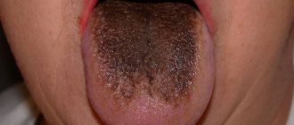
Thyroid diseases in older people
| Figure 1. The cells that make up the crop have increased in size. In elderly patients, the goiter may not be palpable |
How do the manifestations of thyroid diseases differ in old age?
What pitfalls await the general practitioner when interpreting thyroid function tests? How should a general practitioner manage elderly patients with hypothyroidism? Thyroid diseases are common among older people, and accordingly, the mortality rate among this category of population is much higher. In this age group, hypothyroidism accounts for 6% of all diseases, and hyperthyroidism accounts for 2% [1].
The clinical manifestations of thyroid disease in older people are blurred and can easily be mistaken for manifestations of the natural aging process. In addition, numerous concomitant diseases often do not allow the detection of thyroid function disorders. Thus, the diagnosis is usually not established immediately.
Consequently, exceptional care is required from the doctor; he must record the slightest symptoms of a possible thyroid disease.
With age, the function of the thyroid gland does not undergo any significant changes, but a blood test for hormones indicates significant deviations from the norm. This is due to the prevalence of “nonthyroid” diseases, which complicate the interpretation of research results.
Insufficient and unbalanced nutrition, decompensated diabetes mellitus, liver disease, heart disease, cerebrovascular disorders and malignant tumors act as factors that typically reduce the level of total and free serum triiodothyronine (T3), less often reducing total and free thyroxine (T4) and moderately increasing or suppressing thyroid-stimulating hormone (TSH). The interpretation of the results should be treated with caution, and if the clinical picture is not clear, the diagnosis should be postponed until the picture is fully clarified.
Table 1. Common medications that alter thyroid function test results without affecting clinical thyroid status
Reduce free T4 and reduce (or do not affect it) T3 with normal TSH
Increase free T4 and T3 with decreased TSH
Reduce or have no effect on free T4 and T3 with decreased TSH
|
The presence of a whole bunch of diseases in old age determines the need to take into account the effect of drugs on the results of thyroid tests (Table 1) [2]. For example, a number of medications, especially those containing iodine (for example, the antiarrhythmic amiodorone), increase thyroid function (Table 2). Medicines also change the picture of thyroid diseases - for example, b-blockers can smooth out tachycardia and tremor in hyperthyroidism [3].
- Hypothyroidism
The main causes of decreased thyroid function in old age are autoimmune diseases (autoimmune thyroiditis). In addition, cases of iatrogenic hypothyroidism due to previous thyroid surgery, treatment with radioactive iodine, antithyroid drugs, and irradiation of the neck and head can be noted.
Classic symptoms of hypothyroidism may be absent, and some symptoms (fatigue, chills, dry skin, decreased appetite, mental and hearing impairment, heart failure) may be attributed to the natural aging process. As in young patients, these indicators are weakly specific for hypothyroidism.
Neurological manifestations, such as cerebellar dysfunction and neuropathy, as well as mental disorders, including depression, behavioral and cognitive dysfunction, are more common in the older age group. Elderly patients are more susceptible to myxedematous coma, which occupies an important place in the differential diagnosis of comas, especially those accompanied by hypothermia.
| Figure 2. Face of an elderly patient with hyperthyroidism. She presented with weight loss and a feeling of sand in her eyes. |
Hypothyroidism may be suspected when unexplained hyponatremia, elevated creatinine phosphokinase, lactate dehydrogenase, macrocytosis, and anemia are detected. Patients may also suffer from other autoimmune diseases, such as insulin-dependent diabetes mellitus, vitiligo, or Addison's disease.
Taking amiodarone may negatively affect thyroid function. All patients with cardiac problems or weight loss should also undergo thyroid function tests. Data from an objective examination of elderly people have a low diagnostic value, and goiter is often not detected in old people for a long time.
The diagnosis of primary hypothyroidism is made on the basis of a decrease in total serum thyroxine and an increase in TSH. Antibodies to thyroid peroxidase and thyroglobulin also indicate this etiology.
If the level of thyroid hormones is reduced and TSH is normal, one should think about a “non-thyroid” disease and the consequences of drug therapy (see above), although the cause may also be secondary hypothyroidism due to diseases of the pituitary gland or hypothalamus.
Table 2. Drugs that cause hyper- or hypothyroidism in older people
Hyperthyroidism
Hypothyroidism
|
Treatment. The initial replacement dose of thyroxine depends on the clinical condition and the presence of concomitant diseases, such as heart disease. Many patients reach euthyroid levels within 6-8 weeks on 50-100 mcg T4 daily. The euthyroid state is indicated by normalization of serum thyroxine and TSH levels.
There is evidence that the risk of osteoporosis and atrial fibrillation increases when TSH falls below normal levels.
Elderly people, especially those suffering from coronary heart disease, need constant monitoring, since thyroxine can provoke an attack of angina or myocardial infarction in patients with coronary artery disease. However, there is an undoubted connection between hypothyroidism and hyperlipidemia, which increases the risk of developing coronary heart disease, which dictates the need for correction of the hypothyroid state.
Most patients require lifelong therapy and annual TSH testing. It is necessary to ensure that the patient continues to take T4 replacement. The exception is patients with moderate hypothyroidism in the first six months after treatment with radioactive iodine who have undergone partial thyroidectomy, who may experience a transient increase in TSH.
People with subclinical forms of hypothyroidism may have an increase in TSH without a decrease in thyroxine. Such patients with antithyroid antibodies or previous radioactive iodine therapy typically subsequently develop overt thyroid dysfunction.
Patients with antithyroid antibodies have a 5% chance of developing overt hypothyroidism each year, so most experts recommend not delaying treatment in this group [5]. Others have a progression rate of less than 3% and rather require close monitoring to ensure that overt hypothyroidism is not missed.
It has been shown that patients with subclinical forms of hypothyroidism have mild impairments in cognitive function and myocardial contractility.
Most people with hypothyroidism should be closely monitored by a general practitioner. Patients with special problems, such as coronary artery disease or those receiving lithium or amiodarone, are referred to a specialist [6]. Any indication of pituitary disease also requires consultation with a specialist.
- Hyperthyroidism
In old age, hyperthyroidism is less common than hypothyroidism. Most cases are caused by Graves' disease, nodular toxic goiter, and solitary toxic adenoma, with some predominance of nodular toxic goiter. Other causes include subacute thyroiditis, asymptomatic thyroiditis, and iodine-induced thyrotoxicosis [7].
In regions where there is iodine deficiency (for example, in parts of Germany and Western Europe), nodular goiter is more common, and the risk of developing hyperthyroidism due to enlarged follicles in long-standing nodules increases.
Drugs such as amiodarone, radiographic contrast media, or consumption of foods containing excess iodine can provoke the development of thyrotoxicosis, and patients with pre-existing thyroid disease such as nodular goiter or subclinical Graves' disease are more likely to suffer. In addition, amiodarone can cause destructive thyroiditis, which releases already synthesized hormones, but is usually clinically indistinguishable from amiodarone-induced Graves' disease or nodular toxic hyperthyroidism.
“Apathetic hyperthyroidism” is a term used to describe the condition of elderly patients with hyperthyroidism who exhibit few signs of the disease. In old age, eye symptoms, as a rule, do not develop, and their presence indicates a more severe course of the disease.
Weakness, shortness of breath, anxiety, congestive heart failure (CHF), palpitations, and atrial fibrillation are more common in the elderly. CHF is common in older patients with hyperthyroidism and is found in 60% of cases, and red flags include heart failure with high cardiac output, a normal ECG, and persistent atrial fibrillation.
Instead of increased appetite, there is more likely to be weight loss with anorexia, depression, drowsiness, agitation, dementia and confusion. All these symptoms may persist for up to a year.
Physical examination reveals goiter in approximately 25-50% of patients. It is much more difficult to palpate a goiter in old age due to age-related changes in the chest and kyphosis, which leads to displacement of the thyroid gland under the sternum.
Elevated total or free thyroxine levels and low TSH levels support the diagnosis of primary hyperthyroidism.
In rare cases, hyperthyroidism maintains normal thyroxine levels with decreased TSH. It is necessary to evaluate the serum level of T3, since in this group of patients it is more typical for patients with adenoma and nodular toxic goiter.
In addition, serum TSH levels in older adults should be interpreted with caution because of their possible decline with nonthyroidal disease or glucocorticoid use, and as a variant of the normal aging process.
A normal TSH level excludes the diagnosis of hyperthyroidism, except in extremely rare cases of excessive pituitary TSH secretion. Radioactive iodine uptake testing by the thyroid gland may be performed, as a low TSH may indicate asymptomatic thyroiditis or iodine-induced thyrotoxicosis.
Treatment. For proper diagnosis, patients with hyperthyroidism should be referred to a specialist. One of the main treatment methods is thionamides, which are used to achieve a euthyroid state until more specific therapy is selected.
β-blockers are useful adjunctive agents for the symptomatic relief of tremor, anxiety, and palpitations, but they should be used with caution in patients with asthma or heart failure, even if due to hyperthyroidism. Thus, before receiving specialist advice, thionamides and b-blockers are used in general practice.
With long courses of taking thionamides, relapse of Graves' disease is 50%; with nodular toxic goiter, remission is not achieved at all, so all elderly patients need specific treatment. Only in isolated cases, when specific treatment is unavailable for some reason, long-term drug therapy is carried out.
Surgery in old age is associated with a fairly high risk, so the administration of radioactive iodine is preferable, except in cases of Graves' disease complicated by ophthalmopathy. After three months, you need to check the TSH level, an increase in which indicates iatrogenic hypothyroidism and the need to reduce the dose of the drug.
The most serious side effect of thionamides is agranulocytosis. It is more common among patients over 40 years of age, but anyone prescribed these drugs should be warned about the need to urgently do a complete blood count if a sore throat or any other infectious disease develops. Itching, rash, jaundice and hepatitis may also be among the side effects.
Treatment with radioactive iodine begins when a euthyroid state is achieved, it leads to a moderate development of hypothyroidism: by 15-20% during the first two years, and then by 1-3% per year, depending on the dose.
At least four days before the administration of radioactive iodine, you should stop antithyroid drugs and return to them 4-7 days after the first dose of iodine.
During treatment with radioactive iodine, thyroid function is checked every 4-8 weeks.
When a euthyroid state is achieved, antithyroid drugs are discontinued, and thyroid function is examined again after 4-6 weeks. If hyperthyroidism is detected, treatment with radioactive iodine is continued, usually for another 4-6 months.
Radioactive iodine has virtually no side effects, except for the risk of transient hyperthyroidism due to radiation thyroiditis, which also rarely causes pain and swelling of the thyroid gland.
| Figure 3. Severe erythema of the legs of an elderly patient who did not agree to thyroxine replacement therapy |
The possibility of surgical treatment is considered only in patients with a large goiter and if the patient himself prefers this method of treatment. Patients with atrial fibrillation secondary to hyperthyroidism may return to normal sinus rhythm, but the older the patient, the less often this occurs, so even in the absence of structural abnormalities or other causes of fibrillation, anticoagulation therapy should be prescribed. It is important to ensure long-term monitoring of the patient.
Subclinical hyperthyroidism is defined by low serum TSH and normal levels of free thyroxine and T3. There is currently no consensus on the treatment of subclinical hyperthyroidism, and it remains unclear whether this condition increases morbidity due to an increased risk of atrial fibrillation in patients over 60 years of age and due to decreased bone density, which increases the risk of fractures.
- Nodular goiter and thyroid cancer
The volume of the thyroid gland and the number of nodules increase with age, and according to mortality statistics, about 90% of women over 70 years of age and 60% of men over 80 years of age have thyroid nodules.
Nodular goiter is often autonomous and can lead to subclinical and severe hyperthyroidism. In the absence of thyroid dysfunction, goiter is diagnosed without taking into account age characteristics. Palpable nodes undergo aspiration biopsy followed by cytological examination, especially if they manifest themselves in some way or rapidly increase in size.
The incidence of thyroid cancer does not change with age, but the percentage of malignant forms increases.
Management of patients with differentiated thyroid cancer in old age has no special features. A total thyroidectomy, irradiation with radioactive iodine, followed by thyroxine-suppressive therapy is performed.
References
1. Chiovato L., Marrioti S., Pinchera A. Thyroid disease in the Elderly // Balliere's Clinical Endocrinology and Metabolism. 1997; 11(2): 251-270. 2. Davies P. H, Franklyn JA The effects of drugs on tests of thyroid function // Eur. J. Clin. Pharmacol. 1991; 40: 439-451. 3. Cittoes NJL, Franklyn JA Drug-induced thyroid disorders // Drug Safety. 1995; 13(1): 46-55. 4. Toft AD Thyroxine therapy // N. Engl. J. Med. 1994; 331: 174-180. 5. Vanderpump MPJ, Tunbridge WMG, French JM et al. The incidence of thyroid disorders in the community: a twenty-year follow-up of the Whickham Survey // Clin. Endocrinol. 1995; 43: 55-68. 6. Vanderpump MPJ, Ahlquist JAO, Franklyn JA et al. Consensus statement for good practice and audit measures in the management of hypothyroidism and hyperthyroidism // BMJ 1996; 313:539-544. 7. Franklyn JA The management of hyperthyroidism // N. Engl. J. Med. 1994; 330(24): 1731-1738.
How to respond to a patient
- What are the functions of the thyroid gland?
The gland releases hormones that control the speed of cells in the body and, consequently, the intensity of metabolism.
- What is an autoimmune disease?
This disease occurs due to a malfunction of the immune system, which normally fights foreign agents such as bacteria and viruses. Immune cells begin to mistakenly perceive a particular tissue, such as the thyroid gland, as foreign, and strive to get rid of it.
- Do I need to see a specialist?
Hypothyroidism is a stable condition that does not require hospitalization, but you should monitor your condition with blood tests carried out by your GP. Excessive thyroid function sometimes requires numerous studies to determine the cause and closer monitoring, so sometimes a consultation with an endocrinologist is necessary.
- How long do I need to take thyroxine?
Such therapy should be followed throughout life, since the suppressed thyroid gland does not restore its lost functions. Cessation of treatment leads to relapse of the disease. Long-term use of thyroxine has no harmful side effects.
- Why do I need radioactive iodine and is it harmful?
Radioactive iodine is safe and almost always cures an overactive thyroid without surgery.
Note!
- The clinical manifestations of thyroid diseases in older people are not obvious and can easily be mistaken for normal aging processes. Numerous concomitant ailments often do not allow the detection of thyroid diseases
- With age, the function of the thyroid gland does not undergo any significant changes, but a blood test for hormones indicates significant deviations from the norm. This is due to the prevalence of “non-thyroid” diseases, which make it difficult to interpret research results
- It is necessary to approach the interpretation of tests with the utmost caution, and if the clinical picture is not clearly expressed, the diagnosis should be postponed until the picture is completely clarified
- The initial replacement dose of thyroxine depends on the clinical condition and the presence of concomitant diseases, such as heart disease. Many patients reach euthyroid levels within 6-8 weeks when taking 50-100 mcg T4 daily
- Autoimmune diseases are the main causes of hypothyroidism in the elderly. Classic symptoms of hypothyroidism may be absent, and some symptoms (fatigue, chills, dry skin, decreased appetite, mental and hearing impairment, heart failure) may be attributed to the natural aging process
- “Apathetic hyperthyroidism” is a term used to describe the condition of elderly patients with hyperthyroidism who exhibit few signs of the disease. Weakness, shortness of breath, anxiety, congestive heart failure (CHF), palpitations, and atrial fibrillation are more common in the elderly. Instead of increased appetite, there is more likely to be weight loss with anorexia, depression, drowsiness, agitation, dementia and confusion. Elevated total or free thyroxine levels and low TSH levels support the diagnosis of primary hyperthyroidism. Patients with hyperthyroidism should be referred for specialist advice