Diagnosis of acute pancreatitis
Acute pancreatitis remains the most severe disease, the incidence of which varies. The authors of the work “Acute pancreatitis. Pathophysiology and treatment" (Kharkov, 2002) provide the following data: "Patients with acute pancreatitis make up 10–12% of the total number of surgical patients. The incidence is steadily increasing from year to year (according to world statistics, from 200 to 800 patients per 1 million people per year). In 15–20% of cases, the development of acute pancreatitis is destructive, and mortality in the systemic toxic phase varies between 20–45%. Infectious complications in 80% of cases lead to the death of patients with destructive pancreatitis, and currently infection is considered the main cause of death from pancreatic necrosis.”
The cause of the development of acute pancreatitis can be any of the following factors. The basis of the action of each of them is the effect on the pancreas with a subsequent increase in its function.
- Disturbances in the outflow of pancreatic juice in cholelithiasis, strictures and inflammation of the major duodenal papilla.
- Trauma (including operating room).
- Nutritional factor (intake of excess food, extractives, meat, fats, especially of animal origin, alcoholic beverages).
- Acute circulatory disturbance in the pancreas: vascular thrombosis, compression.
- Intoxication, severe allergic reaction.
- Endocrine disorders arising from hyperparathyroidism, pregnancy, long-term treatment with corticosteroid hormones, congenital or acquired disorders of fat metabolism (severe hyperlipemia).
- Infectious and viral diseases (mumps, viral hepatitis).
Prognosis of acute pancreatitis
The overall mortality rate is 3–7%, the mortality rate for pancreatic necrosis is 20–50%, and for pancreatic necrosis that requires surgical treatment, it is from 30 to 85%.
How does acute pancreatitis develop?
In the pathogenesis of acute pancreatitis, there are two main phases of the disease. The first phase of the disease is caused by the formation of a systemic inflammatory reaction during the first day from the onset of the disease, when autolysis, necrobiosis and necrosis of the pancreas and retroperitoneal tissue are abacterial in nature. During these periods of the disease, depending on the severity of pathomorphological disorders, the formation of interstitial (edematous) pancreatitis or sterile pancreatic necrosis is possible.
With the progression of the pathological process in the 2-3rd week of the disease, there is a natural transition to the second phase of acute pancreatitis, which is characterized by the development of late post-necrotic complications associated with infection of necrotic tissues. Under these conditions, inflammatory mediators are released. The basis for the development of septic complications is infection with the release of toxins.
It has been established that with the prevalence of necrotic lesions, a high degree of infection occurs. The addition of infection in conditions of an extensive necrotic process is accompanied by the development of widespread infected pancreatic necrosis, and with limited necrosis, an abscess is formed.
As the disease develops, a number of processes occur, knowledge of which is the basis for planning the choice of type of therapeutic nutrition.
Scheme 2. Stages of development of acute pancreatitis
Pathophysiology of the development of acute pancreatitis:
Swelling of the pancreas and hemorrhages into the gland tissue, retroperitoneal tissue:
- systemic reaction of the vessels of the pancreas and other organs, first in the form of narrowing, then expansion;
- increased permeability of the vascular wall;
- slowing down blood flow due to the release of the liquid part of the blood and blood cells into the surrounding tissues from the lumen of the vessels.
Disorders of local circulation contribute to thrombus formation. In conditions of vascular thrombosis, as a result of disruption of tissue metabolism and the direct action of enzymes on cells, foci of necrosis are formed in the pancreatic parenchyma. Lipases are released from destroyed cells, under the influence of which fat necrosis of the pancreas, omentum, retroperitoneal tissue, etc. occurs.
General changes in the body are caused initially by enzymatic and then tissue (from foci of necrosis) intoxication. Isolation of enzymes from damaged gland cells, under the influence of which trypsinogen turns into trypsin, which in turn activates kallikrein. The latter, acting on kinogen, forms a highly active peptide kallidin, which quickly turns into bradykinin. Under the influence of trypsin, histamine and serotonin are released from the gland cells, and the Hageman factor and plasminogen are activated in the blood. Through the lymphatic and circulatory routes, pancreatic enzymes enter the general bloodstream - a syndrome of systemic “evasion” of pancreatic enzymes is observed.
The syndrome of systemic “evasion” of pancreatic enzymes has laboratory confirmation - the study of amylase and lipase in blood serum and urine. It is the level of these enzymes that is a criterion for the activity of acute pancreatitis and a criterion for abstinence from eating foods per os.
Exposure to the vascular bed of vasoactive substances leads to circulatory disorders at all levels: tissue, organ and systemic, which is the cause of dystrophic, necrobiotic and necrotic changes. Exudation into tissues and cavities causes severe disturbances in water-electrolyte, carbohydrate, protein and fat metabolism.
Pathological changes in the pancreas.
With pancreatic necrosis, changes in pancreatic tissue primarily depend on the duration of the process. Swelling and necrosis of the pancreas and retroperitoneal tissue usually develop in the coming hours of the disease.
1–3 days
In the initial stage, the gland is significantly enlarged, compacted, has a dark red color on the section, wear of the lobular structure is noted, but there are no pronounced necrotic changes. Scattered small yellow foci of fat necrosis are found only under the parietal peritoneum covering the pancreas, in the lesser and greater omentum, renal capsule and intestinal mesentery. A serous or serous-hemorrhagic effusion is detected in the peritoneal cavity.
If the above processes do not undergo reverse development, i.e., the treatment is ineffective, the process moves into the next phase: systemic reactions are formed that trigger the development of the pathological process.
3–7 days
Necrosis of pancreatic tissue develops: diffuse-focal, large-focal, subtotal or total. The outcome of such pancreatic necrosis is diffuse focal fibrosis and lipomatosis of the pancreas. With a large focal lesion, one or several areas of necrosis are identified, extending to the peritoneum covering the gland. The spread of necrosis depends on the depth of the lesion and the localization of the pathological process.
In most cases, the development of acute pancreatitis stops at the stage of edema or necrosis, without moving into the stage of sequestration.
Melting of necrotic foci of the pancreas and retroperitoneal tissue begins no earlier than 3–5 days from the onset of the disease, and sequestration begins 2–3 weeks or later from the onset of the disease.
As the pathological process progresses, suppuration often occurs, which initially has an aseptic character. Foci of fat necrosis undergo earlier melting. When a large number of foci are formed not only under the peritoneum, but also in the retroperitoneal tissue, as a result of melting, large cavities are formed filled with pus-like contents. Purulent inflammation of the gland can become diffuse. In this case, leukocyte infiltrates spread in the stroma of the gland like phlegmon (phlegmonous pancreatitis), which usually indicates the addition of an infection.
Criteria for prescribing nutritional therapy for acute pancreatitis
The appointment of therapeutic nutrition and specialized diets is possible only with the complete elimination of signs of systemic “evasion” of pancreatic enzymes, the subsidence of endogenous intoxication and the resolution of multiple organ failure.
Clinical picture
With the development of acute pancreatitis in the clinical picture of the disease, it is necessary to distinguish three periods, each of which is characterized by a number of symptoms and syndromes, in accordance with the characteristics of the pathophysiological processes occurring in the body.
To assess the stage of development of the process, it is necessary to distinguish the initial stage, characterized by enzymatic toxemia, the stage of temporary remission and the stage of sequestration and purulent complications.
Enzyme toxemia stage
Characterized by severe pain in the upper half of the abdomen of a girdle nature, radiating in some cases to the sternum and to the heart area. Patients are usually restless due to severe pain and are characterized by constant changes of position.
Nausea and vomiting are the second most important symptom of acute pancreatitis. Vomiting can be uncontrollable, painful and bring no relief. Jaundice may appear as a symptom of compression of the common bile duct by the head of the pancreas enlarged due to inflammatory infiltration.
Body temperature in the first hours of the disease is normal or reduced. A high temperature that does not tend to decrease is often a sign of pancreatic necrosis. At the beginning of the disease, bradycardia is observed, later, with increasing intoxication, the pulse rate increases. With acute swelling of the pancreas, arterial hypertension is possible, with pancreatic necrosis - hypotension up to collapse. With pancreatic necrosis, especially in persons suffering from alcoholism and the elderly, delirium occurs, lasting several days.
Palpation in the epigastric region is characterized by severe pain. Subsequently, with the development of paresis of the gastrointestinal tract, the abdomen increases in size and does not participate in breathing.
The patient is given only parenteral nutrition with limited fluid intake per os.
Temporary remission stage
The patient’s well-being may temporarily improve: pain in the epigastric region becomes intermittent, nausea and vomiting disappear. Body temperature is low-grade, pulse is normal. Upon palpation in the epigastric region, an infiltrate without clear boundaries is determined. The general blood test shows leukocytosis.
In the phase of temporary remission, therapeutic measures are aimed mainly at preventing infection of the sequestra and the appearance of effusion in the omental bursa and necrotic retroperitoneal tissue.
At this stage, the process in the pancreas does not stop; destructive changes in the pancreas persist; according to laboratory data, the syndrome of systemic “evasion” of pancreatic enzymes persists. The patient is given only parenteral nutrition with limited fluid intake per os.
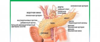
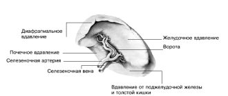
What are the functions of the pancreas?
The pancreas performs two important functions.
- It produces enzymes (digestive enzymes) and releases them into the duodenum. Enzymes in the digestive tract break down carbohydrates, proteins and fats. This is the so-called exocrine function.
- Another function is endocrine , which is performed by beta cells of the islets of Langerhans, producing insulin (hormone), and alpha cells, producing glucagon. Insulin controls the level of glucose (sugar) in the blood. It acts against hyperglycemia (high blood sugar) and glucagon corrects hypoglycemia (low blood sugar). Insulin also promotes the absorption of glucose in the liver, where it is stored in the form of glycogen, and then used during stress and exercise. When the islets of Langerhans produce little insulin, glucose levels rise and there is a risk of developing diabetes, etc.
Sources
- Guyton, A.K. Medical physiology / A.K. Guyton, J.E. Hall / Per. from English; Ed. IN AND. Kobrina. - M.: Logosphere, 2008. - 1296 p.: ill.
- Prives M. G., Lysenkov N. K., Bushkovich V. I. / Human Anatomy. — 12th ed., revised. and additional - St. Petersburg: Publishing house SPbMAPO, 2006. - 720 p., ill.
- Surgical diseases: Textbook / M. I. Kuzin, O. S. Shkrob, N. M. Kuzin, etc.; Ed. M.I. Kuzina. — 3rd ed., revised. and additional - M.: Medicine, 2002. - 784 p.: ill.
- Erika F. Brutsaert, MD. Diabetes mellitus // MSD Handbook - 2019.
- Imaeva A.K., Mustafin T.I., Polovinkina S.R. MORTALITY AND MORTALITY INDICATORS IN ACUTE PANCREATITIS AS AN INDICATOR OF THE STATE OF MEDICAL CARE AT THE REGIONAL LEVEL // Problems of social hygiene, health care and history of medicine. 2021. No. 6.
Prevention of pancreatic diseases
It should be remembered that the functioning of the gland is most negatively affected by alcohol, smoking, irregular meals, fried, spicy and fatty foods. All this should be avoided. The diet should be healthy. You need to eat four to five times a day, and moderation in food is also important.
Pancreatic diseases should be taken seriously and be thoroughly examined in order to receive optimal treatment.
Our clinic offers modern diagnostic methods with consultation of experienced specialists for the treatment of pancreatic diseases. Don’t delay until later and make an appointment with a gastroenterologist right now.