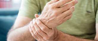
Many patients, having received information about the presence of osteophytes, begin to associate the presence of back pain with osteophytes in the spine. However, bone growths (osteophytes) themselves are only a sign of degeneration in the spine and the presence of osteophytes does not necessarily mean that they are the actual cause of back pain.
In principle, osteophytes are a radiological marker of degenerative changes in the spine, and their appearance only means involutional changes in the spine. Over the age of 60, osteophytes (bone spurs) in the spine are quite common.
Causes
According to the mechanism of their development, osteophytes are divided into 8 main types:
Post-traumatic: occur after trauma to the periosteum with its detachment, bone fractures. The formation of osteophytes is especially stimulated by infection in the wound during an open fracture.
Osteophytes of degenerative-dystrophic origin are formed in those joints where, for various reasons, there is active destruction of articular cartilage and damage to the bone under the cartilage. An example of such a process is deforming arthrosis of the knee joint, in which osteophytes of the knee joint develop as a response to damage to the subchondral layer of the bone and limit movement in the joint, thus slowing down further destruction of the cartilage and deformation of the joint.
Osteophytes formed during inflammatory processes. For example, with osteomyelitis, tuberculosis, brucellosis, rheumatoid arthritis.
Osteophytes that occur during certain systemic changes in the skeleton as a result of endocrine disorders. For example, with acromegaly.
Osteophytes with damage to the central nervous system: osteophytes can develop in various diseases of the nervous system, when the innervation (receipt of nerve impulses into the tissue) of the tissues is disrupted.
Osteophytes, formed as a result of excessive physical exertion, in places where the periosteum is irritated by strong and frequent contractions of the muscles attached to it.
Osteophytes formed as a result of the formation of microtears of the joint capsule or its pinching between the articular surfaces of the joints during sudden movements in them. An example of such an injury is “footballer's foot,” in which, in response to damage to the joint capsule, osteophytes of the ankle joint appear along the anterior edge of the articular surface of the tibia.
Types of osteophytes
In addition to the etiology of osteophytes, the criterion for classifying growths is their location. It not only has implications for the selection of therapeutic measures, but can also give clues about the causes of the pathology.
Osteophytes of the spine
Spinal osteophytes are an integral part of spondylosis
. At these stages, they respond well to treatment with physical therapy and physiotherapy, orthopedic correction (wearing a corset) and drug therapy.
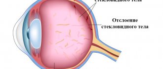
The role of spinal osteophytes is to maintain sufficient distance between the vertebrae. Normally, this function is performed by intervertebral discs - the natural shock-absorbing pads of the ridge. However, with age or under the influence of stress, their height may decrease, which leads to contact between the protruding parts of the vertebrae and compression of the nerve roots. This situation without treatment of spinal osteophytes is fraught with disturbances in tissue trophism and their further destruction. In an attempt to contain the destructive effects, the body builds “protective barricades” in the form of osteophytes with a clearly defined marginal localization.
Signs of osteophytes in the spine are:
- a feeling of stiffness in the spine, especially after sleep or a long stay in one position (patients find it difficult to straighten);
- weakness of the back muscles and a feeling of numbness in some areas along the ridge (so-called clamps);
- back pain when changing position, prolonged standing, certain movements (at first episodic, but can become more and more prolonged; the nature of the pain is stabbing, sharp or pulsating);
- burning and tingling sensation caused by irritation of nerve endings;
- swelling of the back;
- radiating pain;
- changes in gait (especially with twisting of the foot).
Depending on which part of the spine the osteophytes are located in, patients may experience specific symptoms - for example, bowel and urination disorders, sexual dysfunction, numbness and weakness of the upper and lower extremities, tremors in the hands, visual disturbances, surges in blood pressure and other. Sometimes spinal osteophytes are visible under the skin or can be easily felt.
In some patients, the formation of osteophytes in the spine is practically asymptomatic, which makes it difficult to fully assess the prevalence of this pathology. However, doctors are sounding the alarm: the human spine is anatomically poorly adapted to the loads associated with upright posture, and a sedentary lifestyle only aggravates this problem
.
In addition to improving metabolism, when treating spinal osteophytes, it is very important to relieve swelling and inflammation of tissues, since they can lead to complications.
Spinal osteophytes are a common type of osteophyte.
Osteophytes of the knee joint
Three faithful companions of osteophytes in the knee joint are pain
,
impaired mobility
and
joint deformation
(in later stages). People over 40 years of age are especially susceptible to the problem, however, professional dancing and sports, injuries and other factors can shift the formation of growths to an earlier date. As a rule, osteophytes grow on the lateral surfaces of the knee joints and in the periarticular tissues.
Initially, pain due to osteophytes of the knee joint may only appear when standing for a long time or getting up from a seat or bed. Often patients believe that they have failed to stand on their leg, but over time the pain becomes more and more prolonged and background. They are often accompanied by characteristic clicks and a dry, rough crunch. Gradually, the pain becomes intense, intensifies in the evening, and subsequently begins to bother you at any time of the day or night.
In the morning, patients with osteophytes of the knee joint experience characteristic stiffness, which may disappear after warming up. Then the amplitude of voluntary movements is reduced: it becomes difficult for patients to fully bend or straighten the leg at the knee. It becomes hot, swollen and red, which is why patients require constant use of anti-inflammatory drugs
.
If the osteophyte of the knee joint grows in the form of a long thin spike, over time it can break off under stress (for example, when squatting). In this case, a fragment can fall into the joint cavity and completely block it - treatment of osteophyte of the knee joint, which has become the so-called. articular mouse, performed only surgically
.
Remember: at the very beginning (when the size of osteophytes of the knee joint is 1-2 mm), the disease is amenable to local drug treatment, physiotherapy and therapeutic exercises
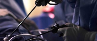
, so you shouldn’t delay seeing a doctor if you have discomfort in your knees. In the later stages, arthroscopy or endoprosthetics are indicated for the treatment of osteophytes of the knee joint.
Osteophytes of the cervical spine
The formation of osteophytes in the cervical spine often causes postural disorders. Being quite mobile, this part of the spine is quite easily injured, and the vertebrae are displaced. The presence in the tissues of the neck of a large number of nerve endings and large blood vessels, on which the normal functioning of the brain, spinal cord and sensory organs depends, makes even small osteophytes of the cervical spine very dangerous. For the same reason, it is difficult to remove them surgically. Therefore, it is so important to monitor the first symptoms of osteophytes in the cervical spine:
- discomfort in the neck, a feeling that it has become harder for the patient to hold his head;
- feeling of stiffness, tension, especially at the end of the working day or after sleep (patients need a warm-up);
- crunching of the vertebrae, feeling as if they are rubbing against each other;
- increasing difficulty turning the head (may be accompanied by nagging pain);
- a feeling of burning, numbness and tightness in the cervical-collar area;
- the development of inflammation, which is accompanied by edema and local fever.
Patients often complain of secondary symptoms of cervical osteophytes - headaches, increased fatigue, weakness, trembling and numbness in the extremities of the shoulder girdle, impaired vision and hearing, spots in the eyes, arterial hypertension, dizziness, deterioration of cognitive abilities.
Symptoms
Osteophytes grow to a certain size, stop growing and, as a rule, remain unchanged for a long time.
Initially, osteophytes have a cartilaginous structure with foci of ossification. Then they acquire the structure of soft spongy bone. Osteophytes complete their development with the formation of dense, compact bone tissue.
Cases of the development of osteoporosis and processes of destruction of osteophyte bone tissue, which can lead to partial or complete resorption of the bone outgrowth, have been described.
The exception is osteophytes that lead to compression of nerve trunks or penetrate deeply into muscles. Such osteophytes can cause pain of varying degrees of intensity.
In some cases, osteophytes are a protective mechanism during the development of a pathological process in the joint, limiting movement and, thus, protecting the joint from further destruction of the articular surfaces of bones and articular cartilage under load.
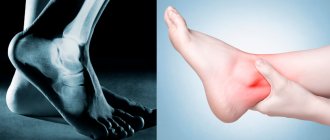
In some cases, osteophytes themselves become a source of tissue injury and only aggravate the processes of joint destruction. For example, osteophytes of the ankle joint contribute to pinching of the ankle joint capsule between the bony outgrowth and the articular surface of the talus, which leads to increased pain, swelling and inflammation in the joint.
Diagnostics
Diagnosis begins with a clinical examination. The doctor should first conduct a detailed examination, a neurological examination to evaluate the functioning of the nerve roots and look for signs of compression of the nerve roots or spinal cord. Based on the examination, medical history, and patient complaints, the doctor prescribes the necessary examination plan, including the following research methods:
ENMG allows you to determine the conduction disturbance along the nerve fiber and determine both the degree of damage and the level of damage to the nerve fibers. Radiography is often prescribed primarily for the diagnosis of osteophytes and allows visualization of osteophytes in the spine . In addition, radiography can detect other changes in the bone tissue of the vertebrae.
Computed tomography (CT) or MRI can provide more detailed information about changes in the spinal structures, both bone and soft tissue, and detect the presence of compression of the nerve roots or spinal cord.
Neuroimaging data allows the doctor to choose adequate treatment tactics, both conservative and, if necessary, surgical, depending on the presence of signs of compression of nerve structures in correlation with clinical data.
Treatment
The detection of an osteophyte in itself has no clinical value. It is necessary to establish the disease that led to the appearance of osteophytes, otherwise treatment will be ineffective and can only worsen the situation.
If osteophytes do not cause pain and do not significantly limit movement in the joints, then treatment is not required.
A modern and effective method of treating osteophytes is Shock Wave Therapy. Due to its unique properties, the shock wave destroys the structure of the osteophyte and gradual resorption of the osteophyte occurs. A typical example is the treatment of heel spurs. The course of treatment is 5 procedures, usually once a week.
MRI or CT for diagnosing osteophytes
According to most doctors, CT and MRI of the spine are the method of choice for detecting osteophytes. For magnetic resonance imaging of the spine, special techniques are used:
- MR myelography;
- kinetic MRI;
- gradient T1 VI;
- 3D reconstructions;
- diffusion-weighted MRI.
The MRI procedure of the spinal region for suspected degeneration generally does not require contrast enhancement and can be performed on any model of MRI machine. In addition to MRI, computed tomography will clearly show the presence of osteophytes. CT scan of the spine is a priority method for diagnosing bone pathologies and is especially indicated for spondylosis and accompanying bone growths.
| Service | Price according to Price | Discount Price at Night | Discount Price During the Day |
| from 23.00 to 8.00 | from 8.00 to 23.00 | ||
| MRI of the cervical spine | 3300 rub. | 2690 rub. | 2990 rub. |
| MRI of the thoracic spine | 3300 rub. | 2690 rub. | 2990 rub. |
| MRI of the lumbosacral spine | 3300 rub. | 2690 rub. | 2990 rub. |
| MRI of the craniovertebral junction | 3300 rub. | 2690 rub. | 2990 rub. |
| MRI of the sacroiliac joints | 4000 rub. | 3190 rub. | 3690 rub. |
| MRI of the coccyx (MRI of the sacrococcygeal region) | 3300 rub. | 2690 rub. | 2990 rub. |
| MRI of the cervicothoracic spine | 6600 rub. | 5380 rub. | 5980 rub. |
| MRI of three parts of the spine (MRI of the cervical, thoracic and lumbosacral regions) | 9900 rub. | 6900 rub. | 7900 rub. |
| MRI of the entire spine / MRI of the back | 9900 rub. | 6900 rub. | 7900 rub. |
| MRI of the central nervous system (MRI of the brain, MRI of the cervical, thoracic and lumbosacral regions) | 13200 rub. | 9590 rub. | 10890 rub. |
| Appointment with a neurologist | 1800 rub. | free after MRI | free after MRI |
| Comprehensive body diagnostics (MRI of the thoracic spine, MRI of the lumbar spine, ultrasound of the abdominal organs, ultrasound of the kidneys, ultrasound of the bladder, consultation with a neurologist, consultation with a therapist) | 11700 rub. | 7000 rub. |
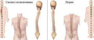
Spondylolisthesis of the vertebral segment
Spondylolisthesis is an abnormal displacement or slipping of a vertebra. As a result, the spinal segment loses stability. The consequences can be traumatic and dangerous - sometimes the displacement of one vertebra in relation to another (upper to lower) reaches 75%.
Spondylolisthesis of the spinal segment can be a consequence of injury, surgery, improper bone development, systematic improper loads, and diseases of the osteochondral tissue. Causes pain (passes after rest) with neuralgic symptoms, dysfunction of the pelvic organs.
To stabilize the spinal segment, the patient may be indicated for neurosurgical surgery with the installation of an implant, metal structure, or interbody cage.
Advantages of treatment at Top Ikhilov
- Qualified doctors with extensive experience.
- Advanced medical equipment for the diagnosis and surgical treatment of spinal diseases.
- Minimally invasive and robotic spine surgeries, after which patients recover in a matter of days.
- Loyal pricing policy, the possibility of stage-by-stage payment for each service performed.
- Extensive experience working with foreign patients. Availability of an international department and a staff of professional translators (if necessary).
- Accompanying a foreign patient at all stages of diagnosis and treatment of the disease.
- The opportunity to combine treatment with a holiday in Israel.
- 5
- 4
- 3
- 2
- 1
(11 votes, average: 3.7 out of 5)
A CT scan revealed a degenerative disease of the spine - what to do?
If back pain does not bother you, but the “accidental finding” on a CT or MRI turned out to be “degenerative-dystrophic disease of the spine,” you should clarify what kind of disease it is and assess the possible health risks in the future.
Timely consultation with a neurologist or osteopath will help develop measures for effective treatment of DDSD and prevention of complications. The worst option would be to ignore the problem and “let go” of the situation to the point of traumatic consequences and the need for surgical intervention.
If there is severe pain associated with an intervertebral hernia, stenosis or other type of DDSD, the patient should consult a neurologist, algologist or neurosurgeon. The pain must be relieved, as it makes it difficult to move and provokes the development of depression, thus aggravating the situation. The patient will also be prescribed therapy (NSAIDs, therapeutic blockades, exercise therapy, radiofrequency ablation, physiotherapy, etc.) or surgery will be recommended.
How to fight osteophytes today
The fight against osteophytes is part of the general treatment regimen for coxarthrosis, gonarthrosis or arthrosis of another joint. It is almost impossible to get rid of bone growths. The goal is to reduce pain and expand the range of motion. For this purpose, depending on the stage of arthrosis, the patient is prescribed:
- ointments to reduce unpleasant symptoms, such as hydrocortisone;
- non-steroidal anti-inflammatory drugs in the form of tablets or injections into the joint;
- physiotherapy - laser, magnets, electrophoresis, shock wave treatment.
One of the physiotherapeutic methods of combating pain due to arthrosis is magnetic therapy. What is it based on and why it helps, says the specialist:
What it is
Osteophyte is a word that is unclear to most patients. In fact, there is nothing complicated here. Osteophytes are pathological growths of bone tissue where it is normally absent. This condition is not an independent disease, but it accompanies many ailments of the musculoskeletal system. Essentially, this is a compensatory reaction of the periosteum to any adverse effects on the articular joint or the result of fractures.
The growths can have different shapes and sizes. For a long time, due to their small size, they do not cause inconvenience to the patient, but over time they make themselves felt. They often take the form of spikes and hooks.
The process of osteophyte formation is as follows:
- the bone structures of the ankle are deprived of joint fluid, or it becomes insufficient for normal functioning;
- friction increases, surfaces begin to collapse;
- gradually, in places of particularly active friction, compensatory ossifications appear, differing in shape and size from what is needed;
- The bone growth is fully formed.
Osteophyte is not a disease, but the result of a previous injury or other pathology.
Principles of recovery after surgery
After surgery to remove osteophytes using arthroscopy (or open methods), a rehabilitation period is required. It is much shorter than during rehabilitation after open surgery, but it cannot be neglected. The recommendations are, in principle, quite standard. need to:
- in the first week, limit physical activity on the operated limb;
- medications to relieve pain and reduce swelling;
- Postoperative discomfort can be alleviated by applying cold for short periods of time;
- you need to use compression garments, which will relieve swelling and help alleviate pain;
- during the rest period, it is recommended to keep the leg above the level of the heart;
- It is necessary, based on medical recommendations, to return to an active life, increasing the load gradually, without sudden jumps.
In our clinic, patients can undergo all stages of treatment: arthroscopy or open surgery, preoperative, and postoperative. This is important for a full and quick recovery, which cannot be perfect without medical supervision.
Osteophytes are an unpleasant pathology that can reduce the quality of life of any person. They often accompany the development of other problems of the musculoskeletal system, which, if ignored, can even lead to disability!
The appearance of signs of pathology is a reason to visit a doctor for diagnosis and choice of therapy! Do not ignore the first symptoms of the disease!
Patient complaints
Osteophytes are an insidious pathology. For a long period of time they do not remind anyone of themselves. Only when they reach a significant size will the first symptoms appear.
The patient will present the following complaints:
- the appearance of pain in the affected area, which may intensify with movement, sudden uncontrolled actions such as coughing or sneezing, or taking an uncomfortable position;
- negative changes in the mobility of the affected ankle;
- the appearance of crunching, creaking in the affected joint;
- the onset of an inflammatory process in surrounding tissues due to their damage;
- swelling of the problem area.
Ignoring pathological changes leads to a gradual but steady worsening of symptoms. Over time, a person may even become disabled if a diagnosis is not made in time and therapy is not started.
How to detect
Before starting treatment, you need to go to the clinic and consult a doctor. He will conduct the necessary examinations on site to assess the presence of reflexes and the patient’s condition. However, by palpation it will only reveal already expanded osteophytes. Small growths, but in the process of growth, are detected during other stages of the examination.
A. Radiography
Radiography is used to diagnose various pathologies of internal organs and the spine. X-rays penetrate dense opaque media and are absorbed by them to varying degrees, resulting in an image that reflects the state of the imaged area.
Varieties and most common localization
There are 4 main types of these formations.
- Post-traumatic
Mainly affects the lower part of the musculoskeletal system, but can also involve the area of the elbow and wrist. This is explained by the fact that these areas are most often injured even in everyday life not associated with major sports. It is important to remember that bone damage is not a necessary factor in the formation of a growth. A simple rupture of the periosteum is sufficient.
- Periosteal
They develop when there is a constant inflammatory process in the ankle joint area. It is impossible to predict which areas will undergo ossification and how the process as a whole will develop in this case.
- Degenerative-dystrophic
Most often it is a consequence of the development of arthrosis. Accompanied by impaired mobility in the foot area.
- Massive osteophytes
A. b
A. massive osteophytes. b. osteophyte of the talus
Volumetric growths that can even change the configuration of the joint are mainly a consequence of the impact of tumor pathologies on the body.
Additionally, there is a classification of osteophytes based on cellular structure. Highlight:
- spongy defects formed from spongy bone tissue;
- metaplastic defects resulting from a violation of the bone structure;
- compact, formed from the outer layer of bone, affecting mainly the area of the feet and fingers;
- cartilaginous, appearing under excessive loads instead of cartilaginous tissue.
The growths can be localized on any joint of the human body. Most often the elbow joint, knee area, hip joint, spinal column, and ankle are affected.