Home Eye diseases
Visitors come to our forum with the same problem. Usually the cause of anxiety is described as follows: “If I look at light-colored things without glasses, I see various transparent threads, small circles, dots, and they seem to flow down like glass.” “A week ago, when he woke up in the morning, he discovered in one eye a feeling like a haze was floating in front of his eyes, he explains how cigarette smoke floats like a cobweb, but his vision has not deteriorated.” “Almost 3 years ago, small circles appeared in both eyes, translucent hairs, and over the course of 3 years, more and more of them gathered into clouds.”
To summarize, most often people see the following picture: floating dots before their eyes; floating spots before the eyes; midges before eyes; black spot before the eyes; spots before the eyes.
All these “objects” are usually best seen against a light background and in good lighting. They move smoothly when the eyes move and continue to move after the gaze is fixed.
Sometimes these visual effects may be accompanied by sparks and lightning. A well-established name for this effect has emerged - flying flies. In medicine, the term “destruction of the vitreous body”, abbreviated as DST, is used for this pathology.
Depending on the degree of severity, these floaters can either not interfere with a person at all, or bring psychological discomfort, and in especially serious cases, significantly interfere with vision. What is destruction of the vitreous body?
Vitreous body
The vitreous is a gel-like substance that fills the cavity of the eye between the retina and the lens. More than 99% of it consists of water and less than 1% of collagen, hyaluronic acid and other substances. Despite being present in such small quantities in the eye, collagen and hyaluronic acid are extremely important components. Hyaluronic acid provides the gel-like structure of the vitreous body. Collagen serves as a framework for it. In addition, collagen, hyaluronic acid and proteoglycans form a complex that also affects the structure of the vitreous body.
The vitreous body is normally absolutely transparent and this is achieved due to the strictly defined structure and composition of the molecules of the substances that make up its composition. Under the influence of various factors, these molecules can disintegrate into fragments, which leads to a qualitative change in the composition of the vitreous body, and its volume also changes. This process is called destruction of the vitreous body. As a result, particles appear in the vitreous body that do not have optical transparency; it is these that our vision perceives as flying flies.
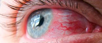
In some cases, changes in the structure of the vitreous body can lead to a mechanical effect on the retina, “irritation” of the photoreceptors occurs and, as a result, the person sees sparks or lightning. However, it is necessary to differentiate the causes of the appearance of floaters. Flies are not always DST. The ingress of blood, drugs and other substances that should not normally be in the vitreous body can cause a visual effect similar to that seen during the destruction of the vitreous body.
Sometimes the fly-fly effect can be associated with increased blood pressure. In this case, blood pressure control is necessary, especially when floaters appear. Seeing a therapist may clear things up.
Destruction of the vitreous body: causes, symptoms
The vitreous body occupies ⅔ of the volume of the eyeball and is located between the lens and the retina. It consists of 99% water and has a transparent gel-like structure. It performs several important functions:
- gives the eye the correct shape;
- provides incompressibility of the eyeball;
- participates in the refraction of light rays entering the retina.
Destruction of the vitreous body is a pathological process of disruption of the structure of the substance that fills the space between the inner membrane of the eye and the lens. During the development of this pathology, opacities are formed that affect the quality of vision.
In a healthy person, the vitreous body is absolutely transparent and thick with many fibrils - proteins that have a thread-like shape. They are very thin but can support the shape of the eye. Destruction leads to thickening of these thread-like elements. They lose their transparency and become noticeable in the field of view. This symptom is called “floaters”.
Destruction of the vitreous body takes various forms:
- Thread-like. It often develops in older people, patients with progressive myopia or its high degree, as well as in patients with atherosclerosis.
- Grainy. Destruction of this type occurs when the inner layers of the retina become inflamed.
- Crystalline. This form of pathology is quite rare and appears due to the accumulation of calcium, cholesterol and tyrosine in the vitreous body.
According to the degree of prevalence, destruction is divided into 2 types:
- Complete, in which the pathological area gradually occupies the entire space between the retina and the lens. This leads to deformation of the eyeball, the formation of adhesions, and detachment of the retina.
- Partial, which is accompanied by the formation of a cavity in the central part of the vitreous.
A little anatomy
The vitreous body is a gel-like colloidal substance that occupies about 85% of the total volume of the eye and is 99% water.
It has an integral structure due to the presence of the finest fibrillar network. Its main component is hyaluronic acid, the molecule of which has a spiral ornate structure and gives the vitreous fluid a gel-like consistency. On the outside, the vitreous body is surrounded by a membrane consisting of collagen fibers tightly adjacent to each other. Normally, the vitreous body is an optically transparent light-conducting medium. It helps maintain the shape of the eye and also lies in the path of the light ray from the cornea, iris and lens to the retina. The three main attachment points of the vitreous membrane are the area of the optic nerve head, the area of the lens, and the so-called base, where the membrane is attached to the ciliary epithelium.
Sometimes, looking at the sky on a sunny day, any healthy person can notice small “snowflakes” or “cobwebs” slowly floating before their eyes. These are the collagen fibers of the vitreous body that a person can see normally. However, with age, the structure of this part of the eye can change greatly, causing patients significant visual discomfort.
Causes of destruction of the vitreous body
This pathological process occurs as a result of changes in the physical and chemical properties of the contents of the vitreous body. A variety of reasons can lead to this:
- myopia;
- age-related changes in the structure of the eye;
- inflammatory eye diseases - keratitis, endophthalmitis, dacryocystitis, blepharitis;
- disorders of the blood supply to the brain and retina;
- eyeball injury;
- eye surgery;
- long-term visual stress;
- atherosclerosis;
- diabetes;
- acute respiratory diseases;
- deficiency of vitamins and minerals;
- hormonal disbalance;
- severe pathologies of internal organs - liver, kidneys;
- constant stress.
With a high degree of myopia, the eyeball has too large a diameter. All its structures are deformed. Metabolism in the eyes is disrupted, which becomes a trigger for a destructive process. Previously, this disease was most often diagnosed in older people and was associated with age-related changes. Over the years, the properties of the colloidal gel, which is located in the vitreous body, change. At the periphery, this substance is compacted, and in the center it is deformed.
Today, destruction is detected in schoolchildren and people whose work involves visual stress. The number of patients is growing in economically developed countries, which is explained by the massive use of electronic devices, increasing life expectancy and a number of other factors.
Destruction of the vitreous body is opacification of the fibers of the vitreous body of the eyeball, which manifest themselves in the form of inclusions of various shapes (threads, dots, grains) and accompany eye movement.
The main reasons for the development of destructive processes in the vitreous body of the eyes are ophthalmological diseases, diseases of the circulatory system and natural physiological changes that occur in the body over time. Treatment of the disease is aimed at eliminating the symptoms accompanying destructive phenomena or removing inclusions through surgery.
The presence of destruction of the vitreous body does not affect the quality of life and the level of a person’s ability to work. In advanced cases, partial or complete loss of vision is possible, but most often the prognosis of the disease is favorable.
General information
The vitreous body is presented in the form of an avascular transparent gelatinous substance that fills the cavity of the eyeball between the lens and the retina. Its presence ensures the preservation of turgor and the correct shape of the eyeball, compensates for changes in intraocular pressure, and carries out light impulses to the retina.
In a healthy person, this substance is completely transparent and does not contain any inclusions. It consists of hyaluronic and ascorbic acids, whey proteins, salts and other substances and is supported by a framework consisting of protein fibrils.
Destruction of the vitreous body occurs as a result of thickening of some fibers and loss of their transparency, which leads to a change in its mesh structure. Destructive processes manifest themselves in the form of liquefaction of the vitreous body, its wrinkling and peeling.
The vitreous body loses its homogeneity: fibers stick together, and weaves of various shapes are formed, which float freely in the liquefied gelatinous substance (filamentous, or filamentous, destruction of the vitreous body). Simultaneously with liquefaction, strands and films can form in the vitreous body of the eye, which vary in size and density. In some cases, these formations are fixed to the fundus of the eye, which leads to serious pathological changes.
Vitreous opacities
Wrinkling of the entire vitreous body or part of it is the most severe form of destruction of the vitreous body. As a result of this process, there is a decrease in volume and a change in the shape of the gelatinous substance, and tension in the vitreoretinal connections is observed. With a severe degree of the disease, these connections can rupture, which can result in hemorrhage into the vitreous body, its detachment, or retinal rupture. Photopsia phenomena are often observed. Ultimately, complete destruction of the vitreous body may occur.
In addition to the “floaters” characteristic of the destruction of the vitreous body of the eye, “lightning” or “flashes” appear in the field of vision, which indicates the presence of “optical cavities” in the eyeball. Thus, the brain perceives an abnormal response of the optic nerve to the presence of voids. Cloudy particles are difficult to see because they follow your eye movements.
Opacities are best seen when looking at a clean, bright surface (clear sky, white ceiling, snow), when squinting your eyes, or in coherent rays. In conditions of low lighting of the environment, as well as when it is heterogeneous, opacities, as a rule, are not visible.
Destruction of the vitreous body can manifest itself in the form of golden or silver rain. This phenomenon is observed in the presence of crystalline inclusions of tyrosine, cholesterol, phosphorus, calcium and magnesium compounds. It is typical for older people suffering from cholesterol metabolism disorders, as well as diabetes mellitus.
Biomicroscopy allows you to detect shiny particles (“rain”) or “dancing snowflakes” that pendulum-like oscillate and move simultaneously with the movement of the eyeballs. Such crystals can be of various shapes (plates, spheres, points), colors (golden, snow-white, brown) and sizes.
Causes
Destruction of the vitreous body of the eye most often occurs due to various physiological and pathological reasons:
- age-related changes in the structure of the eyeball;
- the presence of chronic inflammatory processes in the eye;
- diabetes;
- diseases of the circulatory system (atherosclerosis, arterial hypertension, dystrophic changes in blood vessels);
- severe myopia;
- dystrophy;
- compression of arterial vessels in the presence of cervical atherosclerosis;
- hormonal changes that occur during pregnancy, menopause, puberty, and when hormonal therapy is prescribed;
- injuries to the eyes, nose, head (including surgery);
- helminth infestation (toxoplasmosis);
- frequent and prolonged visual stress;
- psycho-emotional stress, depression;
- physical exhaustion;
- some diseases of internal organs;
- deficiency of vitamins, macro- and microelements;
- toxic or radiation effects on the body.
The cause of destruction of the vitreous body of the eye can be a disruption of the internal organs that regulate the composition and balance of colloids of the vitreous body (endocrine glands, kidneys, liver). This provokes changes in the structure of the colloidal gel (coagulation and precipitation processes). In addition, the occurrence of “flying spots” may indicate the beginning of the process of retinal detachment, which can ultimately lead to complete loss of vision.
Symptoms
The main symptomatic sign of the presence of destruction of the vitreous body is the floating of various visual effects before the eyes - “spots”, “floaters”, “cobwebs”, “opacity”. These optical elements differ from the effects that occur as a result of blows to the head, sudden jumps in blood pressure, or when lifting heavy objects.
Symptoms of vitreous destruction:
- the presence of “floaters” and opacities is permanent;
- visual phenomena have a constant shape and size;
- the effects are noticeable only in good lighting conditions (especially on a white surface).
The more clearly the floating elements are visible and the thicker they are, the greater the destruction of the vitreous body. If the opacities acquire a clear filamentous structure, a person may be diagnosed with atherosclerosis or a severe form of hypertension. The presence of “flashes” and “lightning” is a sign of vitreous detachment or other serious complications.
With filamentous destruction of the vitreous body, disorganized floating of fibrils is dispersed throughout the entire volume of the eyeball, while they twist and stick together, forming into formations resembling balls of yarn.
As a result of eye injury, previous illness, or in the presence of tumor-like formations, destruction of the vitreous body manifests itself in the form of small accumulations of small grains. In the absence of timely treatment, complete or partial loss of vision cannot be ruled out.
Diagnostics
The following methods are used to diagnose the disease:
- ophthalmoscopy (examination of the fundus of the eye);
- visual acuity test;
- ophthalmological examination using a slit lamp;
- compiling an anamnesis.
The data obtained during the examination allows us to conclude about the presence or absence of destructive processes in the vitreous body of the eyeball.
Treatment of the disease
Treatment of destruction of the vitreous body of the eye is ineffective in most cases. Sometimes minor opacities and small fibers can resolve on their own, but large formations, crystal deposits and fragments of connective tissue fibers remain for the rest of life.
Questions about how to treat destruction of the vitreous body of the eye and whether it needs to be done are decided in each case individually. The need and effectiveness of therapy depends on the presence or absence of optical effects, visual impairment, the area of damage to the vitreous, as well as the influence of these pathological factors on the person’s condition and his ability to work.
Currently, methods for specific treatment of vitreous destruction that allow effective and safe elimination of crystal deposits and massive fibrillary formations have not been developed. The main therapeutic measures are aimed at eliminating the causes of the disease, reducing visual stress and using symptomatic drug therapy.
In some cases, the disease is treated through surgery:
- vitreolysis – splitting of opacities existing in the vitreous body using a YAG laser;
- vitrectomy – partial or complete replacement of the vitreous body with an artificial medium (silicone oil, gas bubbles, saline solution).
The use of surgery as a method of treating vitreous destruction has an ambiguous prognosis, since there is a high risk of serious complications (cataracts, hypotension, retinal detachment, hemorrhages). Often the use of such methods is unjustified, especially for older people, since due to progressive age-related changes, vascular problems worsen over time.
It is worth noting that if the vitreous body is destroyed, you can play sports, but preference is better given to activities that do not require significant physical activity and stress.
Forecast
The prognosis for the development of the disease is favorable in most cases. Opacities stabilize relatively quickly after the onset and development of the disease. The occurrence of remissions during destructive processes is extremely rare, and floating opacities in terminal form remain in the cavity of the eyeball.
Destruction of the vitreous body of the eye, manifested in a mild form, does not have a noticeable effect on a person’s ability to work and does not cause serious complications. The development of severe forms of the disease can significantly worsen the patient’s quality of life. The constant movement of floating elements obstructs the viewing of various objects and interferes with the performance of work duties.
Destruction of the vitreous body: symptoms, treatment
The main sign of a pathological process in the vitreous body is “flying flies”. This visual phenomenon is called photopsia. It may not affect the image quality. Cloudiness, spots, dots and other similar formations are visible on a light background. Gradually they grow, there are more and more of them. This leads to the loss of transparency of the vitreous body, which is reflected in the refraction of light rays.
The pathology is also accompanied by hemophthalmos - hemorrhages into the thickness of the substance between the lens and the retina. As the disease progresses, other signs appear, including decreased visual acuity. Other changes inside the eyeball can only be detected during examination using devices. Let's find out how to diagnose and treat eyes with destruction of the vitreous body.
Therapeutic measures and prevention
Floating clouds in the field of vision or so-called floaters most often do not require specialized treatment. Over time, older people get used to their presence and even stop noticing. This situation requires routine follow-up and a visit to the doctor once a year.
Unfortunately, the disease does not always have such a favorable course. Cloudiness can cause severe visual discomfort and significantly reduce visual acuity. But even in such a situation, doctors are in no hurry to resort to surgery, since it carries risks and complications. Treatment of destruction of the vitreous body with the help of surgery - vitrectomy, is performed only in very severe advanced cases. After extraction, the cavity of the eyeball is filled with a special saline solution.
More often, patients are taught to cope with opacities on their own, to look through them and try not to pay attention to these visual phenomena. The indication for radical intervention is retinal detachment caused by degeneration and detachment of the vitreous body.
There are no specific preventive measures that can prevent destructive changes in the vitreous body.
Doctors recommend a healthy lifestyle, proper nutrition, and maximum eye protection from ultraviolet rays. On the Internet today it is easy to find photos of the destruction of the vitreous body with a detailed description of the processes occurring in the eye. Regular visits to the ophthalmologist will help keep the disease under control and prevent more serious complications. Making an appointment Today: 24 registered
Diagnostics
To confirm the diagnosis of vitreous destruction, several diagnostic procedures are required:
- Ophthalmoscopy is a research method that allows you to identify empty cavities and examine in more detail opacities in the vitreous body.
- Ultrasound of the eye in B-scan mode. It makes it possible to identify crystalline formations and the focus of hemorrhage.
- Biomicroscopy with a slit lamp allows you to study changes in the consistency of the gel.
- Optical coherence tomography. It is prescribed when it is impossible to obtain accurate data using other methods. OCT helps to detect heterogeneity in the structure of the vitreous body, changes in its shape or size. This diagnostic procedure is not used for massive hemophthalmos.
- Visometry - determination of visual acuity.
- Tonometry is the measurement of intraocular pressure, which can increase when the vitreous body is destroyed.
After the diagnosis is made, a treatment method is selected. Let's find out how to treat this pathology and how dangerous it is.
Diagnostic measures
If the symptoms described above occur constantly or even periodically, you should visit a doctor. The ophthalmologist will prescribe an examination to assess the condition, structure and structure of the visual organs.
Diagnosis of DST includes the following procedures:
- Ophthalmoscopy is a thorough examination of the human fundus using specialized instruments - a fundus lens or an ophthalmoscope. This method allows you to examine the vitreous in detail and detect areas of opacities or voids in it.
- Visometry. The method involves the use of special tables to identify the real degree of visual acuity.
- Tonometry. It involves assessing the level of eye pressure (this indicator changes with DST).
- Ultrasound of the visual organs, performed in conjunction with B-scanning. Such an ultrasound examination helps to detect areas of hemorrhage, as well as crystalline accumulations.
- Biomicroscopy. A slit lamp is used to analyze the consistency of the gel-like substance.
- Coherence optical tomography. This procedure is prescribed when other diagnostic procedures are uninformative or impossible. This type of tomography is carried out using a special medical tomograph apparatus and detects changes in the shape, size and structural uniformity of the vitreous body. But with significant hemophthalmia, this diagnostic method is not used.
Specific diagnostic procedures are prescribed by the doctor. After a comprehensive examination, the ophthalmologist makes an accurate final diagnosis and issues a conclusion.
How can destruction of the vitreous body be treated?
Destruction is treated in various ways. The choice of method depends on the form of the disease and the degree of its development.
Typically, therapy includes:
- use of medications;
- performing special exercises for the eyes;
- use of folk remedies.
In some cases, surgery is prescribed - vitreolysis or vitrectomy.
Drug treatment
Minor opacities may resolve spontaneously. Sometimes they remain with a person for life. To avoid this, you need to consult a doctor immediately after the appearance of floaters.
Treatment is aimed primarily at eliminating the causes of their occurrence. The patient is advised to reduce eye strain. Drugs are prescribed that treat the underlying pathology. It is quite difficult to get rid of the symptoms of destruction.
For this purpose, medications are prescribed that have absorbent properties:
- locally - potassium iodide;
- orally - drugs to improve blood circulation in the brain.
How to treat destruction with massage and gymnastics?
Eye gymnastics and massage can stop the development of the destructive process and shift floating points in the vitreous body to its periphery. The methods of Bates, Zhdanov, and Norbekov are considered effective. Let's describe them briefly:
- Palming is an exercise aimed at relaxing the eyes. Sit at the table, place your elbows on the tabletop and cover your face with your palms. Light should not penetrate through hands. Sit in this position for 5-10 minutes.
- Shifting your gaze. Move your eyes left and right, up and down.
- Turns. Make circular movements with your eyeballs in different directions.
Palming can be performed by all patients. Other exercises are contraindicated in case of retinal detachment. You can complete your eye exercises by massaging your eyelids. It is performed with the fingers in light circular movements.
Destruction of the vitreous body: treatment with folk remedies
Folk remedies are also used for destruction. They can be used after examination and with the approval of a doctor. This is not to say that they help get rid of floating spots in the vitreous. However, this method of treatment allows you to relax your eyes and prevent the condition from getting worse. As a rule, for destruction the following is used:
- honey drops prepared from water and honey;
- solution of honey and aloe;
- water infusion of propolis.
Solutions are instilled three times a day, 2-3 drops. You can also make compresses with chickweed. Grind the herb, pour boiling water over it, let it brew and prepare lotions. Cool them and apply to the eye for 15 minutes.
Directions of therapy
With a diagnosis such as DST, one cannot delay too much; it is extremely important to begin effective and competent treatment in a timely manner. And although the destruction of the vitreous body does not directly threaten life, it can lead to a decrease in visual functions, as well as to partial or even complete loss of vision, that is, blindness.
There are different methods of treating DST, and one or another method of treatment after a comprehensive examination of the patient is prescribed by an ophthalmologist. When selecting suitable methods and an individual regimen, the doctor takes into account various factors: the degree and form of destruction, the patient’s health and age, lifestyle and professional activity, identified contraindications, as well as the impact of DST on performance and quality of life.
Therapy may include the following areas:
- Conservative drug treatment. In ophthalmology, different groups of drugs are used: local and broad-spectrum for oral administration.
- Special gymnastics. Exercises allow you to stop the processes of destruction and improve blood circulation.
- Massage techniques.
- Surgery. Surgeries are prescribed for complex and severe forms of CTD.
- Ethnoscience. Folk remedies are used in combination with basic traditional therapy and only after consulting a doctor.
All areas are discussed in detail below.
Medicines
Drug therapy is aimed at eliminating and resolving formed formations, normalizing metabolism and blood supply, as well as eliminating the main symptoms.
The following groups of drugs are used:
- Vitamin. For local therapy, drops “Ifiral”, “Octilia”, “Okovit” are prescribed. Broad-spectrum vitamin preparations taken orally include Aevit, Vitrum Vision, and Blueberry Forte.
- Normalizing and improving metabolic processes (“Taufon”).
- Improving blood supply and strengthening the blood vessels of the eyes: “Emoxipin”, “Cavinton”.
- Medicines prescribed for cataracts, such as Quinax. In case of DST, these medications also improve the condition of the visual organs and stop degenerative processes.
- Preparations for resorption of formations, for example, “Potassium iodide”.
Important! Any medications are used under the supervision of a doctor and as prescribed.
Eye gymnastics
Some eye exercises allow you to move the gel-like substance of the vitreous body and redistribute the inclusions and voids it contains in order to free the field of vision from “floaters” and spots.
The following exercises are useful and effective:
- Focusing the gaze on different objects located far and near.
- Circular rotation of the eyeballs, moving them from side to side, down, straight and up.
- Shift your gaze: up, forward, down, then left, forward and right. The movements must be done repeatedly, but this exercise is contraindicated in case of retinal detachment.
Special visual gymnastics complexes have also been developed, for example, by Zhdanov, Bates, Norbekov.
Traditional medicine
Folk remedies are used for mild forms of DST and usually as an addition to traditional primary therapy.
The following recipes are considered the most effective:
- Chickweed compresses
. First, prepare the infusion: pour two tablespoons of crushed dried chickweed with a glass of boiling water, close and wrap the container and leave the mixture to infuse for five hours. Soak cotton pads in the strained solution and apply them to your eyes for fifteen minutes three times a day. - Drops based on propolis
. It is recommended to infuse propolis in shungite water, and then instill one or two drops of this liquid into each eye three times a day. - Drops with honey and aloe
. Mix part of liquid natural honey with four parts of aloe juice, leave the mixture overnight in a dark place, and then instill a couple of drops 3 times a day for a month. - Eye drops from infusions of medicinal herbs
: chamomile, cornflower, caraway, plantain.
Any remedy for DST should be used after approval by a doctor.
Surgical techniques
Surgical intervention for DST may be the only effective method when conservative therapy has proven ineffective and ineffective. The operation is prescribed for rapid loss of vision, rapid destruction of the vitreous body.
For DST, the following surgical methods are used:
- Vitreolysis
. The accumulations and formations formed in the vitreous body during DST are removed with a laser. First, the doctor instills drops to maximize the dilation of the pupils, then places a lens equipped with mirrors in front of the eye to better focus the laser beam. Next, local local anesthesia is performed, after which the laser destroys the seals through a targeted effect. After an outpatient procedure and short-term observation, the patient can be sent home. The operation is quite painstaking due to the constant movement of formations in the gel, so it is performed by experienced specialists and is not yet often practiced in Russia. - Vitrectomy
is the complete removal of the destroyed, heavily clouded and pathologically altered vitreous body. The intervention is carried out under anesthesia and lasts about 30-60 minutes. To preserve visual functions, a synthetic substance with a structure and optical properties close to the characteristics of the lost natural element is placed in the space between the retina and the lens instead of the vitreous body. Typically, silicone oil, gas compounds, polymer fillers or saline solutions are used.
If the operation for DST is performed successfully, vision becomes normal and clear after a few days. The prognosis for a successful outcome of surgical intervention is positive: the functioning of the visual organs is completely restored and preserved for many years.
The success of an operation for DST depends on the professionalism and qualifications of the surgeon, the quality of the equipment, the condition of the patient and his compliance with medical recommendations. But in some cases complications develop:
- cataract;
- hemorrhages;
- partial or complete detachment of the retina;
- purulent inflammation localized in the inner membranes of the eyeballs;
- corneal edema;
- glaucoma.
The presence of certain consequences indicates that recovery after DST and surgery occurs with complications, and medical assistance is required. The lack of timely medical intervention often leads to damage to all elements of the organ structure and loss of vision.
Surgical treatment of vitreous destruction
If the eye is severely damaged, surgical treatment is performed. Vitreolysis or vitrectomy may be prescribed. The first method is a laser procedure. The need for it arises when there is a sharp decrease in vision. During the operation, a YAG laser and a Goldmann lens are used, which is installed on the eyeball. There is no need to hospitalize the patient. Treatment is carried out on an outpatient basis under local anesthesia using mydriatics that dilate the pupil. During the procedure, the patient sees red flashes from the laser beam.
Vitreolysis can lead to complications:
- increased intraocular pressure;
- retinal detachment;
- cataracts;
- choroidal microhemorrhage.
The procedure is not prescribed for:
- clouding of the optical media - the lens and cornea;
- neovascularization - proliferation of blood vessels;
- detachment of the retina or choroid;
- displacement of the lens into the vitreous body;
- high risk of hemorrhage.
Vitrectomy is a radical method of treating destruction, which involves removing the vitreous. The main indication for surgery is extensive clouding affecting the quality of vision.
The procedure is performed under general anesthesia. It lasts on average 1 hour. Instead of the removed vitreous body, a substance that imitates it is implanted into the eye - silicone oil, artificial polymer, saline solution, gas bubble, etc. The operation is quite complicated. Tissue healing takes a long time. Vision returns only after a few days. Possible complications:
- cataract;
- retinal disinsertion;
- endophthalmitis;
- corneal edema;
- hemorrhage.
What is laser vitreolysis?
The vitreolysis procedure is performed using a neodymium YAG laser. For successful treatment, precise focusing is required - at least 6 microns. The energy used is 4-6 mJ. 200-600 flashes are performed. Therapy will require an average of 1-4 procedures.
The technique for performing vitreolysis has some features. Unlike procedures that use a YAG laser (iridotomy, posterior capsule discision), vitreolysis technology is more complex because it involves working with moving objects.
Such laser therapy is carried out if vitreous opacities cause decreased vision or there is a risk of developing vitreoretinal tractions.
The most common complications of vitreous destruction
One of them is the wrinkling of the vitreous substance, which leads to a severe decrease in vision and in some cases ends in blindness. Another complication is retinal detachment. It occurs predominantly in patients with a high degree of myopia. With the rapid growth of the eyeball, all its structures increase, except for the retina. It stretches and is in constant tension. This leads to its thinning, rupture and detachment. This pathology is accompanied by the following symptoms:
- a veil before the eyes;
- blurred vision;
- photopsia;
- shadows in the field of view;
- distortion of the outlines of objects.
When these signs appear, it is urgent to perform laser coagulation (soldering) of the retina. This procedure is safe and is prescribed even to pregnant women.
Destruction and detachment of the vitreous body
These are two different pathological processes with similar symptoms. Detachment is a pathological process that occurs against the background of degenerative changes in the vitreous body. It is located next to the retina. Detachment of the body can lead to damage to the macula, the central zone of the inner lining. This complication is always accompanied by deterioration of vision.
This pathological condition develops with a high degree of myopia. Old age is also a contributing factor. After 50 years, the quality of collagen in the vitreous humor in the eye greatly decreases. Inflammatory eye diseases can provoke detachment. The risk of detachment increases with diabetes mellitus, thyroid pathologies, and hormonal disorders. Often the cause of this disease of the vitreous body is penetrating wounds into the eyeball or its surgical treatment.
Thus, the factors contributing to the development of destruction and detachment are similar. The symptoms of these ailments are not very different.
When the vitreous body is detached, the following are observed:
- “spots”, floating opacities;
- flashes and lightning in the eyes.
Black dots move when you shift your gaze. They are especially noticeable on a white background. In severe cases of the disease, a sharp decrease in vision occurs. It is treated with vitreolysis and vitrectomy.
Clinical manifestations
The main signs of DST are:
- Photopsia
. So-called non-objective images appear in the field of vision, for example, floating “flies”, flickering spots, moving dots, lightning, blurry figures, sparkling sparks. Often visible elements shine and glow and do not have clear boundaries. Most often, “floaters” or spots appear when a person looks at the sky or any light, plain background. When you move or focus your gaze, all figures disappear. - Hemophthalmos
is a decrease in normal visual acuity. A person begins to see worse in the distance. - Hemorrhage into the vitreous region
. It can occur mildly, but is sometimes accompanied by clouding or blurred vision, a veil or cobweb floating across the visual field, a reddish color of the visual field (a person sees as if through a thin red fabric), covering the image with a dark stripe or spot, light flashes in peripheral vision (usually On the sides).
Prevention of vitreous pathologies
In general, the prognosis is almost always favorable. Timely prescribed treatment allows you to stop the destructive process. Even with severe destruction of the vitreous body, surgery helps restore visual functions to the patient. To prevent the development of detachment and destruction of the vitreous body, it is recommended:
- regularly visit an ophthalmologist, especially if there is a predisposition to pathology (myopia, age over 50 years, etc.);
- reduce visual stress;
- do gymnastics daily to relax your eyes;
- eat right, taking vitamins for the visual organs;
- treat all eye diseases in a timely manner;
- Monitor your sugar levels if you have diabetes.
How is the vitreolysis procedure performed?
Laser treatment of vitreous destruction is carried out on an outpatient basis, using local instillation (drip) anesthesia, which does not create a load on the heart, blood vessels and other internal organs. First, the patient is instilled with special drops that dilate the pupil. After anesthesia, a special lens (three-mirror Goldmann lens) is installed on the eye, which allows you to focus the laser beam on the desired area of the vitreous body. Laser vitreolysis is carried out without blood loss and incisions, local anesthesia eliminates pain. The patient can feel the touch of the lens and also hear the characteristic clicks of the laser unit. After the procedure, it is recommended to remain under the supervision of a specialist for some time. After the doctor is sure that everything is in order, you can go home.