Osteochondropathy
name a group of chronic diseases of the musculoskeletal system, characterized by necrotic changes in the apophysis, spongy substance of short and epiphyses of long tubular bones. They appear as a result of excessive mechanical load of a point nature, which causes a malnutrition of the bone tissue.
At the initial stage, especially at a young age, there are no obvious clinical manifestations. This prevents the timely diagnosis of osteochondropathy of bones and the prescription of treatment, which is fraught with microfractures and the development of arthrosis. But, with a competent approach to treatment, the prognosis is generally favorable. Osteochondropathy does not pose a threat to human life. With properly selected therapy, deformation and contracture do not occur.
General information
Keller's disease is a disease that affects the bones of the foot. As a rule, it is diagnosed in adolescence and childhood. The development of this disease is expressed by osteochondropathy - a gradual process of destruction of bone tissue and its subsequent restoration.
This disease was first described by radiologist Keller from Germany; this happened at the beginning of the last century. When a person develops Keller's disease, the body experiences disruption of blood flow to the bones of the foot. As a result, the feet do not receive the required amount of oxygen and a number of nutrients necessary for the normal functioning of tissues. The process of bone tissue dying begins, which in medicine is commonly called aseptic necrosis . Death of foot tissue in Keller's disease occurs without the participation of infectious processes. Among the variety of ailments of the human skeletal system, osteochondropathy accounts for approximately 3%.
What is osteochondropathy of bones?
This disease is the result of embolism or thrombosis, leading to impaired blood circulation and the development of aseptic necrosis. As it progresses, the strength of the damaged bone tissue decreases - the slightest load, cramps, muscle strain intensifies the pathology. Pain sensations appear, mobility of the affected area is limited, joints lose their healthy configuration, which leads to deforming arthrosis. With aseptic necrosis, which occurs with osteochondropathy of the joint, bone cells are destroyed and die, while the intercellular substance is preserved.
The first description of osteochondropathy of the joints appeared at the beginning of the twentieth century. In terms of clinical manifestations, it resembles osteoarticular tuberculosis, but is distinguished by a benign course with the absence of such somatic manifestations as inflammation, fever, and exhaustion. According to the International Classification of Diseases (ICD-10), osteochondropathy is assigned the code M93.9; it is classified as a disease of the musculoskeletal system and connective tissues.
In most cases, osteochondropathy is diagnosed in children and adolescents (3-18 years old) - during the period of active growth. Each nosological form is characterized by development within certain age limits and distribution by gender.
Causes
The disease manifests itself as a consequence of a number of reasons that lead to deterioration of blood circulation in the foot area. First of all, these are various foot injuries, constant wearing of tight and uncomfortable shoes of the wrong size. Keller's disease can develop in people suffering from arthritis , arthrosis , as well as some diseases associated with endocrine and hormonal disorders. Another important factor in this case is the hereditary predisposition to the development of this disease. Acquired or congenital foot defects (most often flat feet ) can also lead to the manifestation of this disease. An important factor is also the disruption of metabolic processes in the human body. However, experts note that the exact causes of osteochondropathy are still not completely known.
Pathogenesis
To date, the pathogenesis of some forms of the disease remains poorly understood. Scientists have put forward numerous theories about the mechanism of development of the disease and its causes. Currently, the development of the disease is explained by the influence of a number of pathological factors. These include the resulting microtraumas, metabolic disorders, dysfunctions of the nervous and cardiovascular systems, too much mechanical stress, etc. In the pathogenesis of aseptic necrosis, local circulation is impaired due to mechanical damage to blood vessels, embolism , thrombosis , etc.
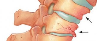
During the development of the disease, a dystrophic-necrotic process occurs, which is conventionally divided into five stages. At the first stage, as a result of circulatory disturbances in the area of the epiphysis or apophysis, necrosis occurs. At the second stage, at the slightest load, a secondary impression fracture occurs. At the third stage, fragmentation is noted due to the resorption of some areas of the cancellous bone that has undergone necrosis. At the fourth stage, repair occurs due to the fact that the connective tissue grows. The fifth stage - consolidation - develops as a consequence of ossification. In this case, the epiphysis is deformed or completely restored subject to timely and correct therapy.
Stages of osteochondropathy
Forms of the disease
Doctors who diagnose Keller's disease define two types of disease, depending on which bones are affected.
If a person is diagnosed with Keller's disease 1 , then we are talking about damage to the navicular bone of the foot. This bone is located at the inner edge of the foot. Keller's disease 2 is manifested by pathological changes in the heads of the second and third metatarsal bones of the foot. These bones are connected by articular surfaces to the phalanges of the fingers. Stage 2 Keller's disease most often develops in adolescents.
In addition, when determining the symptoms that manifest osteochondropathy, doctors divide the course of the disease into several stages. At the stage of necrosis, the patient’s bone beams, which are the structural elements of the bones, die. Such changes are clearly expressed, they can be seen even in the photo. At the stage of a compression fracture , new elements of bone tissue are formed, but now they do not yet have sufficient strength. During this period, bone beams often cannot withstand heavy loads. As a result, their fractures occur, and the beams can wedge into each other. This is followed by the fragmentation stage , in which osteoclasts (those cells that destroy bone) promote the resorption of bone beams. The final stage of the disease is the process of restoring the shape and structure of the bone. The answer to the question of how to treat a disease directly depends on what stage of the disease is currently occurring. Therefore, treatment of Keller's disease can only begin after a thorough professional diagnosis.
Diagnostics
Diagnostic measures for suspected bone osteochondropathy begin with an analysis of complaints and collection of anamnesis.
Clinical examination reveals characteristic signs of the disease. Usually they are not accompanied by deterioration in general condition, laboratory parameters and obvious inflammation (hyperemia, hyperthermia). Subsequently, the patient is prescribed:
- Ultrasonography. With its help, the condition of the bone surface is assessed.
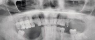
- X-ray.
- Computed tomography. It is effective from the 2nd stage of osteochondropathy. The use of multispiral tomography with 3D modeling allows one to obtain a three-dimensional image of the affected segment. The method is the only possible alternative to MRI when locating metal structures in the patient’s body.
- Magnetic resonance imaging. It is used to evaluate the hard and soft structures of the affected area and the efficiency of the blood supply to the bones.
If osteochondropathy of the joint is suspected, arthroscopy is recommended - a minimally invasive video-endoscopic manipulation to assess the surface of the joint, cartilaginous covering, and identify areas of dead tissue. The technique is used not only for diagnosis, but also for treatment.
Symptoms
Keller's disease I is most often diagnosed in preschool boys aged 3 to 7 years. Initially, the child notices pain and signs of swelling on the back of the inner edge of the foot. Due to the pain, there is a constant limp in the child, who tries to lean on the opposite, outer part of the foot while walking. Typically, the disease affects only one foot. It lasts about a year, after which the pain gradually stops.
Keller II disease is most often diagnosed in teenage girls. It was first described by traumatologist Freiberg from the USA, and Keller studied this type of disease in more detail and gave its description. This form of the disease is often bilateral. As a rule, the onset of the disease goes unnoticed. Initially, pain develops in the area of the head of the II or III metatarsal bones. They immediately appear in cases where a load is placed on the forefoot, and later the patient feels pain even at rest. The appearance of lameness is gradually noted, the patient cannot walk in shoes with thin soles, it is very difficult for him to walk barefoot, especially if the surface is uneven. Where the pathological process develops - on the dorsum of the foot - swelling appears. The finger that is located next to the head of the affected bone becomes shorter, and movements in the joint are limited. All these symptoms are present in the patient for about two years, after which the pain gradually begins to subside. However, if changes have occurred in the joint during this period, the pain may soon reappear. Sometimes familial cases of foot involvement are diagnosed. Most often, the disease is localized at the head of the second or third metatarsal bones, but damage to several bones rarely occurs.
Treatment methods
For osteochondropathy, treatment is complex, along with eliminating the root cause.
Mode
Treatment measures begin with compliance with the orthopedic regimen to eliminate stress on the affected area. Depending on the location of the pathology, it is possible to use bandages, knee pads, plaster splints, rigid reclining corsets, etc. For osteochondropathy of the calcaneal tuber or femur, it is recommended to use crutches or a cane.
Diet
With osteochondropathy of bones, the patient’s diet should be dominated by foods saturated with Omega-3 fatty acids, vitamins A, E, C, zinc, magnesium, selenium, and copper.
Recommended use:
- seafood (especially salmon, mackerel, etc.);
- nuts;
- poultry meat;
- chicken eggs;
- legumes, soy products.
It is advisable to avoid canned meat, fatty dairy products, animal fats, and smoked meats.
Drug therapy
When treating osteochondropathy, it is advisable to prescribe the following medications:
- analgesics to eliminate pain;
- chondroprotectors - they prevent the development of degenerative changes in bone tissue, restore its structure, and reduce pain.
- osteoprotectors – vitamin and mineral complexes for restoring bone and cartilage tissue and maintaining their health;
- disaggregants to prevent thrombosis.
Artracam, a drug with chondo- and osteoprotective effects, has proven to be highly effective. It not only relieves pain and inflammation, but also restores cartilage and joint tissue, preventing their destruction. The convenient form and dosage make the treatment comfortable, effective, and with prolonged results. Remember that all drugs must be prescribed by a doctor - even Artracam has contraindications. Self-medication is unacceptable.
The effectiveness of exercise therapy and massage
The main goal of physical therapy is to strengthen joints, bone and muscle tissue, and restore physical activity. Exercise therapy is prescribed at any stage of the disease and includes two types of exercises:
- active – performed by the patient independently;
- passive - performed by a specialist.
A set of exercises is developed by the doctor depending on the stage of the disease and the patient’s condition.
The effect of the massage is aimed at relieving pain, contractures, improving the nutrition of bone tissue, and correcting blood circulation.
Physiotherapy
Physiotherapeutic effects allow:
- improve blood circulation;
- reduce pain;
- increase regeneration of the affected area;
- reduce the severity of dystrophic changes;
- restore joint function.
In the treatment of osteochondropathy, it is advisable to prescribe: electro-, phonophoresis, heliotherapy, thalassotrapy, air baths, ultrasound, peloid therapy, radon and sodium chloride baths, etc.
Prevention
To ensure the right approach to prevention and prevent the development of both forms of Keller's disease, parents must, first of all, ensure that children always wear only comfortable and appropriately sized shoes. It is also important to reduce the mechanical load on the feet. To ensure that this condition is met, serious physical activity should not be allowed in preschool children. If you receive any foot injury, you must consult a doctor and undergo the tests prescribed by him. It is also important to pay attention to the presence of foot deformities and be sure to consult a specialist about this.
Diet
Diet for osteoarthritis of the knee joint
- Efficacy: no data
- Timing: constantly
- Cost of products: 1700-1800 rubles. in Week
Proper nutrition is very important for the full development of a child and adolescent, as well as for protection against the likelihood of developing osteochondropathy. During the period of growth, the body needs to provide a sufficient amount of vitamins, microelements, and minerals. Particularly important components for the prevention of osteochondropathy are vitamins A, D, C, group B, as well as collagen, magnesium, phosphorus, and calcium. Therefore, the child’s menu must include the following products:
- Food of animal origin: lean meat, offal, dairy products, fish, seafood.
- Plant products: any greens, cucumbers, tomatoes, cabbage of all types, zucchini, radishes, carrots, beets, celery, pumpkin, berries and fruits.
- Porridge.
- Legumes.
- Nuts, seeds, dried fruits.
- Vegetable oils, olives.
- Bread with bran.
- Sweets – jelly, jelly, unsweetened cookies, sponge cake.
Preference should be given to consuming fresh vegetables and fruits; steamed, boiled and baked dishes.
The following foods should be excluded from the menu:
- Smoked meats, dried fish.
- Fatty broths.
- Fried foods.
- Baking with margarine, pastry cream.
- Spicy and salty foods.
- Caffeine.
- Mayonnaise.
List of sources
- Traumatology and orthopedics / Guide for doctors: In 3 volumes T.Z / Ed. SOUTH. Shaposhnikova. -M.: Medicine, 1997.
- Movshovich I.A. Operative orthopedics. M.: Medicine. 1994;
- Sorokin, S.A. Diagnosis of the second Köhler disease / S.A. Sorokin // Restorative treatment of children with diseases and injuries of the musculoskeletal system: collection. scientific tr. / ed. B.L. Andrianova. - St. Petersburg, 1991.
- Traumatology and orthopedics. Multi-volume guide for doctors. Ed. N.V. Kornilov. SPb.: Hippocrates. 2006; III;
- Varshavsky G.I. On the differential diagnosis of Keller II disease / G.I. Varshavsky, I.M. Varshavsky // Annals of Traumatology and Orthopedics. 1994. -№2;
Summary
Although Keller's disease lasts for many months and sometimes with minimal symptoms, treatment should begin when its first signs appear.
This will help avoid serious complications such as joint contracture, the development of osteoarthritis, worsening flat feet, and the formation of irreversible foot deformity due to a fracture of the scaphoid bone or its destruction.
Buying comfortable shoes of the right size, timely treatment of flat feet and pathologies of the endocrine system, and avoiding injuries will help prevent the occurrence of Keller's disease.
Attention!
The information on the site does not constitute a medical diagnosis or a guide to action and
is intended for informational purposes only.