A ruptured eardrum is a hole in the thin tissue that separates the ear canal from the middle ear.
Author:
- Borzenkova Inna Andreevna
otolaryngologist
3.79 (Votes: 19)
- Diagnostics
Section: symptoms and causes
Review
A tympanic membrane rupture (or tympanic membrane perforation) is a hole or tear in the thin tissue that separates the ear canal from the middle ear.
A rupture can lead to hearing loss and also make the middle ear vulnerable to infection or injury.
Perforation of the tympanic membrane usually heals within a few weeks without treatment. Sometimes, however, a ruptured eardrum requires surgery to close. The middle and inner ear consist of delicate mechanisms that are sensitive to injury and disease. Prompt and correct treatment is important to preserve hearing.
Operation process
Myringotomy.
Photo: Dr Paulose FRCS - ENT SURGEON / YouTube The puncture of the eardrum is carried out in several stages:
- removal of wax from the ear canal;
- rinsing the ear with an antiseptic solution;
- administration of anesthetic;
- a linear incision of the eardrum with a sterile paracentesis needle;
- antiseptic treatment;
- installing a thick gauze swab (turunda) with medicine.
Causes
Causes of rupture or perforation:
- Inflammation of the middle ear (otitis media). A middle ear infection often causes fluid to accumulate in the middle ear. The pressure of this fluid can cause perforation.
- Barotrauma. Barotrauma is stress on the eardrum when the air pressure in the middle ear and the air pressure outside are out of balance. If the pressure is severe, the tympanic membrane may rupture. Barotrauma is most often caused by changes in air pressure associated with air travel. Other events that can cause sudden changes in pressure—and possibly rupture of the membrane—include scuba diving and a direct blow to the ear, such as a car airbag.
- Loud noises or explosions (acoustic trauma). A loud sound, such as an explosion or gunshot, or a blast—essentially an overwhelming sound wave—can cause the membrane to rupture.
- Foreign objects. Small objects such as a cotton swab or hairpin can puncture or rupture the tympanic membrane.
- Severe head injury. Severe trauma, such as a skull fracture, can dislocate or damage the structures of the middle and inner ear, including the eardrum.
4. Postoperative rehabilitation
Postoperative rehabilitation includes a number of recommendations that must be strictly followed:
- You can’t blow your nose sharply;
- Intensive breathing through the nose and any other actions that increase pressure in the middle ear are prohibited,
- Swimming is not recommended. You should try to avoid getting liquid into the ear;
- You should also avoid traveling by air;
- You should try to avoid any infectious diseases of the ENT organs.
Treatment effectiveness
(both therapeutic and surgical) largely depends on the degree of violation of the integrity of the eardrum, the location of the rupture and the patient’s compliance with recommendations during treatment and in the postoperative period. When transplanting a skin area, the blood supply to the restored area is important for the engraftment of the graft. Therefore, research in this area is always carried out before surgery. Transplant rejection, as a rule, occurs due to weak blood microcirculation, which slows down or completely eliminates the possibility of fusion of the patch with the eardrum. In general, surgical treatment of significant ruptures of the eardrum gives a good effect: restoration of hearing and elimination of the risk of infection of the inner parts of the ear.
Complications
The eardrum has two main functions:
- Auditory. When sound waves strike, the eardrum vibrates—the first step by which structures in the middle and inner ear translate sound waves into nerve impulses.
- Protective. The eardrum also has a barrier function, protecting the middle ear from water, bacteria, and other foreign substances.
Complications may occur as the membrane heals or if it fails to heal.
Possible complications:
- Hearing loss. Typically, hearing loss is temporary, lasting only until the tear or hole heals. The size and location of the tear can affect the degree of hearing loss.
- Inflammation of the middle ear (otitis media). A perforated eardrum allows bacteria to enter the ear. If a perforated eardrum does not heal or repair itself, you may be vulnerable to ongoing (chronic) infections that can cause permanent hearing loss.
- Middle ear formations (cholesteatoma). Cholesteatoma is a growth in the middle ear consisting of skin cells and other substances. All accumulated impurities and dead skin cells in the ear canals usually come out with earwax. If the membrane is ruptured, skin cells can pass into the middle ear and form a cholesteatoma. Cholesteatoma provides a favorable environment for bacteria and contains proteins that can damage the ossicles of the middle ear.
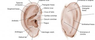
Rupture and perforation of the eardrum
Tympanoplasty
The eardrum is a thin, funnel-shaped skin that separates the ear canal from the middle ear. The role of the eardrum is to transmit air vibration - sound - to the hammer. Its vibrations are transmitted to this auditory ossicle, and further through the system of auditory ossicles - the incus and stapes - to the inner ear.
If the eardrum is ruptured or has a hole in it, its vibrations may be disrupted, which in turn leads to hearing impairment. A perforated eardrum is said to occur when there is a hole or tear in the eardrum. In addition, the presence of a hole in this membrane allows infection to enter the middle ear cavity, which is fraught with its inflammation - otitis media. The reasons that lead to perforation (or injury to the eardrum) are various. These can be inflammatory processes in the ear, as well as ear injuries, including noise trauma.
Causes of eardrum perforation
Inflammatory process in the middle ear. When there is inflammation in the middle ear— otitis media —discharge accumulates. This discharge may also be purulent. Due to the rather small volume of the middle ear cavity and due to the disruption of the outflow of this discharge through the Eustachian tube (since it is also blocked in this disease), the fluid that accumulates in the middle ear cavity puts pressure on the eardrum. In addition, the membrane is subject to purulent melting. As a result, they become thinner and break. This is manifested by the separation of pus from the ear. In this case, the membrane no longer serves as a barrier between the external environment and the middle ear.
Barotrauma, or acoustic trauma (Latin baro - pressure). As stated above, when fluid accumulates on the inside of the eardrum, it can rupture. However, pressure from its outside can also lead to rupture. This happens, for example, when an open palm is suddenly applied to the ear; sometimes a rupture of the membrane can also occur in flight during the ascent or descent of the plane, when a change in pressure occurs. It is not for nothing that they advise opening your mouth or sucking candy to equalize the pressure on the eardrum, since in this case air enters the middle ear through the Eustachian (auditory) tubes with each sip.
Noise trauma . A sudden loud noise (such as an explosion) can also rupture or perforate the eardrum. In addition to a sharp decrease in hearing, severe noise in the ears (tinnitus) may occur. Over time, the tinnitus goes away and hearing is partially restored.
Foreign bodies . Sometimes when cleaning the ear canal, for example, with a cotton swab or other objects, the eardrum can be injured. In addition, this promotes infection in the middle ear.
Symptoms of rupture and perforation of the eardrum
A ruptured eardrum, especially at the very beginning, can be quite painful. Manifestations of perforation of the eardrum include:
- Sharp acute pain in the ear.
- Clear or purulent discharge from the ear due to perforation of the membrane due to otitis media.
- Bloody discharge from the ear due to the traumatic nature of the perforation - a foreign body, direct trauma or noise trauma.
- Sudden decrease in pain when fluid breaks through the eardrum in otitis media.
- Hearing loss.
- Noise in ears.
Risk factors for eardrum perforation
Risk factors that can lead to perforation or rupture of the eardrum include: accumulation of fluid in the middle ear; independent cleaning of the ear from wax with hard objects (sticks, cotton wool, etc.); Excessive scratching in the ear due to itching in the ears.
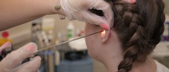
Diagnosis of rupture or perforation of the eardrum
Otoscopy is performed to diagnose membrane perforation. To do this, the doctor inserts a metal or plastic funnel into the ear. Next, the patient's auricle is pulled upward and backward. This technique allows you to even out the course of the ear canal, as a result, the eardrum becomes visible at its end. Light is directed into the ear canal. Perforation marks a hole in the eardrum. If it ruptures, the auditory ossicles of the middle ear may even be visible. In addition, depending on the cause of the rupture or perforation, there may be blood or pus in the lumen of the ear canal. If pus is present, the doctor uses a loop to remove a small amount of it to analyze and identify the pathogen and determine the effectiveness of antibiotics.
Complications of eardrum perforation
Typically, a ruptured or perforated eardrum does not pose a serious threat to the patient's health and usually heals on its own within a few weeks. But there can still be complications.
Hearing loss . Usually this complication is temporary, and it occurs as the membrane rupture heals. Naturally, the larger the gap, the longer it takes to heal and the longer the hearing loss lasts. In addition, the location of the rupture or perforation also influences the degree of hearing loss. With severe traumatic brain injury, which is accompanied by damage to the structures of the middle or inner ear, hearing loss can be severe and permanent.
Recurrent middle ear infection (chronic otitis media ). Extensive perforation of the membrane or its rupture may be accompanied by recurrent infection of the middle ear cavity, resulting in the development of chronic inflammation. This may contribute to permanent hearing loss.
Treatment of eardrum perforation
In most cases, membrane perforation heals on its own without complications within a few weeks. If the membrane does not heal, treatment is necessary.
Eardrum patch
If there is a small tear or perforation, the doctor can close it with a so-called paper patch. Before this, the edges of the tear are treated with a drug to stimulate growth, after which a paper patch is applied to the tear site. Three or four such procedures may be required to completely close the gap.
Surgery
In case of a larger rupture or perforation of the membrane and if the above method is ineffective, surgical intervention may be required. An operation to restore the integrity of the eardrum is called tympanoplasty or myringoplasty . The operation is performed under general anesthesia.
Surgical closure of membrane perforation leads to:
- Preventing water from entering the middle ear while showering, bathing or swimming, and therefore preventing the development of infection in it.
- Improving hearing.
- Eliminate tinnitus.
- Prevention of the occurrence of a special cyst in the ear - cholesteatoma, which leads to chronic infection in the middle ear.
Preventing eardrum perforation
Timely treatment of inflammatory diseases of the middle ear. If you have signs of inflammation of the middle ear: constant dull pain, tinnitus and hearing loss, you should consult a doctor and do not self-medicate. Delay in timely treatment can lead to fluid accumulation in the middle ear and perforation of the membrane. Protect your ears while flying. If you have a cold or an allergic reaction, it is advisable to refrain from flying . It is also recommended to wear ear protection or chew gum or suck on candy during the flight.
Try not to use sharp objects to remove wax from your ears, as they can easily damage your eardrum. Avoid excessive noise.
Prevention
Follow these tips to avoid tearing or perforation:
- Proper treatment of otitis media. Be aware of signs and symptoms of infection, including ear pain, fever, nasal congestion, and decreased hearing. Children with otitis media often have their ears touched or pulled. Contact your ENT doctor for an examination to prevent possible damage.
- Protect your ears during the flight. If possible, avoid flying if you have a pre-flight allergy that causes congestion. During takeoffs and landings, try to keep your ears open. This can be achieved by yawning or chewing gum. Or use the Valsalva maneuver - gently blow air through your nose, pinching your nostrils and closing your mouth. Stay awake during takeoff and landing.
- Do not clean with foreign objects. Do not try to deep clean earwax with cotton swabs, paper clips, or hairpins. These objects can easily rupture or puncture your eardrum. Teach your children about the consequences of putting foreign objects in their ears.
- Protection from excessive noise. Wear earplugs or headphones at work or while relaxing if there is loud noise.
Diagnostics
Photo: kuprevich/freepik.com
Acute otitis media is determined by a combination of signs:
- anamnesis data;
- otoscopy indicators - examination of the outer ear and eardrum using a special device;
- characteristic symptoms of the presence of fluid behind the eardrum: severe ear pain, hearing loss and intoxication: pallor, weakness, lack of appetite, headache and fever.
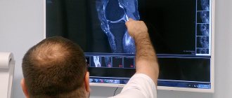
In some cases, the doctor prescribes additional diagnostic procedures: bacterial culture of the exudate and x-ray of the temporal bone.
Section: diagnosis and treatment
Diagnostics
An ENT doctor can determine the presence of a ruptured eardrum by visual examination using a lighting device (otoscope, microscope or endoscope). Your doctor may do additional tests to determine the cause of the tear or the extent of the damage. These tests include:
- Laboratory tests. If there is discharge from the ear, your doctor may take a swab to look for a bacterial infection.
- Study with tuning forks. A tuning fork is a metal instrument in the form of a two-pronged fork that produces a sound when struck. Tuning fork studies help the doctor detect hearing impairment, determine whether it is due to perforation of the tympanic membrane, damage to the sensory cells of the inner ear, nerves, or damage to both.
- Tympanometry. A tympanometer is a device inserted into the outer ear canal that measures the eardrum's response to small changes in air pressure. Some answer choices may indicate perforation or rupture.
- Audiological hearing test. If these tests aren't enough, your doctor may order an audiology test, done in a soundproof booth, that shows how well you hear sounds at different volumes and pitches.
Otoscopy for damage to the eardrum
In case of minor trauma, otoscopy only determines the injection of the vessels of the tympanic membrane. More significant damage is usually visualized in the form of subtotal defects, as well as round and pinpoint perforations, possible slit-like ruptures, up to the complete destruction of the eardrum itself.
A ruptured eardrum is characterized by uneven, scalloped edges. When performing otoscopy in the membrane of the perforation, you can observe the medial wall in the tympanic cavity. Often, otoscopy helps diagnose a hematoma located in the tympanic cavity due to damage to the membrane. If there is acoustic or mechanical damage, the doctor may observe hemorrhages in the tympanic cavity. After a certain time, it is necessary to conduct a control otoscopy, which will be aimed at assessing the reparative processes that occur in the eardrum. It is the control otoscopy that is able to determine scarring or the nature of the perforation hole. Sometimes in the very thickness of the eardrum you can see a dense white formation, which is a deposition of calcium salts on the scar.
Treatment
Most perforations heal without treatment within a few weeks. Your doctor may prescribe antibacterial drops if there are signs of infection. If the tear or hole in the membrane does not heal on its own, treatment will include procedures to close the perforation. These may include:
- Closure of the eardrum. If the tear or hole does not close on its own, the ENT doctor can close it with a special flap. This is an outpatient procedure in which the doctor freshens the edges of the wound to encourage growth and then places a flap over the hole. It may be necessary to repeat the procedure several times before the hole closes.
- Surgery. If the flap does not heal, or the doctor determines that the gap is unlikely to be closed with it, surgery is recommended. The most common surgical procedure is called tympanoplasty. The surgeon takes a tiny piece of your own tissue to close the hole in your eardrum. This procedure is performed on an outpatient basis, meaning you can go home the same day unless the anesthesia required requires a longer hospital stay.
How is the operation performed?
The procedure for performing the operation is determined by the ENT doctor based on a comprehensive examination of the patient, a confirmed diagnosis and the presence of indications for intervention.
To get the best view of the middle ear cavity, a microscope is used. A small incision is then made in the eardrum and the fluid in the middle ear is removed. In most cases, a small tube may be needed to remain in the incision site to allow drainage to continue.
No stitches are required to close the incision - it will heal on its own.
Lifestyle and recommendations
Perforation of the tympanic membrane usually heals on its own within a few weeks. In some cases, treatment requires several months. Until your doctor tells you that the tear has healed, take care of it by doing the following:
- line the perforated ear with a cotton ball and Vaseline when bathing;
- refrain from cleaning;
- Avoid blowing through the nose, as the pressure created when blowing through the nose can damage the healing eardrum.
What you can do before visiting your doctor
Make a list before your doctor's visit. Your list should include:
- the symptoms you are experiencing, including any symptoms, even those that may not seem related to your hearing loss;
- events that could cause ear problems, such as recent injuries or flights, inflammation of the middle ear;
- medications, including any vitamins or supplements you take;
- questions for your doctor.
If you think you have signs or symptoms of a perforated eardrum, you can ask your doctor the following questions:
- Is there a ruptured eardrum?
- What else can cause hearing loss and other symptoms?
- If I have a ruptured eardrum, what should I do to protect my ear while it heals?
- When should I come for a follow-up examination?
- At what point should we consider other treatments?
Ask any other questions you have.
Forecast
In terms of treatment and recovery, traumatic perforations that are localized in the posterior quadrants have a more favorable prognosis. A favorable prognosis is also observed when myringoplasty is performed at the earliest possible date. Damage to the eardrum at the edge is more dangerous than in the central parts. This is due to the fact that there is a possibility of ingrowth of the epidermis into the tympanic cavity with the formation of cholesteatoma . The ingrown epidermis subsequently causes purulent inflammation, destruction of ear structures and severe hearing loss.
The prognosis of myringoplasty in young children may be unfavorable, since recurrence of perforation occurs quickly. This is due to the fact that the plastic membrane is made from the tissues of a small child, which are thin and delicate, so the membrane is fragile. Older children have better results. One of the conditions for a successful result is normal function of the Eustachian tube and good ventilation of the tympanic cavity. Meeting these conditions in young children is problematic due to frequent respiratory diseases and the presence of enlarged adenoids. In this connection, they often resort first to adenotomy, restoring the function of the eustachian tube, and then perform myringoplasty or, if necessary, tympanoplasty.