Compression fractures of the spine are quite common, especially in older people. A compression fracture occurs when there is significant pressure on the spine and simultaneous flexion. The anterior parts of the vertebrae and discs are subject to the greatest damage during compression. The anterior part of the vertebral body is flattened and takes on a wedge-shaped shape. In rare cases, compensatory extrusion of the posterior part of the vertebra into the spinal canal is possible with compression of the spinal cord. Such fractures are very severe and quite rare. Compression fractures of the spine are most often located in the area of the 11th and 12th thoracic vertebrae, as well as the first lumbar vertebra. Localization to other areas of the spine is possible but less common. Treatment for vertebral compression fractures depends on the origin and severity.
Types of compression fractures
Compression fractures according to severity are divided into: complicated and uncomplicated
Fractures without complications, that is, without the threat of damage to the spinal cord, are also divided into subtypes:
- The height of the anterior part of the vertebral body is reduced by less than 50%;
- Compression reduces the height of the vertebral body by half;
- The height of the vertebra is significantly reduced - more than half.
Complicated compression fractures pose health risks in both the short and long term. With a complicated compression fracture, not only the vertebrae are damaged, but also the structures inside the spinal canal. According to statistics, such fractures account for only 5-6% of all diagnosed injuries to the musculoskeletal system. Most often, the cervical vertebrae are susceptible to severe fractures, as they are the most vulnerable and fragile, but such fractures can also occur in the thoracic and lumbar spine. Severe, mechanical trauma to the cervical vertebrae C1 and C2 is considered fatal. The vertebrae of the thoracic spine with a complex form of compression fracture are subject to dislocations and cracks, as a result of which the expanded or broken edges of the vertebra put pressure on the soft tissues in the middle part of the back, and then on the spinal cord. Because the spinal canal in the thoracic spine has very little unfilled space, the deformed parts of the vertebrae damage the spinal cord and cause its dislocation. In the thoracic spine, the vertebrae most susceptible to compression fractures are Th11 and Th12. In the lumbar region, where the axial load is greatest, the L vertebrae (1 and 2) are damaged.
Thoracic region
The main tasks of rehabilitation for compression fractures of the thoracic spine are solved through exercise therapy. By performing certain exercises, physiologically normal mobility is ensured not only of all central segments, but also of the entire back, muscle tone is restored, pain in the scapulothoracic zone is eliminated, and posture is normalized. Here are examples of some useful exercises. The average repetition frequency is 10 times.
- Take a standing position. Place your feet hip-width apart, hands on your waist. As you inhale, bend back, slightly raising your shoulders and pushing them back; as you exhale, round your back, pointing your shoulders forward.
- Now we complicate the task a little by doing the same thing, but with the body turning to the right/left: we turn to one side and spread the shoulder girdle, return to the starting position and round the back, turning the shoulders forward.
- Let's move on to a new task. The position is the same, only the arms are extended along the body. Perform lateral bends: bend to one side, the arm of the bending part slides down the side of the limb, while simultaneously pulling the opposite arm towards the armpit. Similarly, we tilt the body in the other direction. Each side – 10 repetitions.
- Get on all fours with your straightened upper limbs resting on your palms. As you inhale, we bend your back, your pelvis moves back, your head drops down. As you exhale, arch your back upward (like a cat), smoothly throwing back your head, while tightening your stomach.
- We partially change the previous pose - the support in the hands now falls on the forearm area. We lift our right hand off the floor, take hold of the shoulder, slightly turn our chest to the right and make 5 springy movements with this glenohumeral part with the emphasis “up”. We work in a similar way with the left side.
- Lie on the floor with your stomach down, resting on your forearms, chest open. When the thoracic fracture has healed, the “sphinx” pose has a beneficial effect on this line of the spine; ask your orthopedist whether your rehabilitation can include this type of warm-up. Method of execution: pull your head up towards the ceiling (feel your ribs straightening), stay in this position for a few seconds, then relax, lowering your head to the floor.
- Lie on your side (head on the floor), bend your knees (knees together), stretch both arms forward in front of you, clasping your palms. Next, with a sliding movement of the upper hand, as if opening, we pass it along the inner surface of the adjacent arm, then along the chest, and finally place it on the floor. At the moment of the so-called “opening”, turn your head in the direction of movement of the limb, as a result the shoulder blades lie on the plane. The pelvis and legs should remain in position at all times. p. Then “get together” again and repeat the task. After five times, turn to the other side and repeat the exercise by analogy.
- Lumbar part
Rehabilitation for compression fractures of the lumbar spine has similar goals. This is to restore the full functionality of the musculoskeletal system, make it resilient and strong, and balance your posture. What kind of exercise do physical therapy instructors and orthopedic doctors suggest their clients do?
- Lying on your back, perform simultaneous flexion of the knee and hip joints. Thus, the lumbosacral region and abdominal muscles are excellently trained.
- We do not change the IP. Raise your legs straight up. Extending the limbs to the sides followed by bringing them together. When your legs come together, you need to cross them slightly.
- The well-known exercise called “bicycle” brings considerable benefits. We won’t dwell on the technique; we think everyone knows how to simulate riding a bicycle in a recumbent position.
- Roll over onto your stomach, spread your arms to the sides. Lift your chest off the surface along with your arms, hold in this position for as long as your physical fitness allows, then lower and relax. Note that the feet do not come off the floor. If it is more convenient for someone, you can stretch your arms in front of you.
- The position remains the same, but your legs need to be spread slightly and your arms should be extended straight in front of you. Lifting the sternal complex (along with the arms) and legs from the support, lift them simultaneously with a bend in the back. Fix the pose and stay in it for as long as possible. Lower yourself, rest a bit, then repeat.
- Get on your knees. The pelvis and back are strictly vertical, hands on the belt. Walk on your knees in a straight line forward, then backward. We complicate the task: we begin to walk in a circle, first clockwise, and then in the opposite direction.
Causes of spinal compression fracture
- Vertebral compression fractures occur due to the impact of a force vector on the spinal axis, for example, during an episode of trauma (a fall from a height onto the legs or buttocks).
- Impairments in the strength of bone tissue, for example in osteoporosis, when the risk of a compression fracture increases sharply and can occur even with small force loads. These fractures are more common in older people or people with pre-existing problems in the spine that have contributed to weakened bone strength.
- A bone infection such as osteomyelitis or tuberculosis (Pott's disease) may also predispose to vertebral fracture.
- When the bone structure weakens, the vertebra becomes compressed under the weight of the vertebra above, thinning the bone and leading to a fracture. The reason for this may be various degenerative changes in the spine
- Compression fractures of the spine can also occur as a result of automobile accidents.
- Malignancies that lead to vertebral fractures are most often metastases rather than primary bone tumors. Primary foci of cancer, which often give hematogenous metastases to the spine, include prostate, kidney, breast and lung cancer. Melanoma is the less common but most aggressive cause of spinal metastasis. The most common primary cancer of the spine is multiple myeloma and various sarcomas, which can also present with compression fractures.
- Benign tumors such as aneurysmal bone cyst and hemangioma can also cause vertebral compression fractures.
- Diseases of the parathyroid gland can also lead to a decrease in bone strength by increasing the release of parathyroid hormone, which most often occurs with adenoma of the gland.
Possible complications
If you receive a back injury or just sudden pain, you should immediately consult a doctor, especially older people and those who are forced to regularly lift heavy objects as part of their job. Only timely, well-chosen treatment for a compression fracture can minimize the risks of complications and subsequent disability.
Since the spinal cord passes between the vertebral bodies and their arches, damage to it is the most dangerous complication of a compression fracture. It is he who is responsible for a person’s ability to fully move.
With compression fractures, especially when combined with the separation of fragments, it is possible that the nerve roots of the spinal cord, the blood vessels that feed it, and also the spinal cord itself may be pinched. In more severe cases, contusion or even rupture of the spinal cord is possible, leading to irreversible paralysis.
If treatment is not started in time, injury can provoke secondary degenerative and neurological disorders:
- segmental vertebral instability;
- curvature of the spine or the formation of a hump (kyphotic deformity), which is especially typical for older people;
- persistent movement disorders;
- protrusion and herniation of intervertebral discs;
- scoliosis, radiculitis;
- formation of bone structure calluses;
- disruption of the functioning of internal organs, including the genitourinary system, which can provoke urinary and fecal incontinence, and in men, additionally, erectile dysfunction;
- osteochondrosis and the formation of intervertebral hernias.
They tend to progress gradually and also lead to paralysis. Therefore, it is better not to hesitate and immediately contact a traumatologist or vertebrologist.
Symptoms
The main symptoms that accompany compression fractures of the spine:
- Pain at the site of the fracture, usually in the lower back.
- Patients with complex compression fractures usually assume a forced posture with the torso tilted forward.
- Possible pain in the lower extremities
- Numbness, tingling and weakness in the extremities usually indicate the presence of root or spinal cord compression
- Urinary or urinary incontinence or inability to urinate voluntarily is a sign that the fracture is accompanied by compression of the spinal cord.
- Compression fractures of the vertebrae, associated with weakening of the strength of bone tissue, may manifest with slight pain or be completely asymptomatic. Sometimes the pain is localized in the area of the fracture. Changes in vertebral height place excess stress on the spinal muscles, leading to fatigue and muscle pain. As a rule, such pain disappears after a few weeks.
- Traumatic compression fractures can cause severe back pain that radiates down to the legs. If a fracture severely damages the vertebral body, bone fragments can protrude into the spinal canal and cause compression of the spinal cord. In such cases, corresponding neurological symptoms (paresis or paralysis, sensory disturbances, dysfunction of the pelvic organs) will appear below the site of spinal cord compression. Traumatic fractures often lead to the development of kyphosis.
Anatomy of the spine
The spine is one of the main organs of the human body, responsible for maintaining it in an upright position. It is formed by 33 vertebrae, between which are intervertebral discs. They are responsible for cushioning bone fragments under static and dynamic loads.
From the back of each vertebra there are arches shaped like a semicircle and forming the spinal canal. It is in it that one of the most important organs of the human body is located - the spinal cord.
The arches have 7 processes:
- 4 articular;
- 2 transverse;
- spinous.
The articular processes of adjacent vertebrae are connected in pairs to each other, thereby forming intervertebral joints. They are entrusted with the function of protection under heavy loads. Thus, openings are formed between 2 adjacent vertebrae, called foraminal. The nerve roots extending from the spinal cord pass through them. Additionally, the bodies, processes and arches are connected by ligaments that provide stability and strength to the entire spine.
Since the spinal cord runs a few millimeters from the spinal column, and its joints are densely penetrated by nerve fibers, spinal fractures are always dangerous. They require not only lengthy and sometimes complex surgical treatment, but also long-term rehabilitation. Moreover, sometimes, despite all the efforts of doctors, such injuries still lead to disability and death.
MRI
MRI is necessary in cases where there are motor or sensory disturbances in the lower extremities. Radicular pain or suspected spinal cord compression is also an indication for MRI.
Magnetic resonance imaging is important because it provides the best visualization of the neural structures of the spine. In addition, MRI can be performed using contrast, which allows better diagnosis of conditions such as hemorrhage, tumor or infection with greater sensitivity.
Signs of illness and first aid
As already noted, in most cases only a doctor can determine the presence of a spinal fracture. The latter prescribes the patient an X-ray of the spine, computed tomography (sometimes X-ray is sufficient, but if not, CT can confirm or refute the diagnosis), MRI (this is needed if damage to the nerve endings of the spine is suspected). The doctor also conducts 2 more studies on the patient: a neurological examination and densitometry (if the development of osteoporosis is suspected).
As you know, every injury can be mitigated if qualified first aid is provided to the victim in a timely manner. When you receive a compression fracture of the spine, it is important not to hesitate to call an ambulance, and then to properly transport the victim. The transfer of the patient, as well as his transportation in transport, must be carried out with the utmost care, thanks to which it is quite possible to avoid the displacement of fragments.
Medicines
Most patients in the acute stage of fractures receive analgesics. With pain control, the patient can get up and move around easily and avoid problems caused by immobilization.
Corseting
Most patients are prescribed the use of a rigid corset, which is selected individually. A corset can dramatically reduce the range of motion and fix a broken vertebra. Patients who wear a special brace are allowed to move, but strenuous activities such as lifting and bending are significantly limited.
Motor activity control
. Bed rest is usually recommended for several days, since prolonged immobilization leads to weakening of the muscular corset, and in the presence of osteoporosis, to further bone loss and progression of osteoporosis.
Correction of osteoporosis
. Bone-strengthening drugs such as bisphosphonates (Actonel, Boniva, and Fosamax) help stabilize or reverse bone loss. This is a critical part of the treatment and helps prevent further fractures when axial loads are placed on the vertebrae.
Classification of fractures in the lower back
Taking into account the vector of force, several types of lumbar vertebral fractures are distinguished: compression and isolated. Depending on the presence of nerve damage, uncomplicated and complicated fractures of the lumbar vertebrae are distinguished. A distinction is made between injuries caused by trauma and pathological fractures due to tuberculosis and osteoporosis.
Compression fracture
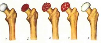
This type is characterized by flattening, compression or fragmentation of the anterior part of the vertebra. There are several degrees of compression:
- the first is a reduction of one third or less;
- second – reduction is up to half the size;
- the third is flattening by more than half.
With such compression, the discs between the vertebrae and the processes of the joints are not deformed. The main cause of compression is a person falling from a height.
Comminuted (comminuted) fracture
The upper vertebra fits like a wedge into the one below. This is accompanied by destruction of the intervertebral disc. The piece comes forward. If it goes backwards, it hits the spinal cord.
Exercise therapy
Physical activity is of great importance in the rehabilitation of a patient after a compression fracture of the spine.
Rehabilitation periods can be divided into 4 stages:
First stage
lasts during the first – second week after injury. The goal of this period is to increase the patient’s motor activity and restore muscles after immobilization.
Second phase
begins a month after the injury. The main goal of rehabilitation is to stimulate blood circulation in the damaged area and increase physical activity. It is possible to include light exercises to restore the muscle corset.
3rd stage
begins several months after the injury. The goal of this stage of rehabilitation is to restore the mobility of the motor segments of the spine. During this period, the intensity and duration of exercise therapy classes increases. Gymnastic exercises can be supplemented with weight-bearing exercises.
4th stage
lasts from the moment the patient resumes independent movements until complete actual recovery. Complexes of exercise therapy are aimed at completely restoring both spinal mobility and posture.
During this rehabilitation period, there is a smooth transition to full axial load of the injured spine.
Rehabilitation after traumatic vertebral fractures can be a fairly lengthy process. As a rule, complete recovery occurs within 6-12 months.
Physical rehabilitation
Rehabilitation for a spinal fracture is the next stage of the treatment process of paramount importance. After providing first aid, even if it involved minimally invasive cementoplasty, the patient must strictly observe a specific physical regimen for a certain period, attend physiotherapeutic sessions, undergo therapeutic exercises, etc. It cannot be otherwise if you have suffered such a serious injury.
Complicated injuries and those provoked by osteoporotic processes require a very careful selection of recovery methods. Therefore, it would be more advisable to take care in advance where to undergo rehabilitation after a spinal fracture; today there is no particular shortage of good specialized institutions.
Prescribing any restorative tactics to yourself is strictly prohibited! Rehabilitation of patients with a spinal fracture is being developed exclusively by the treating doctor together with a rehabilitation specialist. Our recommendations are given in general form and are for informational purposes only, so before using them, consult with a specialist about the possibility of using them for your specific medical problem.
We will talk about that important period when complete bone consolidation has already been achieved, since some patients at this stage stop working on their spine, but in vain. It is after 2-3 months that all emphasis should be directed toward developing and strengthening the musculoskeletal corset, which has become considerably weakened after prolonged immobilization and long-term unloading. In addition, thanks to the unique physical organization, the work of the gastrointestinal tract, cardiovascular system, respiratory system, reproductive, urinary system and many other important components of the body, which are entirely dependent on the health of the spine, will be stabilized.
Surgery
Surgical methods for treating spinal compression fractures are necessary for complicated fractures. They can be using open surgical techniques or using minimally invasive techniques. Open surgeries are performed for severe trauma when bone fragments damage the spinal cord and spinal roots.
Minimally invasive methods are used more widely, since during surgery the damage to muscles and bones is insignificant, which reduces the risks of complications and allows the damaged bone to recover faster. The main methods are:
- vertebroplasty
- kyphoplasty
Cervical area
After removing the plaster, rehabilitation after a fracture of the cervical spine involves restoring the mobility of the corresponding segment, strengthening the muscular-ligamentous apparatus of the neck and upper shoulder girdle. At the same time, coordination abilities are improved, and complete adaptation to social and living conditions occurs.
Surprisingly, all of the above methods are optimally suited after such a spinal-motor body injury as a fracture of the cervical spine; rehabilitation with the inclusion of these techniques in the exercise therapy complex, naturally, must be approved by a specialist. As for the fundamental exercises, they are outlined below.
- First you need to warm up the glenohumeral part, which will increase blood flow in the neck. To do this, take a vertical position, place your hands on your shoulders. Perform calm rotational movements in the shoulder joints - 10 times back, and then the same amount forward. When finished, lower your hands and shake them.
- I. p. is the same as in the first exercise. No. 1. Now spread your upper limbs in different directions, straightening them, and place them on your shoulders again. Make sure that at the moment when you spread your arms, they remain on the same horizontal plane with your shoulders, that is, do not lift them up or lower them. After 10-15 repetitions, lower the limbs that worked hard and shake them a little, and at the same time rest.
- Cross your hands with your fingers, place them on your forehead (with the inside of your palms facing the frontal area). Apply resistance to your head with your upper limbs, continuously pressing your forehead on your palms for 5 seconds. Simply put, imagine that you want to lower your head, but an obstacle prevents you from performing this action. Please note that the head does not drop anywhere, but remains in place, but you feel tension in the back of the neck. On the count of five, relax and remove your hands from your forehead. We do 5-7 approaches.
- The next method of warming up is based on the same principle, but we place the “lock” on the back of the head, pressing the head backwards. Make sure that the spine does not bend and the head does not fall back. Work with this type of exercise, observing the time and frequency of sets according to method No. 3.
- Both for stretching, balance, strength of the neck muscles, and for a beneficial effect on the respiratory center and heart function, shoulder abduction is used. The person stands with his arms along his body. In practice, it looks like this: we tense the shoulder muscles and turn our shoulders forward as if you want to close them, then we open our shoulders, straighten up and try to bring our shoulder blades towards each other.
- We save the original position. Now simultaneously move your right hand along the side of the body to the armpit and turn your head to the right, while the shoulder rises and touches the chin. Now we will carry out an identical manipulation with the reverse side.
Such fruitful rehabilitation after a fracture of the cervical spine, or rather its biomechanics, will lead to an amazing state. Of course, training should be implemented systematically, and not just during this difficult period, but preferably throughout life.
Vertebroplasty
This procedure is most useful for reducing pain. It also strengthens damaged bone, allowing patients to recover faster. Using a needle, under X-ray control, a cementing substance, polymethyl methacrylate (PMMA), is injected into the area of the vertebral fracture. The cement hardens within 15 minutes. This fixes the bone and prevents further destruction and reduces pain in more than 80% of patients.
First aid
It is necessary to prepare the victim for transportation to a medical facility. The help of three people is required to place the patient on a stretcher without allowing the vertebrae to become displaced. The fracture area is fixed with a corset or a rigid bandage to prevent movement during transportation.
Basic actions before the doctors arrive:
- prevent movement of the victim;
- provide peace;
- call an ambulance;
- stay with the patient until the doctors arrive.
Forecast
- Overall, the prognosis for simple compression fractures is good for most patients who recover fully or with minor back pain. Vertebroplasty and kyphoplasty have improved recovery options for patients with chronic back pain who have failed conservative therapy. These procedures have proven their effectiveness and are used quite widely.
- The prognosis for patients with traumatic compression fractures is largely based on neurological symptoms and is related to the degree of impact of the damaged vertebra on the root or spinal cord.
- Patients with compression fractures caused by spinal infections have a generally favorable prognosis. According to statistics, two thirds of patients with fractures associated with purulent osteomyelitis have a good chance of recovery. At the same time, with tuberculous genesis of compression fractures, in some patients, neurological symptoms do not disappear and the patients lose their ability to work.
- Most vertebral compression fractures caused by osteoporosis stabilize within eight weeks. With adequate drug correction and fall prevention, recurrent fractures can be avoided.
Consequences
The consequences of a compression fracture of the spine can be very serious. Their severity depends on the severity of the injury, the extent of damage to the spine, and the adequacy of the therapy performed. Complications after a spinal fracture can occur due to severe trauma, improper transportation of the victim, or the provision of unprofessional assistance to him. Therefore, if you suspect a spinal fracture, you must call an ambulance and not touch the victim (of course, if his situation is not life-threatening).
The consequences of a spinal fracture include:
- Instability of the vertebrae in the spinal column;
- Impaired conduction of nerve fibers as a result of compression of the nerve roots of the spine;
- Radiculitis;
- Rachiocampsis;
- Formation of kyphosis (hump);
- Constant back pain;
- Breathing disorders;
- Callus;
- Intervertebral hernia;
- Infection of the damaged area;
- Inflammation and suppuration;
- Paralysis of limbs.
Urological consequences of a spinal fracture include disruption of urination and the functioning of the urinary system. In some cases, patients experience erectile dysfunction.