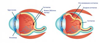
The eyeball consists of several membranes. Its innermost shell, lining the cavity of the eye from the inside, is called the retina, or retina. On the outside, the choroid, or choroid, is adjacent to it; on the inside, the retina borders on the vitreous body. The thinnest layer of retinal tissue contains light-sensitive cells (photoreceptors) that are responsible for image perception. The processes of these cells, folding into nerve fibers, carry information about the image to the corresponding parts of the cerebral cortex.
Retinal diseases can occur both due to direct damage to its structures and as a result of systemic diseases of the body. Changes in the retina are diagnosed in patients with diabetes mellitus, hypertension, pathologies of the kidneys and adrenal glands, after traumatic brain injuries, and with some infectious diseases. Major retinal pathologies include:
- Dystrophic and vascular lesions (angiopathy, retinopathy, maculopathy, hemorrhage)
- Retinal tears and detachments
- Inflammatory changes (retinitis)
- Retinal tumors
There are groups of patients whose risk of developing retinal diseases is increased. These include people with moderate and high myopia, elderly patients with diabetes and high blood pressure, and pregnant women.
At an early stage, many retinal diseases may not cause any discomfort. So if you or a loved one is at risk, don't skip your annual eye exam with a qualified ophthalmologist.
Retinal hemorrhages
Hemorrhages in the retinal area can occur as a result of injury to the eyeball, vascular pathologies or dystrophic diseases of the retina. Retinal hemorrhages are found in diabetes mellitus, thrombosis of the central retinal vein, defects of the cardiovascular system, blood diseases, and burns.
The blood in the cavity of the eyeball has a toxic effect on the internal structures of the eye, changes the structure of the vitreous body and reduces the transparency of the optical media of the eye, which is accompanied by a deterioration in the quality of vision. Severe complications of retinal hemorrhage are increased intraocular pressure and secondary glaucoma, retinal detachment, degenerative changes in the retina, etc.
To treat hemorrhages in the retina, conservative methods are used (injections of drugs that accelerate the resorption of hemorrhages), laser treatment (laser vitreolysis, laser coagulation), and surgical techniques (vitrectomy with replacement of the clouded vitreous).
Read more about the treatment of retinal hemorrhages >>>
Main “risk groups”
- People with moderate and high degrees of myopia;
- pregnant women;
- elderly people with diabetes.
The initial stages of the disease may not be accompanied by any symptoms , so if you are at risk, be sure to undergo vision diagnostics using modern equipment. This examination will reliably determine whether you need treatment. If you are scheduled for surgery, do not put off surgery for too long. Before surgery, you need to protect your eye from possible damage in every possible way.
If diseases of the retina of a dystrophic nature are detected, with its thinning and rupture in the periphery, it is strengthened using a laser. Otherwise, any sufficiently strong tension can lead to detachment, requiring immediate surgical intervention. It is better to prevent such a situation. Moreover, detachment can occur when urgent provision of qualified ophthalmological care is impossible (at the dacha, on a trip, etc.)
Retinal inflammation
Inflammation of the retina of the eyeball (retinitis) can occur as a unilateral or bilateral process. The causes of retinitis are varied: infectious diseases of a bacterial and viral nature (including tuberculosis, syphilis, AIDS, herpes), toxic eye damage due to other pathologies, allergic diseases, chronic heart and kidney diseases.
Manifestations of retinitis can be different and depend on in which parts of the retina the pathological process is localized. Common symptoms for any form of retinitis include changes in visual fields (scotoma) and a decrease in its acuity. Scotomas are focal loss of visual fields; their size and location directly depend on the location and size of the inflammatory focus. If retinitis is untimely or incompletely treated, persistent vision deterioration occurs, which is especially noticeable in poor lighting conditions.
Treatment of retinitis should be aimed at identifying and eliminating the cause that caused the inflammation of the retina. The most effective is an integrated approach, including drug treatment, injections of anti-inflammatory and antibacterial drugs, and laser coagulation of retinal vessels.
Read more about the treatment of retinal inflammation >>>
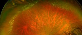
Retinal detachment (detachment)
It is a separation of the retinal layer and the underlying tissues of the choroid. In the event of a tear or rupture of the retina, intraocular fluid leaks between it and the choroid, leading to disruption of the normal nutrition of photoreceptor cells. If you suspect retinal detachment, you must immediately contact an ophthalmologist to provide qualified assistance. The ability to maintain the patient’s visual acuity will directly depend on how quickly and completely assistance is provided for retinal detachment. Delay in this case threatens irreversible loss of vision and blindness!
When retinal detachment is detected, surgical treatment methods are used: extrascleral filling, laser coagulation of tissue around the area of tissue detachment, vitrectomy (replacement of the vitreous body of the eye) to remove blood from the cavity of the eyeball and restore the transparency of the optical media of the eye.
Read more about the treatment of retinal detachment >>>
TsSHRD (TSSKHRP, TsSH)
Central serous chorioretinopathy (CSC) is one of the most common pathologies of the posterior segment of the eye. The cause of the disease is considered to be distress, a situation of “stress” that is extremely common in the modern world. Patients are young people, under 50 years old, mostly men. Complaints: the appearance of a translucent spot in front of the eye, a decrease in the size of objects, impaired color vision - everything seems a little yellowish-brown.
This disease of a non-inflammatory nature, in 50–70% of cases, resolves on its own, but requires dynamic observation and active intervention for a duration of more than 3–6 months.
The observation is based on regular optical coherence tomography of the macular zone in order to assess the dynamics of the height and extent of exudative retinal detachment in the macula.
To identify the point (or points) of filtration - sources of subretinal fluid, FA (fluorescein angiography) is used. The treatment method is laser coagulation of points of fluid leakage under the retina according to FA data. An outpatient painless procedure, after which fluid is reabsorbed under the retina.
Retinal reattachment after laser photocoagulation of leakage points.
Tumors of the retina of the eye
Retinal neoplasms can be benign or malignant, affect one or both eyes, and are detected in young (usually children) and adulthood. Gradually growing, the volumetric formation of the retina occupies most of the eye, and therefore the patient develops an asymmetric protrusion of the eyeball noticeable to the naked eye. Vision in the affected eye disappears, the eye loses mobility.
For timely detection and treatment of retinal diseases, contact the Retina Center. Our specialists will conduct a qualified eye examination using modern diagnostic equipment and will accurately determine whether you require eye treatment. If your doctor recommends surgery, do not delay the decision for long; this will help avoid progression of the disease and preserve your vision.
Treatment of retinal tumors is carried out in specialized medical institutions; delay in seeking qualified help can lead to irreversible consequences.
Causes
There are three reasons for the formation of retinal diseases:
- Infectious. If the provocateur is an infection, then inflammation develops, affecting the visual apparatus.
- Acquired or congenital. Associated with changes in the structure of photoreceptors. The shell cracks, breaks, or becomes thinner.
- Vascular. As a rule, pathology is a consequence of existing vascular diseases (for example, atherosclerosis, hypertension).
Most often, vision impairment due to retina diseases occurs in patients suffering from myopia, since their retina is stretched.
Retinal dystrophies
The cause of dystrophic changes in the retina is usually disorders in the vascular system of the eye. Most often, dystrophies are found in elderly patients, as well as with moderate and high myopia. The myopic eye is usually extended in the anteroposterior direction, and the resulting overextension of the posterior parts of the eyeball is accompanied by the appearance of dystrophic changes in the fundus. Retinal dystrophies can also occur with various vascular and inflammatory diseases of the eye.
The main method of treating retinal dystrophies is photocoagulation of altered tissues using an argon laser to strengthen thinned areas of the retina and delineate the area of detachment, if one has been identified.
Read more about the treatment of retinal dystrophies>>>
Symptoms
The developing pathology of the retina does not cause pain or other physical discomfort, since its constituent tissue is devoid of sensitive innervation. Most often, gradually developing retinal dystrophy is subjectively felt as a thickening veil before the eyes. Objects become blurry, and even in good lighting the image is blurry, like at dusk. If the pathology is localized in a certain area, then vision is impaired only in the focal area, which, without proper treatment, expands over time.
In some cases, sudden visual phenomena may be observed: lightning, flashes of light, glare, sparking. Black spots and flies appear in the visible area, the field of vision narrows or is lost in a certain area. Some patients note micropsia or macropsia, as well as a sharp deterioration of vision in twilight and dark times of the day.
Age-related macular degeneration (AMD)
The disease is most often detected in people over 50 years of age and is the main cause of irreversible vision loss. The risk of the disease increases with a genetic predisposition, as well as in patients with diabetes mellitus, hypertension, and atherosclerosis. Symptoms of the disease are associated with damage to the macula area of the retina.
The initial manifestations of AMD do not have any clinical manifestations and can only be detected during an examination by an ophthalmologist. As the disease progresses, symptoms such as distorted outlines of objects, blurred vision, and blurred vision appear.
The dry form of AMD does not require special treatment. To treat the wet form of age-related macular degeneration, intravitreal injections (injections into the vitreous cavity) of drugs that block the growth of defective vessels under the retina (anti-VEGF drugs) are used. In addition, photodynamic therapy and laser photocoagulation of the retina are indicated.
Read more about AMD treatment >>>
Retina: diseases and their treatment
The retina is the inner and most sensitive layer of the eye. Its main function is to transmit images to the brain. Due to its structure, it is often susceptible to various diseases. The danger is that the initial stages of retinal damage are not accompanied by clinical signs. Timely detection and treatment of pathology allows you to restore vision. This is why it is so important to undergo regular examinations with an ophthalmologist.
The most common diseases of the retina
All pathological conditions of the retina are divided into the following main groups:
- dystrophic;
- inflammatory;
- vascular.
Dystrophic diseases are among the most common. The pathology is based on the death of cells in the inner lining of the eye. Most often occurs in older people. There are several methods for correcting this type of pathology, including drug treatment and physical therapy. Inflammatory diseases of the retina often affect the choroid of the eye. It is extremely rare that pathology occurs without involvement of the choroid. Therapy depends on the pathogen. Angiopathy is considered the most diagnosed form of vascular diseases. It occurs as a complication of many somatic diseases.
Many forms of retina pathology are asymptomatic in the early stages. This is the reason for late consultation with a doctor and the progression of diseases. In severe cases, complete loss of vision occurs with no possibility of its restoration. In order to reduce the risk of complications, it is necessary to regularly visit an ophthalmologist.
Retinal dystrophy
This form of pathology is one of the rare diseases of the inner lining of the eye. Dystrophy is characterized by the gradual death of cells, which is expressed in progressive loss of vision. The pathology is hereditary. Therefore, if one of the relatives has a dystrophic lesion of the retina, subsequent generations are at risk. Clinical signs of pathology are:
- gradual decrease in visual acuity, up to its complete loss;
- photosensitivity and painful reaction to light;
- blurred vision at night and in poor lighting;
- reduction of visual fields;
- change in color perception.
The danger of dystrophy lies in the absence of symptoms in the early stages of the disease. Presence of diabetes mellitus, chronic kidney disease, as well as all types of myopia are identified as provoking factors. Regular examination by an ophthalmologist can reduce the risk of progression of the process.
How does the treatment work?
To get rid of the symptoms of retinal dystrophy, an integrated approach is used. Depending on the severity of the symptoms, the following are prescribed:
- drug therapy;
- physiotherapeutic procedures.
The main task of medications is to nourish and improve blood supply to the inner lining of the eye. The argon laser is widely used as a means of physiotherapy. With its help, the retina is strengthened, as well as the prevention of its rupture. To achieve the best result, it is recommended to perform complex treatment of retinal dystrophy 2-3 times a year.
Retinal tumor
According to statistics, retina tumors account for a third of all eye tumors. The most commonly diagnosed malignant pathology is retinoblastoma. The disease is hereditary and appears from an early age. Retinoblastoma is characterized by a bilateral location. For a long time, the tumor may not be accompanied by clinical signs. As the formation grows, the following symptoms appear:
- glow of the pupil in bright light;
- decreased visual acuity;
- the appearance of strabismus;
- pain syndrome;
- swelling and redness;
- change in pupil color to light yellow.
These signs are not specific for tumor formation. However, the appearance of any of the above symptoms requires additional diagnostics. This is especially true for children who have a history of visual cancer in their family. Methods for diagnosing retinoblastoma include ophthalmoscopy, ultrasound, and molecular genetic analysis. This complex is carried out to determine the type and extent of spread of the tumor process.
Treatment of retinal tumor
The choice of treatment tactics for retinoblastoma depends on the stage of its development. The main task is to remove the tumor within healthy tissue. The following methods are used for this:
- surgical intervention;
- radiation and chemotherapy.
The operation is performed when the tumor is large, when it is necessary to remove the tumor along with the eye. Radiation or chemotherapy is used as an organ-preserving method of combating retinoblastoma. They can only be carried out if the pathology is detected in time. The sooner you see a doctor about the disease, the higher the chance of complete preservation of vision.
Occurs due to a violation of the integrity of blood vessels. Even a small hemorrhage negatively affects the condition of the retina. Provoking factors for this condition are injuries and some somatic diseases. Among the main reasons are:
- bruise or blunt trauma;
- increased blood pressure;
- hyperglycemia;
- systemic diseases;
- blood pathology;
- oncological formations;
- inflammation of the choroid;
- high degree of myopia;
- excessive physical activity;
- severe stress during childbirth.
Retinal hemorrhage may occur unnoticed. This is the main danger of pathology. The very first symptom may be slight blurred vision. The leading signs of the disease are a decrease in visual acuity, a sensation of “floaters” and impaired movement of the eyeballs. Ophthalmoscopy allows diagnosing retinal hemorrhage. Depending on the extent of the lesion, doctors choose drug or surgical treatment of the disease. In case of extensive hemorrhage, surgical removal of blood clots and part of the vitreous body of the organ of vision is indicated. Drug therapy can cope with minor lesions.
Vascular diseases
Angiopathy is the most diagnosed form of retinal vascular pathology. Occurs due to impaired blood circulation in the inner lining of the organ of vision. Angiopathy is regarded as a complication of many somatic diseases. The following are identified as provoking factors for the development of retinal vascular lesions:
- degenerative-dystrophic diseases of the musculoskeletal system;
- arterial hypertension or hypotension;
- all types of diabetes;
- occupational hazard;
- age over 60 years;
- autoimmune diseases;
- injuries;
- long smoking history;
- inflammatory diseases of the vascular wall.
Angiopathy has no specific manifestations. It is accompanied by a drop in visual acuity, narrowing of its fields, blurred vision, throbbing pain, and minor hemorrhages.
Treatment
Therapy for retinal angiopathy depends on the etiological factor of the disease. An integrated approach is considered the most effective. For this purpose, medication and physical therapy are prescribed. The main means in the fight against vascular pathologies of the retina are:
- hypoglycemic drugs or insulin therapy;
- antihypertensive drugs;
- drugs that lower blood cholesterol levels;
- magnetic therapy;
- laser irradiation.
Moderate physical activity, therapeutic physical training, and a rational and balanced diet have a positive effect on the vascular wall. To prevent the development of angiopathy, it is recommended to monitor the level of glycemia, blood pressure, and cholesterol levels in the blood.
Inflammatory diseases of the retina
Retinitis belongs to this group of retinal pathologies. Inflammation of the inner lining of the eye most often occurs with the involvement of the choroid. The cause of the disease is the introduction of infectious agents into the retina, which occurs in the following conditions:
- Violation of the integrity of the retina due to injury.
- Exposure to ionizing and ultraviolet radiation.
- Burns.
Clinical symptoms of retinitis are decreased visual acuity, blurred vision, blurred vision of objects, and flickering “spots.” The signs are not specific, so it is important to diagnose in time to determine an accurate diagnosis.
Treatment
Treatment for retinitis depends on the type of pathogen. As treatment, anti-inflammatory, antibacterial or antiviral agents are used that act on the cause of the disease. Therapy is supplemented with vitamin therapy, which improves nutrition and restoration of the visual organ. Prevention of retinitis involves timely treatment of inflammation of any location.
Injuries
They are one of the most common reasons for visiting a doctor. Retinal injury occurs under the following conditions:
- psycho-emotional stress;
- a sharp increase in blood pressure;
- excessive physical activity;
- head injury.
A retinal tear can exist for a long time without symptoms. This is the reason for late seeking help from an ophthalmologist. A serious complication of the pathology is complete loss of vision. In most cases, retinal injury is accompanied by the following symptoms:
- sensation of flashes of light;
- flashing “flies”;
- decreased visual acuity and reduced visual fields;
- blurred vision;
- feeling of a veil.
The appearance of any of the above symptoms requires specialized diagnostics and appropriate treatment. Therapy depends on the degree of retinal damage. Medicines are prescribed to improve nutrition, blood circulation and restore the inner lining of the organ of vision. Laser therapy successfully treats retinal tears. The method is one of the bloodless interventions, which is its advantage.
Age-related diseases
People over 50 have an increased chance of developing age-related macular degeneration. The center of the lesion is the macula of the retina. The following are identified as provoking factors of pathology:
- age;
- presence of heart and vascular diseases;
- hereditary predisposition;
- female.
Due to the fact that the disease is progressive, its first stages may be missed due to the absence of symptoms. As the pathology develops, a decrease in visual acuity and blurred vision appear. In severe cases, some areas of the visual field are lost. This indicates a worsening of the disease and requires immediate medical attention. Therapy uses drugs whose action is based on blocking retinal neovascularization. Physiotherapeutic methods enhance the positive effect of medications. An integrated approach to combating age-related macular degeneration increases the chance of rapid vision restoration.
You can undergo a full examination by an ophthalmologist at the Zdorovye clinic. Modern equipment allows our specialists to determine the initial stages of development of eye pathology. High accuracy and speed of diagnosis play an important role in the treatment of ophthalmological diseases.
Trust us with your vision, and we will give you the happiness of seeing the world in all its colors.
Diabetic retinopathy
Eye changes in diabetes mellitus cause disruption of oxygen delivery to the retina and lead to the development of diabetic retinopathy. The disease does not develop immediately, but 5–10 years after the onset of diabetes.
Depending on the form of the disease (diabetes type 1 or 2), retinal lesions will differ. A patient with type 2 diabetes often exhibits signs of diabetic maculopathy, leading to decreased visual acuity. In the insulin-dependent form of diabetes, changes in the retina are much more pronounced and are manifested by proliferative changes: the growth of new defective vessels (retinal neovascularization), hemorrhages and scarring of the retina, macular edema.
To treat diabetic retinopathy, laser coagulation of the retina, intravitreal injections of anti-VEGF drugs, and in severe cases, surgical treatment (vitrectomy surgery) are used.
Read more about the treatment of diabetic retinopathy >>>
Diagnostics
To assess the condition of the retina, identify signs of its damage and make a diagnosis, the following is carried out:
- Assessment of visual acuity.
- Determination of visual fields.
- Examination of the fundus, retina and other structures of the eye.
Refractometry, perimetry, visometry, ultrasound of the eyeballs and other diagnostic methods are carried out, which allow a comprehensive assessment of the condition of the eye and visual impairment.
At the appointment, the ophthalmologist may recommend that the patient contact other clinic specialists and undergo examinations to find the causes of the disease. This could be diabetes, hypertension and other pathologies that negatively affect the eyes. This will make it possible to prescribe treatment not only for the consequences, but also for the root causes of diseases, and to avoid further progression and relapses.