Table of contents
- What is the coccyx and do we need it?
- What is the difference between a fractured tailbone and a bruise?
- What are the causes of a coccyx fracture?
- What is the prevention of a coccyx fracture?
- Symptoms of a coccyx fracture
- What is the treatment for a fractured coccyx?
Problems associated with the coccyx occur in people of completely different ages. These could be bruises or (which is much more unpleasant) fractures. Depending on how the coccyx was damaged, the type, location of the fracture and further actions related to its restoration are determined. For example, if there is a fall on the buttocks, then, as a rule, the tip of the tailbone suffers. Children often experience dislocations of the coccyx.
Anatomical features
The coccyx is a small bone consisting of 4-5 interconnected vertebrae. In children, the vertebrae are inactive due to the softness of the cartilage connecting them. As cartilage ages, the cartilage becomes thinner and the vertebrae fuse together, losing their ability to move.
At the base, the coccyx is connected by cartilage tissue to the lower sacral vertebrae. In women, the bone in this area remains somewhat mobile and tilts outward during labor, allowing the pelvis to expand and making it easier for the baby to come out.
Basic information
The coccyx is located at the very bottom of the spinal column; it consists of 3 to 5 vertebrae that are fused together. The bone is beak-shaped, its base is directed upward, and its upper part is directed downward and forward. Its first largest vertebra is connected to the sacrum by the coccygeal horns, thus forming the sacrococcygeal joint.
In children, a cartilage pad is placed between the vertebrae of the coccyx, so this area is mobile. Over time, these elements fuse and form a single bone.
Reference. The tailbone is an almost motionless area. Movements in it appear in women during childbirth, then the bone bends back, expanding the birth canal.
The functions of the coccygeal bone are very important; muscles and ligaments are attached to it, which are necessary for the normal functioning of the organs of the genitourinary system, the most distant parts of the colon. In addition, bundles of the gluteus maximus muscle, which is involved in hip extension, are attached to its outer surface. The tailbone reduces the load on the pelvic bones when the torso is tilted back. This area is surrounded by nerve fibers, so pain occurs when it is damaged.
In most cases, coccyx injuries are diagnosed in patients 33–45 years old (more often in men). Children and adolescents commonly suffer from dislocation of the vertebrae of the coccygeal bone.
Often a fracture of the coccyx is combined with the following injuries:
- fracture-dislocation (often combined with a fracture of the pelvic bones);
- ligament rupture.
In most cases, the following injuries to the lower spine are observed:
- Injury to only the vertebrae of the coccygeal bone.
- Damage to one or more elements, in which the integrity of the intervertebral joints above or below the injured vertebrae is disrupted.
- A fracture of the coccygeal bone, in which the nerve fibers are torn or torn.
- Combined fracture of the coccyx and other segments of the spinal column.
More often, a closed fracture occurs, in which the integrity of the skin over the injured area is not broken.
The weakest area that is usually damaged is the ossified sacrococcygeal joint. Synchondroses (the joints between the vertebrae of the coccyx) are also often damaged.
The most severe are comminuted fractures, in which the intervertebral disc is destroyed and its fragments move.
With an isolated injury, the spinal cord is not damaged, but the risk of pinching or rupture of the cauda equina (spinal nerve roots) increases.
Depending on the severity, there are uncomplicated and complicated coccyx fractures. The latter type of injury is accompanied by damage to nerve structures.
Coccyx fracture: causes
Most often, a tailbone fracture occurs when a person falls on his buttocks. Injury can also occur in cases such as:
- Poorly developed muscles;
- Calcium deficiency in the body where bones are fragile;
- Road traffic accidents (RTA);
- Direct blows to the buttocks;
- Strong shocks in vehicles while driving.
A fractured tailbone can occur as a result of a car accident.
Also, in women, a fracture of the coccyx can occur during difficult childbirth, with a large fetus or with great effort.
Compression fracture of the spine
Any spinal injury is a danger to humans! If, after a fall from a height, a bruise, an unsuccessful jump into the water or an injury during sports training, you feel discomfort and pain in the back, radiating to the arms and legs - symptoms that do not go away for a long time, then most likely the cause of the discomfort will be a compression fracture spine.
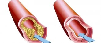
A compression fracture is an injury to the spinal column that occurs during simultaneous compression and flexion. Under pressure, the vertebra is compressed and flattened, causing it to crack. An injury such as a compression fracture often occurs after landing from a great height on straight legs or on the buttocks, during forced bending of the body, or a car accident. A vertebral fracture is also possible if the cartilage of the intervertebral disc loses its shock-absorbing properties as a result of the development of various diseases. The bone structure is unprotected and with a relatively small impact a vertebral compression fracture can occur. Physiological problems in bone tissue and intervertebral discs, for example, osteoporosis, cancer metastases can also cause a vertebral fracture.
A compression fracture occurs, most often, in the lumbar and lower thoracic spine, when the eleventh and twelfth thoracic and 1st lumbar vertebrae are affected, and as a result of a compression fracture, the spinal nerve roots are compressed.
Compression fractures of the cervical spine occur much less frequently and can be the result of a strong blow to the head; they also occur in car accidents and falls from a height.
Types of coccyx fracture
Depending on the type of injury caused, injuries are divided into the following types:
- Fracture of the coccyx without displacement and with displacement;
- Closed or open fracture of the coccyx;
- Compression fracture. The individual vertebrae that make up the bone compress and damage each other;
- Fracture dislocation. Damage in which part of the bone not only breaks off, but also bends unnaturally;
- Tear in the coccyx. This occurs when the bone in a given vertebral segment is completely torn;
- Transverse.
Displaced open fractures are one of the most dangerous conditions, as they can only be treated with surgery. No less dangerous is a dislocation with a fracture, which is difficult to diagnose.
In addition to fractures, the integrity of the coccyx is violated in the form of fractures, as well as complete and incomplete fractures.
Coccyx fracture: symptoms
Symptoms of a tailbone fracture rarely depend on the type of injury. Symptoms of all types of injuries are:
- Acute, severe pain at the time of injury, which gradually passes;
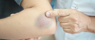
- The appearance of swelling and bruising at the site of injury;
- Increasing pain when trying to move the sciatic muscles or during bowel movements;
- Discomfort that turns into pain when trying to sit or stand, as well as when sneezing or coughing.
Acute severe pain at the time of injury is one of the symptoms of a coccyx fracture.
A distinctive feature of closed coccyx fractures is that walking after an injury is rarely accompanied by severe discomfort. However, the victim always tries to limit his movements for fear of pain.
A sign of an open fracture of the coccyx is the presence of a bleeding open wound in which a small area of bone tissue is visible.
In women, the symptoms of a coccyx fracture are much more pronounced. If the injury occurred during childbirth, very often blood is observed in the victim's stool.
Recovery prognosis
It is quite difficult to give a prognosis for recovery after a fracture of the coccyx . Most likely, this can only be done by a specialist who observes the picture of the disease and treatment. The process depends on many factors.
These include the severity of the injury, the course of treatment, the rehabilitation period, etc. If the fracture is not too complex and the victim has completed a full course of treatment and rehabilitation, then, as a rule, the tailbone heals properly, the pain goes away, and the ability to work fully returns. But at the same time, for some time you can only do light work.
Coccyx fracture: first aid
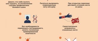
First aid for a fractured tailbone should be provided in the following order:
- Place the victim on the side or stomach and call an ambulance.
- Apply ice to the damaged tissue. This will help reduce bleeding. Ice for a cold compress should always be wrapped in a clean cloth, especially if the crack is open. Avoid direct contact with skin.
- Painkillers, preferably non-steroidal anti-inflammatory drugs (NSAIDs), should be given. Diclofenac, ibuprofen, Voltaren or generic analgin may work.
If medical personnel cannot arrive, the victim must be taken to the hospital. Transport should be done without pushing and lying on your side. You must contact a medical facility.
How to protect your tailbone?
Instead of treating a bruise, it is better to try to avoid situations where injury may occur or soften the force of the blow. It is worth taking care of comfortable non-slip shoes for winter and not going down any inclined surfaces or icy steps.
When skating or roller skating, you need to redistribute your center of gravity so that when you fall, you group yourself and fall to the side or forward.
And general physical development, activity, healthy eating and good sleep will help maintain balance, agility and flexibility.
Coccyx fracture: diagnosis
Diagnosis of the patient’s condition and the degree of damage to the body is carried out comprehensively. First, the traumatologist collects complaints and examines the patient. This includes both external examination and palpation of the rectum or vagina. The patient is then given an x-ray to confirm the diagnosis.
Computed tomography is one of the diagnostic methods for coccyx fractures
In some cases, x-rays do not show existing damage. Therefore, to clarify the diagnosis, computed tomography (CT) of the spine is recommended.
If there is an open fracture of the tailbone, it is imperative that a doctor examine and treat the injury before making a diagnosis.
Other restorative directions of the Osteopathy Clinic
Osteopathic treatment does not imply instant healing, although the first sessions often bring relief.
It is not at all necessary to continue to endure pain while the injury is still making itself felt. Homeopathic preparations have a good restorative and analgesic effect: external ointments, substances for oral administration, lotions. A competent homeopath selects treatment individually for each patient.
If osteopathic methods are gentle and suitable even for expectant mothers and newborns, then manual therapy involves a more severe impact.
The manual doctor realigns the coccygeal bone using the rebound technique, pre-heating the lower spine. Reduction can be done through the anus.
Kinesio taping is a treatment by applying special tapes to a bruise. They reduce pain, swelling, and other symptoms.
If there has been a fracture of the coccyx, kinesio tapes will be useful after completing the general treatment course.
Coccyx fracture: treatment
Treatment options for a fractured tailbone largely depend on the type of injury sustained. In this case, therapy can be carried out using the following methods:
- Conservative treatment;
- Surgical intervention;
- Use of traditional medicine.
Only the doctor decides which method will be used to treat the pathology. However, regardless of the nature of the injuries received, the patient’s movement should be limited throughout the entire period of treatment so as not to provoke complications.
Regardless of how a fracture of the coccyx is treated, the attending physician prescribes the patient to take painkillers and anti-inflammatory drugs in the first 2-3 weeks. And within 7-10 days after the injury, the victim needs to defecate, use enemas or laxatives. This will avoid muscle overload and additional damage to the tailbone.
With any type of fracture, the patient cannot sit or walk for the first 1.5 weeks. You can only lie on your stomach occasionally, turning over onto your back to avoid bedsores. However, when lying on his back, the patient must use a special orthopedic pillow under the buttocks.
Conservative
This type of therapy is used for closed coccyx fractures that have occurred without dislocation or dislocation. In this case, the patient can stay at home, periodically visiting a trauma surgeon to assess the effectiveness of therapy.
If a bone injury is accompanied by a dislocation, the patient is hospitalized for several days. In this case, the bone repositioning procedure is performed in the first 24 hours after the injury. Reconstruction of dislocated vertebrae is performed with fingers through the rectum. Manipulations are performed under local anesthesia with a 2% novocaine solution. If the procedure does not produce a positive effect, surgical treatment of the pathology is prescribed.
Conservative treatment includes the following measures and procedures:
- Eliminate symptoms of inflammation and pain with NSAIDs in the form of rectal tablets or suppositories;
- Prescribing calcium and calcium-based drugs to accelerate the healing of damaged bones;
- Using anti-edema ointments. Possible only with a doctor's prescription.
The main physiotherapeutic methods for treating a coccyx fracture include hirudotherapy.
A visit to a physical therapy office usually occurs after the damaged tissue has healed, which takes about 1 month. Basic physiotherapeutic procedures include electrophoresis, quartz treatment, hirudotherapy and ultraviolet irradiation. In addition, during recovery, the patient will need to undergo therapeutic physical therapy (PT) to restore normal muscle function and activity.
Surgical
Open coccyx fractures, bone injuries with fractures, and fibula fractures that cannot be treated by reduction require only surgery. Surgery can also be performed for compression fractures and uneven tissue growth.
The procedure is performed under general anesthesia. During the procedure, damaged fragments and fragments of the coccyx are removed. And after the operation, the patient must be hospitalized for 1.5-2 weeks and be kept on strict bed rest.
If no improvement is observed after surgery or long-term conservative treatment, the entire tailbone should be surgically removed.
Traditional methods
A fracture of the coccyx is treated with traditional medicine. Therapy in this case usually includes the use of ointments and compresses:
- In 10 grams of rose oil, 0.5 grams of powdered mummy is diluted. Gently rub the resulting mixture onto the injured tailbone once a day.
- In 300 ml of boiling water, add 1 tablespoon each of chamomile and crushed quinoa leaves, mixed in equal proportions. The liquid is boiled over low heat for 5 minutes and left to infuse for 15 minutes. After straining the broth, add 1 tablespoon of honey and mix thoroughly until completely dissolved. The resulting infusion is soaked in cotton wool and applied to the damaged area for 10 minutes. After the time has passed, you should put on a new cotton pad for another 10 minutes. The procedure should be carried out daily for 1.5 weeks.
- We grate the peeled raw potato tubers on a fine grater and place them together with the juice on the sore spot. Leave the potatoes for 20 minutes.
Treatment of pathology at home with traditional methods is possible only as maintenance therapy and only after consultation with a doctor.
Diet
When treating a tailbone fracture, it is extremely important to follow a diet. In your diet, you should limit the amount of flour products and sweets, as well as dishes and foods high in starch. All of them can cause constipation, which is very dangerous for the patient.
The following products are recommended for consumption:
- Dairy products and calcium-rich foods;
- Persimmon;
- Hazelnut;
- Greenery;
- Sesame seeds;
- Fish.
It is recommended to eat persimmon if you have a fractured tailbone.
It is also imperative that the victim drinks plenty of fluids throughout the day. This can be still water, juice, tea, compote and a snack.
The most important
A fracture of the coccyx should be treated as early as possible, then the bones will heal faster. Displaced injuries are more dangerous; they are accompanied by severe pain and neurological disorders, and therefore are more difficult to treat. For a mild fracture, conservative methods are used: bed rest, analgesics, diet, exercise therapy, physiotherapy, etc. For comminuted damage, bone fragments are compared through the rectum. If after this the coccyx fragments are not held in the correct position, then surgery is prescribed. Surgical intervention cannot be avoided if neurological disorders are present, the functioning of the pelvic organs is disrupted, or the pain persists for a long time. After the main treatment, rehabilitation is carried out. It is important to follow the doctor’s recommendations at all stages of therapy, then the bones will heal faster and you can return to a full life.
Using an orthopedic pillow for a fractured coccyx
If there is an urgent need to sit until the fracture of the coccyx is completely healed, as well as during the rehabilitation period, you can use a special orthopedic pillow. The device allows the patient to remain in a sitting position without putting pressure on the injured area.
The product is a small, usually inflatable circle with a hole in the center. To use it correctly, the wheel must be positioned so that when sitting, the hole is located where the tailbone is located. The buttocks will be on the sides of the orthopedic pillow.
Rehabilitation
Rehabilitation after a tailbone fracture usually lasts at least a month. It is during this period that the bones grow together and their lost strength is restored. For severe injuries, the rehabilitation period can reach 2-3 months and includes the following procedures:
- exercise therapy;
- massage;
- herbal medicine;
- acupuncture.
In addition, it is important to organize a nutritious diet, avoid increased physical activity and use special orthopedic devices to reduce pressure on the damaged tailbone. With this pathology, it is recommended to sleep and lie on your stomach.
A month after the therapy, the patient returns to his normal lifestyle. However, it is recommended to give up sports and increased physical activity for one year.
Orthopedic pillow
When the coccyx is injured, treatment is mainly carried out using an orthopedic pillow. It resembles a wide circle in the form of a lifebuoy with emptiness inside. With the help of such a device, it is possible to ease the load on the musculoskeletal system and achieve a positive result:
- unload the spinal column;
- relieve muscle tension;
- prevent blood stagnation;
- cope with pain.
An orthopedic pillow can be used even after recovery of an injured tailbone. Thanks to it, a person is able to sit for quite a long time without harm to health.
Proper nutrition
If the coccyx is damaged, proper nutrition is important during the recovery period. The patient's diet should be filled with the following products:
- greenery;
- hazelnuts;
- persimmon;
- fish;
- sesame;
- dairy products.
In addition, it is necessary to introduce into the diet foods that provide microelements such as calcium.
Features of a coccyx fracture in childhood
Because the tailbone in children is flexible where it connects to the vertebrae, a fracture of the tailbone is very rare and the injury is actually a bone strain or tear of the cartilaginous joints. Injury can occur when falling on the buttocks from a great height or at high speed.
To distinguish a fracture from a bruise, it is enough to pay attention to the nature of the pain. If a fracture occurs, the pain will increase when you try to stand or sit, as well as when you cough and change your body position.
A fracture of the coccyx in a child can result in problems with bowel movements and the genitourinary system, as well as inflammatory processes in surrounding organs, nervousness and chronic headaches.
Coccydynia - symptoms and treatment
Coccydynia, pain in the tailbone area, usually occurs when sitting. The intensity of the pain varies and is sometimes worsened by sitting. Less severe symptoms can be controlled by changing your sitting position or injecting a local anesthetic and corticosteroids into the painful area. If treatment is unsuccessful, surgical removal of the coccyx may be indicated.
Our clinic has accumulated experience in managing patients with pain in the coccyx. Such patients are treated by orthopedic traumatologists and neurologists.
Sign up for treatment for Coccydynia
Prevalence
Who has tailbone pain more often? Coccydynia is five times more common in women than in men. Although it can occur over a wide age range, the average age of onset is 40 years. Coccydynia has many causes: it can be post-traumatic, starting from a fracture or bruise, or after a difficult vaginal birth. 1-3 Most cases exhibit coccyx subluxation or hypermobility, which can be seen on dynamic radiographs taken with the patient in a standing and sitting position 1-3. The cause of pain in patients with normal coccygeal mobility is unknown. 2
This is what it looks like on an x-ray
Anatomy of the sacrococcygeal region
Five fused sacral and three or four fused coccygeal vertebrae form the terminal end of the spinal column. The lower end of the sacrum is connected to the coccyx through the articular-disc complex. This joint can be a symphysis (slowly moving cartilage) or a synovial joint. The coccyx is a triangular structure consisting of three or four coccygeal vertebrae that are usually fused, although the first coccygeal segment may not be fused to the second. The sacrococcygeal joint can also fuse. The coccyx provides attachment to the gluteus maximus muscle.
IMPORTANT INFORMATION! It is important to understand that there are 3 states of the coccyx:
- Its instability, subluxation, displacement.
- His normal mobility
- His fixation and immobility
- For acute or mild coccydynia, treatment begins with anti-inflammatory and painkillers, load reduction, and soft pillows under the coccyx.
- If this approach is not effective, then physiotherapy is used (ultrasound, high-intensity magnetic stimulation has shown a good effect
- Further, if the effect is insufficient, a targeted blockade of the coccyx is performed under ultrasound navigation. If it has a good effect, the treatment is stopped. If there is an effect, but not enough, then several blockades are performed.
- If the effect is insufficient, a blockade is performed + placing the coccyx in place and stretching the levator rectus muscle (with anesthesia).
- If this does not help, a decision is made about surgery to remove the coccyx.
A, Normal view of the coccyx while standing. B, Increased mobility of the coccyx in flexion when the patient is sitting. C, Posterior subluxation of the coccyx while the patient is sitting. D, Coccygeal spicule is a bony spine (arrow) extending from the posterior surface of the coccygeal segment.
Etiology or causes of coccydynia
Why does my tailbone hurt? Common causes include obesity, previous trauma to the area or childbirth, and instability.
Obesity, which reduces pelvic rotation when the patient is sitting, is three times more common in patients with coccydynia than in the normal population. 2
An interesting fact is that instability of the coccyx occurs equally often in people both with and without a history of trauma. In this regard, according to studies, only an injury less than 3 months old is associated with the development of pain in the coccyx.
Patients with normal coccygeal mobility have an idiopathic (no obvious cause) type of coccydynia, which may be associated with pelvic floor muscle tension and spasm or other pelvic muscle abnormalities.
Coccydynia, which occurs with a stationary coccyx, is often associated with inflammation of the membrane (adventitial bursitis) at the tip of the coccyx. Other suspected etiologies include post-traumatic arthritis of the sacrococcygeal joint and nonunion fractures or dislocations of the coccyx.
Abnormal psychological conditions as a cause of coccydynia have been largely refuted, as behavioral testing of these patients reveals personality profiles similar to other patient groups. 5
Other and very rare causes of pain in the sacrum and coccyx area are lesions of the lumbar discs, arachnoiditis of the lower roots of the sacral nerve, tumors of the coccyx or sacrum, pilonidal cysts and sinuses, perirectal abscesses. 4
Symptoms of coccydynia
The main symptom is PAIN. The onset of pain may be subtle, which can lead to a long delay from onset to diagnosis. Patients typically complain of pain in and around the coccyx without significant low back pain, radiating pain, or referred pain. The pain is localized in the sacrococcygeal joint or mobile segment of the tailbone and can be relieved by sitting on the legs or buttock.
Chronic pain is pain that persists for >3 months. Patients may experience frequent urge to defecate or pain during defecation. Women with a history of vaginitis, discharge or associated pelvic pain should be referred for gynecological consultation. Associated constipation should be treated accordingly. If the patient has blood in the stool, the possibility of a tumor or metastasis should be considered.
Physical examination or what the doctor does during the appointment
The doctor examines the surrounding skin and soft tissue for the presence of pilonidal cysts or fistulas. External palpation or rectal examination (through the rectum) can reveal bone growths and local swelling. The coccyx should be palpated externally, and if there is insufficient information, the segment should be manipulated rectally to detect pain caused by movement of the coccygeal segments.
Diagnosis of coccydynia
Initially, diagnosis consisted of standard x-ray examinations. Unfortunately, there is often no difference on a regular X-ray between sick and healthy people.
They have been replaced by dynamic radiographs - this is the most reliable way to diagnose coccydynia. Unfortunately, in St. Petersburg, such a study is practically not performed anywhere, so for our patients we organized such a study as part of cooperation with trusted radiologists.
In their study, Maigne and colleagues 1 - 3 , 6 compared standing and sitting lateral radiographic images of the coccyx in 582 patients with coccydynia and reported abnormalities in 70%. The normal coccyx rotates slightly (5° to 25°) posteriorly or anteriorly when sitting and returns to its original position when standing (Fig. 2, A). Abnormalities of the coccygeal segments in the sitting position have anterior hypermobility >25° (Fig. 2, B and 3). Subluxation or posterior displacement of the mobile segment of the coccyx is observed when the patient is sitting (Fig. 2, C and 4, A). The spicule of the distal end (Fig. 2 D) is most often visible when the coccyx is stationary (<5° of movement in the sitting position). 2
Anatomical signs of coccydynia. 1 A, Normal view of the coccyx while standing. B, Increased mobility of the coccyx in flexion when the patient is sitting. C, Posterior subluxation of the coccyx while the patient is sitting. D, Coccygeal spicule (arrow) arising from the posterior surface of the coccygeal segment.
A 19-year-old man fell on his buttocks 2 years prior to presentation and immediately developed coccydynia. His symptoms were chronic and disabling. A. Lateral radiograph demonstrates posterior subluxation of the coccygeal mobile segment. B, Sagittal T2-weighted spin-echo magnetic resonance image shows distal coccygeal edema, especially coccygeal subluxation.
Advanced Imaging Techniques
Magnetic resonance images may also demonstrate edema in such areas of inflammation ( Fig. 4
, B). None of the imaging modalities can accurately diagnose coccydynia, and they are not as accurate as compared standing and sitting dynamic radiographs.
Non-surgical treatment of coccydynia in St. Petersburg
Non-surgical treatment is effective in more than 85% of cases. His tactics seem simple and reasonable. From smallest to largest. This is exactly how we treat patients in the clinic.
You can read more about treatment with links to scientific sources by expanding this section
Non-surgical treatment options include nonsteroidal anti-inflammatory and pain medications, rest, hot baths, and a cushion to protect the tailbone area from repetitive trauma. Physiotherapy, consisting of diathermy and ultrasound, can bring temporary relief.
In their study, Wray et al [5] used a staged treatment, with each step being slightly more invasive than the previous one. First, an anti-inflammatory hormonal drug (methylprednisolone (40 mg)) and a local anesthetic (bupivacaine (10 ml 0.25%)) were injected into the side and tip of the coccyx. For persistent coccydynia, the drug mixture was reintroduced and the tailbone was manipulated through the rectum, repeatedly flexing and stretching it for a minute. If the treatment was initially successful, but the pain returned, the injections and manipulations were repeated.
If no effect was obtained, then after 6 weeks a coccyxectomy was performed - surgical removal of the coccyx. The cure rate with a single injection was 59%, but 85% of cures were achieved with manipulation and injection.
Although relapses occurred in the injection group (21%) and manipulation group (28%), retreatment in each group resulted in good success. Coccygectomy was performed in 20% of patients and had a success rate of 91%. Maigne and Chatellier 6 prospectively compared levator ani muscle massage, joint mobilization, and gentle stretching of the levator ani muscle without the addition of injections. At 6 months, treatment success was 29.2% with massage, 16% with mobilization, and 32% with stretching, for a total of 25.7% overall success. When a patient had a satisfactory result, it was invariably achieved within a week. The good results tended to remain stable. The best results were shown by patients with normal coccyx mobility (43% success after 6 months). The worst results were patients with a fixed tailbone (16%). Outcomes for patients with unstable subluxation (22.2%) and hypermobility (25%) were moderately successful. Massage and stretching were found to be more effective than manipulation. When therapy failed, patients moved on to injections or surgery.
For acute coccydynia (duration ≤ 2–3 months), treat with 8 weeks of rest using a stool softener, adjustable seating, and nonsteroidal anti-inflammatory drugs. If this does not relieve symptomatic coccydynia or the patient develops chronic symptoms (duration >2–3 months), evaluation is performed, including standing and sitting radiographs of the coccyx in addition to MRI to evaluate injury swelling, tumor, or other conditions. Stretching, massage and injections are usually started at this time. If these treatments are unsuccessful or the pain returns, a coccyxectomy may be suggested.
Surgery
Surgery consisting of complete coccygectomy or simple removal of the mobile segment should be performed only after conservative treatment has failed.
Our clinic does not provide surgical treatment.
Summary
Dynamic radiographs can help identify causes and abnormalities in most patients with coccydynia, and patients with normal coccygeal motion can also be successfully treated. Most patients benefit from injections of corticosteroids and anesthetics combined with massage or stretching of the levator ani muscle.
Doctors at our clinic have experience treating patients with coccydynia in St. Petersburg. Within the clinic, you can perform the entire range of medical procedures, including blockades. We perform blockades under ultrasound guidance for maximum accuracy! We also refer you to trusted specialists to perform dynamic radiographs.
Coccyx fracture: complications
Most often, complications after injuries occur when DeepL consults a doctor prematurely. Sometimes patients only come to the trauma surgeon's office a few weeks after the fracture. This is due to the fact that a few hours after the bone is damaged, the symptoms of the fracture become less pronounced, and the patient assumes the presence of a bruise.
The main complications of a coccyx fracture include:
- Severe pain in the area of the damaged bone, which makes it impossible to sit down;
- Neurological disorders;
- Diseases of the genitourinary system;
- Delivery difficulties;
- Frequent migraines in the lower back, buttocks and head;
- Spinal cord injury, which can lead to paralysis of the lower limbs and even the entire body;
- Muscular dystrophy, which causes constant pain in the lower back and legs and weakness in the legs.
Severe pain in the area of the injured bone, preventing you from sitting, is a possible complication of a fractured coccyx.
The consequence of an untreated fracture of the coccyx is the development of an acute inflammatory process in the surrounding tissues, as well as their gradual destruction and decay, turning into gangrene.
Postoperative complications include fistulas, purulent conditions, and tissue edema.
Classification of compression fractures
Classification according to the degree of vertebral deformity:
- I degree - the height of the vertebra decreases to less than ½ of its size.
- II degree - the height of the vertebra is halved.
- III degree – height decreases by more than half.
A grade I spinal compression fracture is stable and does not disrupt the stability of the spine.
Compression fractures of grade II and III lead to instability of the spinal column.
Classification of compression fractures according to the presence of complications:
- An uncomplicated type of spinal compression fracture can often occur in a latent form and be accompanied by pain in the area of damage to the spinal column. The patient self-medicates and does not seek help from a doctor. In this case, a compression fracture can cause the development of diseases such as osteochondrosis or radiculitis.
- A complicated form of spinal compression fracture, in addition to pain, may be accompanied by a neurological disorder. Particularly dangerous are complicated compression fractures with the formation of bone fragments that damage the nerve roots. Over time, the sensitivity of the limbs gradually decreases and numbness occurs.