Symptoms of a dislocated joint
A joint dislocation is a displacement of the articular surface of the bones, accompanied by damage to the capsular-ligamentous apparatus.
Dislocation occurs, as a rule, with damage to the soft tissues of the joint. In this case, ligaments with blood vessels, the capsule, and the tendons of adjacent muscles may tear. Damage leads to serious impairment of the functionality of a single joint or the entire limb. It should be noted that correct and timely provision of first aid for joint dislocations is of great importance for the full restoration of function. Types of dislocations:
- by degree of displacement (complete and incomplete dislocation);
- by origin (congenital and acquired);
- open dislocations with the formation of a wound;
- closed without tearing the tissue and skin over the joint.
Read on to learn more about the symptoms of a dislocated joint.
Special classification
The sequence for diagnosing a deviation involves the need to assign a certain status to the dislocation according to several classifications. All such injuries can be sorted according to:
- origin;
- degree of displacement;
- skin condition;
- statute of limitations.
There is no place for pre-medical support for newborns who have become victims of intrauterine anomalies. Instead, help lies in long-term therapy, which is beneficial in the early stages in most situations.
Also, emergency medical care will be useless if the visit was due to pathological displacement, which is caused by a chronic illness. It is most effective to use a summary of pre-medical rules for cases of traumatic injury to a leg or other part of the body.
Orthopedics has its own classification according to the degree of displacement of the bone structure:
- full;
- incomplete.
The second version is also called subluxation, since it involves the divergence of the ends of the joints not completely. Some contact area still remains. At the full stage, the articular ends of the arm diverge completely.
A quick examination of the problem area allows you to make an assessment of the skin. If signs of destabilization include a violation of tissue integrity, then this indicates an open type of dislocation. When the analogue is closed, the top layer remains unharmed.
But you should not assume that the second subtype of injury is safer. In fact, it poses a serious threat as it sometimes involves ruptured blood vessels. And with bleeding without an open wound surface, the average person usually cannot cope on his own.
The statute of limitations also affects the severity of the disease, so it was included in a separate classification. Fresh injuries cover a time interval of up to three days. Stale – from three days to two weeks. Outdated – terms higher than indicated. The latter option is especially characteristic of lesions of the shoulder joint.
But, regardless of what degree the lesion was assigned, it must be certified according to the latest special classification, which covers the division into the following groups:
- paralytic;
- complicated;
- habitual;
- irreducible.
The first point covers muscle paralysis that outweighs the antagonist muscles in strength.
A correctly diagnosed complicated variant helps in further development of therapy, as it allows one to identify complications such as nerve lesions and disorders of the main blood vessels. In the most dangerous scenario, symptoms indicate intra-articular or periarticular fractures.
As for the shoulder, he most often has to deal with the usual class. Its peculiarity lies in periodically repeated dislocation at the same point. This is often caused by anatomical changes due to a number of pathologies, as well as muscle weakness or problems with ligaments.
The irreducible analogue covers old injuries, which are almost always accompanied by penetration of tissue between the articular surfaces of the bone ends.
Types of joint dislocations
Dislocation of the shoulder joint. Depending on which direction the head of the humerus is displaced, the damage may be inferior, posterior, or anterior. The latter occurs in 98% of cases. Pain with any attempt to move the joint, swelling, decreased joint mobility, impaired sensitivity and blood supply are the main symptoms of this condition. Shoulder dislocation should only be treated by a specialist.
The causes of a dislocated knee joint can be a strong blow, a fall from a height, congenital anomalies, and a sharp contraction of the thigh muscle during fast walking. Symptoms of such damage will include pain, swelling, numbness of the limb, changes in the shape of the knee, a specific position of the leg and the inability to move. Since the injury is considered serious, first aid, treatment and rehabilitation after a dislocated knee joint should be supervised by a doctor.
Dislocation of the hip joint can be anterior or posterior. The front one may appear as a result of a fall from a height. The cause of the rear is most often a road traffic injury. Symptoms of injury include severe pain, forced positioning of the limb, changes in joint shape, and slight shortening of the affected limb. Dislocation of the hip joint in newborns will be discussed below.
How to identify characteristic symptoms?
To understand the signs of a possible dislocation, people use tables, physiological diagrams, and even a thematic presentation helps. The latter may include information about adjacent injuries, which occurs with burns and frostbite.
Despite the fact that all types of dislocations have their own characteristics, they can still be combined into a general list. With such injuries, a person complains of:
- sharp pain with increasing intensity when trying to make a movement;
- redness of the skin;
- swelling;
- local temperature, which means an increase in temperature in the affected area;
- articular deformation, covering a change not only in shape, but also in size;
- change in the length of the affected limb, which is more suitable for an ankle injury;
- the articular end, which is palpated in the “wrong place”.
Occasionally, such foot or hand problems are additionally accompanied by a general increase in temperature and chills. Cases of bruising are common. And when the nerve endings are compressed, the victim complains of partial or complete loss of sensitivity.
Diagnostics
After examining and palpating the damaged area, the traumatologist (namely, he deals with such injuries, including congenital dislocations of joints) will refer the patient for an x-ray. After confirming the diagnosis, the doctor can provide full specialized assistance, starting from eliminating the dislocation and ending with the necessary immobilization or fixation, supplemented, if necessary, with medication and restorative treatment. At the end of the immobilization period, a rehabilitation program is prescribed to restore movements and predict the outcome of treatment.
Providing first aid for dislocated joints
The main task of first aid is to completely immobilize the damaged joint in a position of minimal pain (splinting, bandaging to the body/limb, tight bandage, etc.). If there is damage to the skin in the area of injury, it must be treated with hydrogen peroxide and a clean (ideally sterile) bandage applied or the wound bandaged with a bandage. To reduce swelling, apply a cold compress to the joint. It is not recommended to give the victim water or painkillers in the form of tablets in most cases (the measures described above will in most cases be sufficient until the ambulance arrives) to assist in eliminating the dislocation in a medical facility as soon as possible, the main condition of which is the injured person’s empty stomach.
You cannot correct a dislocation on your own!
Treatment of shoulder dislocation
Treatment of any dislocation first of all involves realigning the joint and returning it to its usual physiological position. It is recommended to carry out the procedure as soon as possible under general anesthesia (especially in children) and in some cases under local anesthesia (dislocations of the hand or foot). The decision in this case is made by the doctor, taking into account the patient’s condition and the degree of damage. Surgery for habitual dislocation of the shoulder or knee joint is performed by an orthopedic traumatologist or a surgeon specializing in sports injuries.
In case of untimely treatment, there is a possibility of developing contracture. With long-standing dislocations (more than 3 weeks), the function of the joint may be irreversibly lost.
Reasons for destabilization
In order for assistance to have an almost immediate effect, it is extremely important to understand the original source of the damage that occurred.
In short, a joint is a connection between bones that allows the mobility of a limb. It is formed by the articular ends of both bones, which are covered with a cartilaginous layer. Cartilage is designed to soften and at the same time absorb friction during any movement. If the pathology concerns the knee joints, then the presence of an additional cartilage pad, which is called the meniscus, adds complexity.
Content:
- Reasons for destabilization
- Special classification
- How to identify characteristic symptoms?
- Differences between a fracture and a dislocation
- Basics of first aid
All articular elements of the bone structure are hidden in the so-called articular capsule, which contains a special fluid inside. Additionally, ligaments are attached to the joint, which are designed to fix it securely. The entire mechanism is a fairly strong natural structure, but under a number of circumstances even it fails.
As with bruises, destabilization of joint activity is often caused by various mechanical damage, external pressure, or classic injuries received during physical activity.
But if in case of fractures you can immediately pay attention to the damage due to a number of specific alarming signs, then with a dislocation the situation is more complicated. This is explained by the fact that in clinical practice there are a number of diseases that contribute to the onset of such a condition. Ailments undermine health methodically and slowly, which is why it is not always possible to recognize something wrong instantly, as with sprains.
Among the uniaxial diseases that provoke such a serious deviation from the physiological norm are:
- dysplasia;
- arthritis;
- tuberculosis;
- oncological neoplasms of a benign or malignant nature;
- osteomyelitis.
All of the above applies to the camp of acquired injuries, when medical care is provided on the spot depending on the circumstances. But sometimes dislocations are congenital. This occurs when the intrauterine development of the fetus is abnormal. After birth, the baby is immediately diagnosed with dysplasia. Most often in children
The disease targets the hip area or knee area.
Hip dislocation in newborns
The main symptoms of the disease will be asymmetry of the gluteal folds, limited mobility of the joint during passive abduction of the hip, a click during various movements in the joint and relative (comparative) shortening of the limb (in the case of unilateral pathology). A dislocated hip joint, like a dislocated finger, can be diagnosed by a highly specialized specialist after a clinical examination with the appointment of instrumental studies.
Treatment of hip dislocation in newborns is conservative. Therapy includes exercise therapy, massage, wide swaddling and the use of a fixation device. If all recommendations are followed, the head of the femur is reduced independently in most cases; the success of treatment is determined by the timing of detection of the pathology and the severity of the latter.
Congenital joint dislocations
Congenital hip dislocation most often occurs in girls and is diagnosed immediately after birth. The cause of the disease is underdevelopment of the femoral head and glenoid cavity. The main symptoms of dislocation include asymmetry of the gluteal folds, limited movement in the hip and slight shortening of the limb. The final diagnosis is made after ultrasound and radiography of the hip joints.
Treatment for a sprained ankle depends on the energy level of the injury. In such cases, after examination, a specialist can assess the degree of damage to the joint and determine the most optimal treatment tactics. This can be either symptomatic drug treatment in simple cases with the possibility of loading, or plaster fixation or, in some cases, surgical restoration of the ligamentous apparatus of the damaged joint.
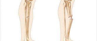
Differences between a fracture and a dislocation
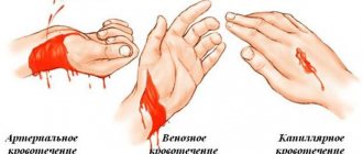
In order to correctly use measures to alleviate the patient’s condition, you will first need to determine whether the victim has suffered a dislocation or fracture. Usually the task presented is beyond the capabilities of a person who does not have the appropriate education. The final verdict can be made only after the victim undergoes an X-ray examination. A black and white photograph will reveal a possible fracture of the bone structure.
But even without radiography, there are primitive methods that make it more likely to identify the exact format of the lesion. The significant difference between a fracture lies in the following aspects:
- The presence of bruising and swelling strictly over the damaged part with subsequent spread to surrounding tissues much later. A dislocation forms a bruise with swelling over the problem joint, followed by “spreading” to the surrounding skin.
- Palpation of bone fragments is characteristic of a classic displaced fracture. With a dislocation, you can only feel the torn articular surfaces.
- A fracture involves intense pain exclusively in the area of destruction of bone integrity. When a dislocation occurs, the pain increases when trying to palpate the damaged area.
Separately, you need to pay attention to the length of the limb. With a dislocation, only this value changes, while a fracture is characterized by a change not only in length, but also in shape.
Rehabilitation activities
The main methods of rehabilitation, including recovery after dislocation of the wrist and elbow joint in adults, include exercise therapy, massage and physiotherapy. Laser therapy, ultrasound, myoelectric stimulation, magnetic therapy and interference therapy are widely used as the latter.
By performing therapeutic exercises after a dislocation of the shoulder joint, you will quickly restore joint mobility, improve blood circulation and increase its flexibility. For the same purpose, different types of massage are prescribed during rehabilitation. Please note that after a joint dislocation, the recovery period is individual for each patient and requires regular supervision by an orthopedist and rehabilitation specialist, in some cases a neurologist, massage therapist and physiotherapist.
Prevention of dislocations can only be prevention of injuries or early diagnosis (professional examination for congenital pathology) in newborns. On our website you can familiarize yourself with the qualifications of specialists who advise in this area. You can make an appointment at any time convenient for you.
Related services: Orthopedics and traumatology Calling an ambulance
What to do if you have a sprained ankle?
Ankle sprain is a fairly common injury at any time of the year. But with the arrival of spring and an increase in people's activity, as a rule, there is a surge in such injuries.
The ankle joint, along with the knees, supports the entire weight of the body during movement. Therefore, weakening of the ligamentous apparatus and decreased muscle tone, especially with inactivity and excess weight, creates conditions for various injuries, including injuries. Sloppy, sudden movements, incorrect positioning of the foot with overload of one part of the joint, threatens with ankle dislocations.
Damage to the ankle joint, especially with ligament rupture or bone fracture, can lead to quite severe pain, limited mobility and temporary disability.
The most common causes of ankle sprains:
- walking on wet floors, ice or slippery surfaces, leading to falls and joint damage;
- wearing uncomfortable shoes, including high-heeled shoes, platforms, mules;
- movement on uneven surfaces, stones, when there is a high risk of twisting your leg;
- sports, outdoor games with running, jumping;
- professional competitions;
- skeletal pathologies with damage to joints, ligaments and bones, muscles, leading to “loose” joints and gait instability;
- lesions of the nervous system, in which coordination and stability when walking suffer, a person places his foot incorrectly on the floor.
Damage to the joint occurs if the foot turns sharply inward or outward during movement. Then the external or lateral ligaments are overloaded, the bones of the joint are displaced relative to each other. In parallel with a dislocation, ligament sprains or ruptures, as well as bone fractures, are possible.
Symptoms of a sprained ankle:
- severe, acute pain that occurs immediately after the injury - it intensifies when trying to move the foot;
- increasing swelling of the tissues at the site of injury, especially pronounced in the lower part of the leg, the junction of the foot - due to swelling and pain, it is almost impossible to step on the leg and walk;
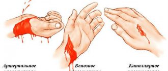
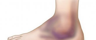
- When blood vessels rupture, a hematoma can form and the skin becomes bluish.
With ankle subluxation, the symptoms are not as pronounced as with dislocation and rupture of the ligament. Pain and discomfort are especially pronounced when walking; they may subside with rest. The joint is swollen, deformed, foot movements are limited, the skin has a normal color.
Treatment of a sprained ankle
If, after an injury (fall, twisted leg), pain and swelling occur, or a cracking or crunching sound is heard, you should immediately go to the emergency room. It is necessary to determine the severity of the damage to the joint and exclude bone fractures, which will be important for the treatment of a sprained ankle. Self-medication for this type of injury threatens subsequent instability of the ligaments and repeated injuries at the slightest damage and the development of osteoarthritis of the joint (a chronic inflammatory lesion with the destruction of cartilage and bones).
1. Diagnostics
If you suspect a sprained ankle, you need to go to the emergency room and/or make an appointment with a traumatologist-orthopedist at the Canon clinic.
The first stage is to clarify the circumstances of the injury and examine the doctor, during which he will find out where the most severe pain is localized, how limited the movement of the foot is, and what additional complaints the patient has. The traumatologist will assess the amount of swelling, the presence of bruises or hematomas, and the position of the foot.
An X-ray of the affected joint is required to rule out bone fractures. In order not to miss possible damage, x-rays are taken in two projections, this makes it possible to assess the condition of the tibia and fibula, the outer and inner ankle.
2. Modern methods of treatment
After making a diagnosis, the doctor administers anesthesia to reduce the dislocation and apply a fixing bandage. If a bone fracture, joint deformity or bone displacement is confirmed, after restoring the correct structure of the joint, the doctor fixes the foot in the correct position with a plaster cast.
If this is a dislocation of the ankle without a fracture, the doctor will apply a U-shaped plaster splint (from the toes, along the bottom of the foot, covering the heel and upper third of the leg) for a period of 12 - 14 days. If the injury is more serious, the entire foot and lower leg are cast for up to 3 to 5 weeks.
To reduce pain and inflammation for the first 3 to 4 days, it is recommended to take non-steroidal anti-inflammatory drugs (in tablets or capsules). They reduce pain, swelling, and discomfort in the affected area.
After removing the bandage, physical therapy and massage of the limb are prescribed, gradually increasing the load on the foot to help restore the functions of damaged joint tissues, ligaments, and improve blood circulation. When walking for the first time, you should not overload the joint; it is possible to walk with a badik. Additionally, a course of physical therapy may be recommended, especially in old age when the healing process is slower.
It is important to choose comfortable and non-slip shoes to prevent re-injury during the first weeks after removing the bandage or plaster, which can seriously damage the tissues of the joint.
Prevention of ankle sprains
There are a number of tips that can help prevent serious leg injuries, including the ankle joint. To prevent ankle sprains, before any sports activities, a warm-up is necessary, a series of warm-up exercises that increase blood flow to the joints and improve the elasticity of the ligaments.
It is important to move carefully on wet floors or slippery surfaces, holding onto objects and handrails. It is necessary to select the most comfortable shoes without heels or with a stable thick heel no higher than 3 - 4 cm and a hard heel.
You should avoid walking on rough terrain where there is gravel or stones, and do not exercise on treadmills covered with pebbles or steep mountain paths.
When it comes to preventing re-injury, at first it is important to use special fixing orthoses with elastic bandages. The doctor will show you how to properly bandage damaged joints.
Rehabilitation for a sprained ankle
In the first 24 - 48 hours after injury, any warming procedures are contraindicated; rubbing, compresses, ointments. Do not take a hot bath or drink alcohol. Since all this dilates the blood vessels and will inevitably lead to an increase in edema. If you did everything correctly and it became easier, it is still better to see a doctor and undergo additional tests - x-rays, ultrasound to rule out more serious damage.
With this type of damage, it is enough to wear a fixing 8-shaped bandage made of an elastic bandage for 10 - 14 days or, for convenience, an elastic bandage; it is recommended to remove the bandage at night. Non-steroidal anti-inflammatory ointments and gels (Dolobene, Artrosilene, Indovazin) are used locally.
Also, in a complex of rehabilitation measures, you can use a modern and well-proven method - kinesio taping. Depending on the application method, it can be used both in the acute period to reduce swelling, and in a later period to accelerate the recovery of ligaments and stabilize the ankle joint.
Pain and swelling usually go away within the first 10 to 14 days. But complete restoration of the ligaments occurs no earlier than 4 - 6 weeks. Therefore, during this time it is recommended to avoid excessive physical activity (running, jumping, dancing, etc.), and ideally undergo a course of physical rehabilitation in order to restore the biomechanics of movement not only of the foot, but of the entire body.
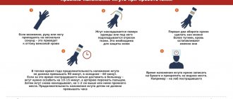
First aid for a sprained ankle:
1. It is necessary to exclude any load and movement in the ankle joint in the first hours, or preferably a day after the injury.
2. Give the limb an elevated position, at an angle of 15 - 30 ° - any pillows that you have at home are suitable for this.
3. Apply cold to the area of maximum pain and swelling. This could be a piece of meat from the freezer, a bag of frozen berries, etc. It must first be wrapped in cloth. To prevent tissue frostbite, the exposure time is no more than 10 minutes, then a 10-15 minute break and repeat this 3 times.
4. Apply an 8-shaped fixing bandage, you can use a regular bandage, but preferably an elastic bandage. The main thing is not to overdo it: the bandage should not increase pain, cause numbness or coldness of the fingers.
Questions and answers
Questions about the treatment of injuries and diseases of the musculoskeletal system that interest you, you can ask the head doctor of the Canon clinic, orthopedist-traumatologist, candidate of medical sciences Leonid Kurzov weekly on Fridays from 10.00 a.m. until 12.00