Etiology and pathogenesis
The development of the inflammatory process in the outer ear is preceded by a violation of the integrity of the skin, which can be caused by many reasons - traumatic injuries, prolonged exposure to a humid environment, skin changes due to metabolic disorders, diabetes mellitus, various dermatitis, eczematous processes. Anatomical structural features predispose to the occurrence of external otitis - narrow external auditory canals, the presence of exostoses, as well as wearing a hearing aid, water getting into the ears, insufficient formation and changes in the composition of earwax, impaired local and general immune status, radiation exposure.
According to the literature, 60–98% of inflammatory diseases of the external ear are of a bacterial nature. The microbial landscape of otitis externa has changed over time. If earlier in 70 - 90% of clinical cases Staphylococcus aureus was cultured, and Pseudomonas aeruginosa was present in 10 - 20% of cases, then recently the role of Pseudomonas aeruginosa has increased to an average of 78%, while Staphylococcus aureus is found only in 9 - 27% of cases . Less commonly, in inflammatory diseases of the outer ear, Staphylococcus epidermidis Streptococcus pyogenes, Streptococcus pneumonia, Enterococcus, Escherichia coli, Proteus, Klebsiella pneumonia, Mycoplasma pneumonia, anaerobes and other microorganisms are also detected. In addition to the bacterial flora, pathogenic fungi play a significant role in the development of otitis externa. Most often, fungal infections of the outer ear are caused by molds of the genus Aspergillus and Penicillium and yeast-like genus Candida, less often by fungi of the genus Mucor, Alternaria, Geotrichum, Kladosporiu. In some cases, bacterial or bacterial-fungal associations act as etiotropic factors. Also, otitis externa can be caused by measles, chickenpox, and herpes viruses.
Anatomy of the ear
[Home] [To volume contents] I.P.
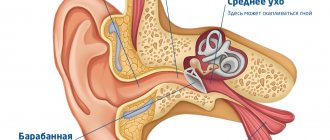
Voloshin (Voronezh) The auditory organ is divided anatomically into the outer, middle and inner ear, and physiologically into sound-conducting and sound-receiving apparatus. The first includes the outer and middle ear, the second - the inner.
The external ear includes: the auricle (Auricula) and the external auditory canal (Meatus acusticus externus); the middle one includes: the tympanic membrane (Membrana tympani), the tympanic cavity (Cavum tympani) with accessory cavities: the Eustachian tube (tuba Eustachii) and the cavities of the mastoid process; The inner ear includes: the vestibule (Vestibulum), cochlea (Cochlea) and semicircular canals (Canales semicirculares).
Rice. 1. Auricle. 1 - cavitas conchae; 2 - anthelix; 3 - helix descendens; 4 - fossa navicularis; 5 - tuberculum Darwini; 6 - helix ascendens; 7 - crura anthelicis; 8 - fossa triangularis; 9-cymba conchae; 10 - crus beliefs; 11 - tragus; 12-antitragus; 13 - incisura intertragica; 14-lobulus auriculae; 15-meatus acust ext. |
Auricle - Auricula
The auricle is used to catch sounds. In general, it represents a peculiarly shaped cartilaginous funnel, the bottom of which passes into the external auditory canal. The shape, size and degree of deviation of the shell from the skull are normally very different. On the shell, there is an outer - anterior surface, concave, and a medial surface, facing the skull - convex. The names of individual parts of the concave surface of the auricle are shown in Fig. 1. The skeleton of the upper two-thirds of the shell consists of elastic cartilage, covered with perichondrium, very rich in elastic and fibrous fibers; it, in fact, gives the auricle its characteristic shape. The lower third - the lobe, or earring (lobulus auriculae), does not contain cartilage, but is a skin sac filled with adipose tissue. The cartilage of the auricle directly passes into the cartilaginous part of the external auditory canal, which has clinical significance in the pathology of the auricle and auditory canal.
The auricle is attached to the temporal bone of the skull by a whole system of fibrous fibers of the perichondrium, in some places collected into cords - ligaments (ligamentum auriculae anterius, posterius et superius). The skin lining the auricle on its concave surface adheres to the tissues so tightly that it cannot be moved or folded; on the contrary, on its convex surface this is done easily. There are hairs, sweat and sebaceous glands all over the surface. There are more of them on the concave surface, especially at the entrance to the auditory canal. Hair is especially strongly developed here, trapping dust, insects, etc. when they enter the ear canal.
The auricle has two muscle groups:
1. A system of cutaneous muscles, numbering 5. They begin and end on the perichondrium. They have no functional significance and are rudiments.
2. System m-li epicranii, number 3. It consists of: m. attolens auriculae seu auricularis super., elevating the auricle; m. retrahens auriculae seu auricularis poster., pulling the shell posteriorly; attrahens auriculae seu auricularis anter., pulling it anteriorly. The functional significance of these muscles in humans is very insignificant, while in animals, for example, in a horse, it is great: the muscles direct the auricle towards sounds.
External auditory canal - Meatus acusticus externus
The external auditory canal is a slightly curved canal, with an average length of 35-36 mm; its direction is from outside to inside, slightly anteriorly and downwards. It begins at the bottom of the auditory shell and blindly ends in the tympanic membrane in the depths. The auditory canal is divided into: the cartilaginous part - the outer part, which accounts for two-thirds of its length, or 21 mm, and the inner - bone part, which accounts for a third of the entire length of the passage, or 14-15 mm (Fig. 2).
Rice. 2. Scheme of a horizontal incision through the external auditory canal. 1 and 2 - cartilaginous part of the auditory canal; 3 - bone auditory canal; 4 - tympanic cavity; 5 - sigmoid sinus; 6 - head of the lower jaw; 7 - parotid gland; 8 - tragus; 9 - mastoid process; 10 - auricle. |
The cartilaginous section of the external auditory canal contains cartilage, in the form of an incomplete cartilaginous tube or groove, only in the anterior and lower walls; the cartilage gradually decreases towards the bony part of the ear canal, and the posterior and superior ones are composed of fibrous connective tissue (Fig. 3). In addition, in the cartilages of this groove, i.e. on the anterior and lower walls of the passage, there are from two to five slits (incisurae Santorini), filled with connective tissue.
Rice. 3. Transverse sections of the external auditory canal. A - not far behind the opening of the external auditory canal; B - through the middle of the cartilaginous auditory canal; S. - near the inner end of the cartilaginous canal, 1,1,1, - cartilaginous groove; 2 - fibrous plate. |
The cartilaginous and bony parts of the ear canal are connected through pliable connective tissue. All this together gives the ear canal a certain mobility both when expanding its lumen and when pulling it back. Incisurae Santorini, as well as the partly fibrous structure of the posterior-superior wall, are of particular clinical importance, since they allow the spread of pathological processes from the ear canal to the outside, to neighboring organs and tissues, and vice versa.
The bony part of the external auditory canal in an adult is formed below and in front from the os tympanicum, and above and behind from the squama resp. pars mastoidea of the temporal bone. On the macerated temporal bone, the outer edge of the auditory canal (porus acusticus externus), in the part formed by the os tympanicum, protrudes sharply in front and below. On the contrary, from above and behind, the outer surface of the processus mastoidei gradually passes into the external auditory canal. In the corner, on the border of its upper and posterior walls, a bone protrusion in the form of a spike (spina supra meatum seu spina Henle) is constantly observed, and now behind it there is a fossa (fossa mastoidea). Both are very important identification points when chiseling the mastoid process, indicating the location of the antrum (Fig. 4).
Rice. 4. Macerated temporal bone of an adult. 1 - spina supra meatum; 2 - crista temporalis; 3 - fossa mastoidea; 4 - porus acusticus externus; 5 - proc. styloideus; 6 - proc. zygomaticus. |
At the inner end of the bony part of the auditory canal there is a circular groove (sulcus tympanicus), into which the eardrum (pars tensa) is inserted and reinforced with a tendon ring (annulus tympanicus). A similar semi-ring-shaped groove, not closed at the top, is present only in that part of the auditory canal that is formed by the os tympanicum, while in the squama it is not present. In the notch that exists here (incisura Rivini), the tympanic membrane (pars flaccida s. Schrapnelli) is attached directly to the squama. The external auditory canal in a newborn differs significantly from that just described (Fig. 5).
At this age, there is only the skin-cartilaginous part. It is compressed from top to bottom in the form of a gap; there is no bone wall yet, but instead there is an os tympanicum in the form of a thin incomplete bone ring, open upward and anteriorly - incisura Rivini; the eardrum is inserted into this ring; subsequently, the bony part of the auditory canal is formed from it. Os tympanicum during this period is attached with its horns almost horizontally to the scales and bent to the base of the skull; therefore, the eardrum at this time is visible from the side of the ear canal, also in an almost horizontal position, as a continuation of its upper wall.
Rice. 5. Temporal bone of a newborn. 1 - processus zygomaticus; 2 - squama; 3 - fenestra ovalis; 4 - pars mastoidea; 5 - fenestra rotunda; 6 - foramen stylomastoideum; 7 - inner wall of the tympanic cavity; 8 - annulus tympanicus; 9 - pars petrosa. The direction of its longitudinal axis is not strictly transverse, but deviates from a straight line in both the frontal and horizontal planes (Fig. 6.). |
Inspecting the eardrum during this period and distinguishing it from the soft parts of the ear canal presents great difficulties even for an experienced otialogist.
Further formation of the bony part of the external auditory canal occurs through the growth of the os tympanicum. Os tympanicum, and with it the tympanic membrane, with its lower edge extends from the base of the skull and from an almost horizontal one passes into a more vertical position, with an angle of 45° to the horizon; This is typical for adults. The posterior and posterosuperior walls are formed partly due to the os tympanicum, and mainly due to the mastoid part and ossification of the posterior fibrous wall. The formation of the bone part, as well as the entire external auditory canal in general, ends by 5-7 years of life (according to Ostmann).
The entire length of the auditory canal is covered with skin, which has a different structure in its different parts. The skin of the cartilaginous part of the passage is a continuation of the outer skin of the body, 1-2 mm thick. It is loosely connected to the underlying tissues and is rich in hairs and sebaceous glands. There is also a special type of deep, large clubbed, so-called. sulfur glands (gl. ceruminales), which secrete a light yellow secretion - sulfur. There are 1-2 thousand such glands.
In the bony part, the skin fits tightly to the bone, thins to 1 mm or less, loses hair, glands and corium, and only the epidermis passes onto the eardrum. Along the upper wall of the bony part of the ear canal and along the eardrum along the handle of the malleus there is a narrow strip of skin that retains all the properties of the outer skin - stria malleolaris.
This histological structure of the skin of different parts of the ear canal is of particular clinical importance.
Rice. 6. Diagram of the direction, shape and size of the lumen of the ear canal at various places along its length. I - at the entrance; II - at the end of the cartilaginous part; III - at the beginning of the bone section; IV - at the end of the bone section. |
The direction, shape and width of the lumen of the external auditory canal vary greatly.
The figure shows two clearly defined bends: at the entrance and at the junction of the cartilaginous and bony parts of the ear canal, due to which its front wall protrudes into the ear canal, and inspection of the front part of the eardrum is difficult even with maximum retraction of the auricle posteriorly (alignment of the ear canal) and expanding it. In the same way, the lower wall is not strictly horizontal, but has a curve upward, so that inspection of the lowest part of the eardrum is also difficult. Here, in front of the eardrum, something like a depression is formed in which small foreign bodies (sinus mae) can be hidden. The upper wall is approximately horizontal and imperceptibly merges with the pars flaccida, the posterior wall merges with the tympanic membrane without a noticeable angle, while the anterior wall forms an acute angle of about 27° with it.
The average length of the external auditory canal is 25.25 mm (according to Bezold). But, due to the double inclination of the eardrum to the horizontal and frontal planes, the length of the walls of the ear canal is not the same: the longest of them is the front, and the shortest is the back wall - the first is longer than the second by 5-7 mm, the upper is shorter than the lower by 4-5 mm.
This is of clinical importance when removing foreign bodies, cerumen plugs from the ear canal and during paracentesis of the membrane.
The width and shape of the lumen of the auditory canal are different along its entire length. In Fig. 6 it can be seen that its cross-section generally has the shape of an oval. The dimensions and slope (oval section), as well as the shape of the oval itself, are different in different places: at the entrance to the auditory canal the slope is greatest and is located so that its long axis is almost vertical, and its transverse axis is almost horizontal. The narrowest place is at the junction of the cartilaginous and bone parts of the auditory canal; then it expands somewhat again, so that at the eardrum itself it narrows (shrinks) again. At the same time, the position of the oval of the initial part of the auditory canal changes: it gradually tilts its long axis, which from almost vertical at first becomes close to horizontal - at the eardrum, and the almost horizontal transverse axis gradually becomes close to the vertical - to the eardrum. As a result of the indicated spiral change in the longitudinal axis of the external auditory canal, the posterior wall of the initial part of it then, at the end, becomes posterosuperior, and the anterior one initially becomes anterior-inferior at the end of the auditory canal at the eardrum.
Topographic relationships of the external auditory canal
In front and below, the cartilaginous, and partly bone, sections of the external auditory canal border on the parotid gland (gl. parotls), adjacent to it directly, since in this part of the gl. parotis does not have a capsule. As a result of this, as well as the presence of incisurae Santorini in the cartilaginous part of the auditory canal, inflammatory processes of the canal can penetrate into the gl. parotis and vice versa.
The articular head of the lower jaw with its inner half is in contact with the bone half, and the lateral half with the medial part of the cartilaginous part of the external auditory canal. This is easy to verify: if you insert a finger into the ear canal, then during chewing movements you can easily feel the movements of the capituli mandibuli. For the same reason, chewing movements are impossible - at least they are very painful - with furunculosis of the ear canal.
The upper wall of the bony part of the external auditory canal borders the tympanic cavity and the middle cranial fossa and is separated from them by a bone plate of varying thickness. It almost always contains pneumatic cells that open into the upper part of the tympanic cavity (recessus epitympanicus); they thin the upper wall so much that suppuration in the middle ear can break into the external auditory canal in this place, without breaking the eardrum, or even penetrate into the middle cranial fossa.
The back wall faces pr. mastoideus; it contains the descending part of the n. facialis in its bony canal. With a strongly pronounced cellularity of the appendix, the back wall becomes thinner, like paper, and pus from the cavity of the appendix, when it suppurates, can break through this wall into the ear canal. Dehiscence of the facial nerve canal is often observed here.
The most medial part of the posterior superior wall of the auditory canal in its upper half is the anterior, outer wall of the antrum mastoideum, aditus ad antrum et atticus, s. recessus epitympanicus.
When suppuration occurs in them, this wall noticeably swells and falls, which serves as one of the reliable and early indications for mandatory surgery on the mastoid process.
Blood and lymphatic vessels of the external ear
The posterior surface of the auricle is supplied with blood from a. auricularis post. - branches a. carotis ext., and the anterior one - from a. auricularis anterior - branches of a. temporalis superficialis and from a. auricularis posterior with its branches. They also supply the cartilaginous part of the external auditory canal in the lateral part. The medial part, the bony part and the skin plate of the tympanic membrane are supplied by a. auricularis profunda - branches of a. maxillaris int. Each artery is accompanied by two veins. The veins of the anterior and posterior surfaces of the tympanic membrane anastomose with each other via vv perforantes.
Lymphatic vessels are divided into anterior, posterior and inferior. The anterior ones flow into the gl. auricularis ant., located in front of the tragus, the lower ones - in the lymph glands located on the gl. parotis, and the hind ones - in gl. mastoideae, located on pl. mastoideum
Nerves
The motor nerve branches of the auricle muscle receive from n. facialis in the form of rami temporales and auriculares post., sensitive - from the third branch of n. trigemini in the form of rami auriculo-temporales and from n. auricularis magnus from plexus cervicalis.
The external auditory canal is innervated from r. auricularis n. vagi and n. auriculo-temporalis, one of the branches of which innervates the eardrum - n. membranae tympani.
[to contents]
Epidemiology
The prevalence of inflammatory diseases of the external ear ranges from 17 to 30% among all otiatric pathologies. The growth of this pathology is facilitated by deteriorating environmental conditions, an increase in the level of resistance of the flora, an increase in the number of people with metabolic disorders, immune status, including allergopathology, irrational treatment of acute inflammatory pathology, untimely contact with an otorhinolaryngologist (ENT doctor) and a number of others moments.
Otitis externa is a fairly common disease, but the epidemiology has not yet been sufficiently studied, including due to the different designations of the same type of pathological process. Inflammatory diseases of the outer ear occur in all countries and regions of the globe, but are most often observed in hot and humid climatic regions; an increase in incidence is noted in the warm season. On average, every 10th person experiences this disease at least once during their lifetime, and 3–5% of the population suffers from a chronic form of otitis externa. On average, 0.4% of the population suffers from acute external otitis every year. The disease is most common among people exposed to high humidity for a long time.
Otitis externa occurs in all age groups, the highest prevalence is observed in older children and young adults, then increases slightly after 65 years. The incidence of inflammatory diseases of the outer and middle ear in men and women is approximately the same. No racial differences have been identified in the epidemiology of otitis externa.
Classification
According to the course of the disease in otorhinolaryngology, acute (up to 1 month), subacute (from 1 to 3 months), chronic (lasting more than 3 months), and recurrent (3 episodes or more acute external otitis within a year) forms of external otitis are distinguished.
The type of disease is determined by the location and nature of the inflammatory process. Among the nosological forms there are diffuse (eczema, dermatitis, erysipelas, herpes, perichondritis, chondroperichondritis, malignant external otitis and a number of other types and limited external otitis (furuncle, abscess).
According to the degree of severity, there is a mild degree (minor discomfort and itching in the ear, minimal swelling of the skin of the external auditory canal), moderate (pain and itching in the ear, narrowing of the external auditory canal due to severe swelling of the skin), severe (severe pain in the ear, external auditory meatus completely closed, periauricular erythema, regional lymphadenopathy and fever)
Read more on the topic “Useful information about hearing”
Why you don't need a hearing aid
The Academy of Hearing has been working with people suffering from hearing loss for over ten years. During this time, we have collected a whole collection of rumors, myths and misconceptions that surround this disease and the means of its correction - a hearing aid. We collected the most popular ones and asked our experts to comment on them in an easy, understandable and ironic way of “bad advice.”
How to choose a hearing aid
Hearing deterioration can be the result of injury, a consequence of a chronic or acute course of the disease, or age-related changes in the structure of the ear apparatus. In any case, to maintain the quality of life, it is necessary to compensate for this deficiency. Today this is possible thanks to the existence of a large number of special devices. If you're considering whether to use one of these, you need to think about how to choose a hearing aid? After all, manufacturers offer the opportunity to choose.
Voice and voice production
The main carriers of information for humans are signals that are generated by the senses, delivered to the brain and processed there. First of all, these are acoustic speech signals of the voice. In this article, we will take a closer look at the mechanisms of speech sound, voice and vocal production, and also find out why parrots can speak almost the same as humans.
Tinnitus: causes, treatment methods, consequences
Tinnitus is a widespread phenomenon and one of the most common diagnoses in the practice of treating ENT diseases. In 1999, in Germany, on behalf of the German League of Physicians, a large study was conducted in the field of auditory acoustics, during which it was found that tinnitus entails psychosomatic disorders such as sleep disturbances, depression and increased audiological stress.
How often should you change your hearing aids?
Do you remember what feelings and emotions you experienced when you first put on your hearing aid? How much brighter and more fulfilling has your life become, filled with clear sounds and voices? If several years have passed since then, are you sure that your hearing is still as good? Often, people who use hearing aids begin to notice over time that they begin to hear “somehow differently.”
Hearing aid or hearing amplifier: making the right choice!
Typically, a person loses their hearing gradually. First, the quietest, most inexpressive sounds and subtlest nuances “disappear.” Because of this, it is difficult for a person to ascertain a change in his ability to hear. Quite often, people try to ignore minor hearing loss until the truly important sounds for everyday life, such as the cry of a child or the sound of a car approaching, begin to disappear from the range of audible sounds.
Read all useful articles about hearing >>>