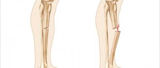
Depending on the intensity of the force acting on the bone, complete and incomplete fractures occur. With incomplete fractures, predominantly longitudinal bone cracks are formed, less often - transverse, subperiosteal cracks in children.
Causes and types of displacement of bone fragments Features of bone fractures in children Features of bone fractures in older people Traumatic disease as the body’s response to injury
With direct trauma, more severe damage occurs directly at the site of application of force than with indirect trauma. Soft tissues and blood vessels are injured, and the bone breaks transversely, sometimes with the formation of several fragments. Indirect trauma is characterized by less soft tissue damage and a spit or spiral line of bone fracture (from torsion), and sometimes by a small, triangular-shaped mid-bone fragment.
More often, the mechanogenesis of a fracture of a long tubular bone consists of its bending, when the force of a mechanical factor acts on the principle of a lever. In an accident, the force can act in one direction, on a limb that is fixed to a certain extent, or in two opposite directions at different points of application.
Torsion fractures occur when one end of a bone segment is fixed, but the other is twisted beyond the limits of functionality and strength of the bone.
Avulsion fractures occur as a result of a strong, unexpected contraction of a muscle. When a muscle contracts, a piece of bone is torn off at the site of attachment of its tendon (the dorsal edge of the distal phalanx of the finger, the olecranon, the base of the fifth metatarsal bone, etc.). In adolescence, avulsion epiphysiolysis occurs - a fracture in the zone of the growth cartilage.
Mechanical damage to the support and movement apparatus and organs can be isolated (monotrauma), that is, damage within one anatomical segment of a bone, joint or internal organ. In addition, this injury is mono-or polyfocal. With polyfocal trauma, multiple segment injuries occur (double or comminuted bone fractures).
There are such concepts as multiple injuries (wounds, bone fractures), that is, similar injuries of different localization of one system; combined injury - damage to various body systems, that is, the skeleton and organs of the cranial cavity, chest or brain (for example, a hip fracture and liver rupture, a tibia fracture and a concussion); combined trauma - damage to the body caused simultaneously by different etiological factors - mechanical, thermal or ionizing (bone fracture and burn, bone fracture and radiation sickness).
With these injuries, which interfere with each other, systemic disorders occur in the body - a traumatic disease (exhaustion), which, depending on the age and protective forces of the patient, can have a different clinical course, and if vital organs are damaged, it can even result in death.
Causes
Depending on the cause of occurrence, traumatic and pathological fractures are distinguished. The cause of traumatic bone fractures is a sharp, sudden action of a mechanical shock force on the bone. Pathological fractures appear when a certain pathological process affects bone tissue. This may be the result of a cyst or the development of a malignant tumor. In this case, the structure of the bone tissue is gradually destroyed and even small loads can lead to a fracture. With the second type, the risk of fracture increases several times. It even gets to the point where a person can also break a leg while walking. Here the reason is that this is a pathology of the bone itself, and not an external influence on it.
Late postoperative complications
1. Slow healing or non-union of the fracture (in the absence of stable fixation of fragments, periosteum in the area of the fracture, poor circulation, suppuration, etc.).
2. Osteomyelitis is a consequence of inadequate or ineffective treatment of the inflammatory process and suppuration of the wound after surgery or an open fracture.
3. Migration or fracture of the fixator (in case of defects in its design and poor-quality metal, presence of micro-movements in the fracture, etc.). A single moment of large acting force leads to fracture of biological clamps and deformation (curvature) of the metal clamp.
Symptoms
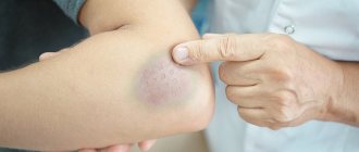
Bone fractures have characteristic symptoms that make it possible to identify pathology when it occurs, which is very important to exclude complications. The following main relative signs of a fracture can be distinguished: - Pain syndrome: sharp pain when a bone ruptures and aching pain in the future, which intensifies with longitudinal load or its imitation. - Swelling: Swelling in the affected area develops gradually. — Hematoma: of different sizes on the affected area; in this case, a hematoma with pulsation indicates continued bleeding. The absolute sign of a fracture occurs as a result of direct destruction of the bone and indicates the completion of the process. These signs of a fracture are: - A characteristic crunch (crepitus): occurs when bone tissue ruptures, and is subsequently audible with a phonendoscope due to the friction of the fragments. — Unnatural direction of a limb or other bone. — Increased mobility in case of joint rupture. — Bone fragments are visible visually during an open fracture. - Shortening of the limb when fragments are displaced, protrusion of the broken bone. Some signs of a fracture without displacement or an incomplete fracture may not appear, which will complicate the diagnosis. The symptoms of a fracture are clearly determined by radiography - the location, type and degree of destruction are recorded.
Structure and functions of the metacarpal bones of the hand
There are a total of 5 metacarpal bones on each hand. They have a tubular structure.
The bases of the metacarpal bones are cuboidal in shape, connecting through ligaments to the distal row of carpal bones.
The body of the metacarpal bone is slightly curved towards the palmar surface and resembles a rocker. Interosseous muscles are attached to the lateral surfaces of the body, which provide fine motor movements of the hand. In case of a transverse fracture of the metacarpal bone, the displacement is difficult to eliminate and maintain precisely because of these muscles, because they constantly pull the fragments onto themselves. The extensor tendons of the fingers are attached to the dorsal surface. The body of the metacarpal bone passes into the neck and then into the head, on which there is an articular surface for connection with the proximal phalanx of the finger. The weakest point of the metacarpal bone can be called the neck; it is at this level that most fractures occur, due to the anatomy and mechanism of injury.
The first metacarpal is unique in that it is shorter and wider than the others and has more extreme angles with the carpus, opposing the axes of the other bones, which characterizes the primary function of the first digit. The second and third metacarpal bones are most rigidly attached to the bones of the wrist with their base. In contrast, the fourth and fifth are more loosely attached and allow the hand to perform more movements and securely grip objects and tools of various shapes.
Diagnostics
For most closed fractures, X-ray diagnosis plays a leading role. This study is necessary not only to confirm the diagnosis of the fracture and document it. It is very important for the traumatologist, based on radiographs, to get an idea of the nature of the displacement of the fragments, the direction of the fracture lines and the presence of additional cracks indicating splitting of the fragment. This information is needed to determine treatment tactics and select the type of osteosynthesis. X-ray examination is also important during the treatment process. It determines the completeness of reposition, the correct position of the fixing structure, the absence of secondary displacement (as swelling subsides), the appearance and formation of callus. The surgeon and traumatologist must follow the rules for taking radiographs for fractures.
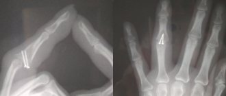
Fracture of the phalanges of the fingers
The cause is a blow with the fingers, an injury while fixing the fingers, or a direct blow to the phalanges. Fractures of the phalanges of the fingers can be:
- intra-articular;
- extra-articular;
- single;
- multiple - within one finger or several;
- combined with dislocations in the metacarpophalangeal or interphalangeal joints.
Symptoms: pain, swelling, hematoma, deformity. The pain intensifies when you try to move your fingers. The diagnosis is established on the basis of complaints, trauma history, objective examination and X-ray results. To treat a fracture of the phalanges of the fingers without displacement, fixation is performed with a plaster cast for 3-4 weeks. In case of fracture-dislocations, joint reduction is performed; in case of displacement of fragments, closed reduction is performed. If it is not possible to compare the fragments using a closed method, skeletal traction or pin osteosynthesis is indicated.
Treatment
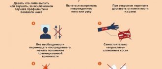
The main objectives of treating a victim with a fracture are saving his life and preserving the limb, restoring the integrity of the bone and the anatomical shape of the joint, the function of the damaged limb and the working capacity of the victim. First aid is aimed at preventing displacement of bone fragments, damage to soft tissues, wound infection, the development of traumatic shock and massive blood loss. Necessary actions: immobilize the damaged area of the skeleton using a splint that captures the joints above and below the site of injury. Stop the bleeding with a tourniquet and apply a sterile bandage to the wound. Give an anesthetic: analgin or promedol. Transport the victim to the emergency room. In case of multiple fractures and spinal injuries, it is not recommended to move the patient until the ambulance arrives. Conservative treatment consists of immobilization and the use of plaster casts after closed reduction of the fracture or without it (if there is no displacement). When applying plaster, the limb must be in the physiologically correct position. Its distal parts (for example, fingers in case of a fractured limb) must be open to be able to determine swelling and prevent disruption of tissue trophism. Sometimes traction is necessary for a fracture—the use of skeletal, cuff, adhesive, or adhesive traction. This method helps to neutralize the action of muscle layers that are attached to bone fragments, prevent their displacement and create conditions for bone tissue regeneration. Skeletal traction gives the greatest effect. A weight attached to a wire that is passed through the bone ensures that the bone fragments are maintained in a position that is optimal for tissue restoration. The disadvantage is the forced immobilization of the patient, leading to a deterioration in his general condition. For cracks in long bones and fractures of small bones, functional methods are used. They involve the absence of immobilization or minimal immobilization of the damaged area and are reduced to providing it with rest. Surgical treatment is necessary for jaw fractures (installation of an external fixation device), restoration of spongy bones (cranial vault), excessive formation of callus, etc. In the postoperative period, the fracture site is immobilized. Recovery time ranges from several weeks to several months. If bone restoration does not occur and a false joint is formed (persistent abnormal mobility at the fracture site), endoprosthetics methods are used (replacement of elements of the musculoskeletal system with implants). After the cast is removed, rehabilitation therapy begins. This is, first of all, a massage. Prescribed 10-45 days after the fracture. It accelerates the process of callus formation, improves blood circulation and tissue nutrition, and prevents muscle atrophy. CRM therapy is a passive development of joints (without the participation of muscles) using a specially tuned mechanical device. Physiotherapy. In the first 10 days, exercises are done for intact joints and limbs. They prevent muscle weakness and joint stiffness. After removing the cast, exercise therapy helps restore the mobility of damaged joints and muscle strength. It is recommended to increase the load gradually, completing rehabilitation with active gymnastics, which helps neutralize the consequences of the fracture.
Types of modern plastic plaster
Plastic plaster is a modern orthopedic product that is made from polymer materials. Plastic plaster is lightweight, easy to use, and provides reliable fixation.
Types of plastic plaster:
- Scotchcast is a lightweight, breathable product; no additional equipment is required to apply such plaster. It is imperative to use lining material (stocking).
- Softcast is an elastic product, used to fix ligaments during stretching, made of polymer fiberglass fabric. It is imperative to use lining material (stocking).
- Primcast - made from polyester fiber, this removable product allows air to pass through well, prevents swelling, and does not cause an allergic reaction.
Advantages of ORDEKT orthoses
The advantages of Ordekt orthoses can be divided into two groups.
For patients:
- High strength of the product with minimal weight - the thermoplastic from which the orthosis is made has a special structure, making the structure lightweight and very durable.
- Reduced rehabilitation time - the orthosis fits tightly to the limb, while the muscles are toned, the mobility of the limb improves, the blood flow does not slow down, which leads to faster healing.
- Resistance to moisture - with the cut you can take a shower, go to the pool, in a word, without changing your usual lifestyle.
- Good breathability - ventilation of the fixed area avoids bedsores, itching, and allergies.
- Lightweight and easy to use - the orthosis can be removed, for example, for hygiene or physiotherapy, and then put on independently.
- Aesthetics - the orthosis, due to its small dimensions, is practically invisible under clothing.
- Allowed for use by children.
For doctors:
- Simplicity of modeling - the new fixation method is easy to master; the entire process takes place on the patient’s injured limb.
- Fast - Thermoplastic heats up and hardens quickly. It will take no more than 10 minutes to fix a joint or bone.
- Full fit - no injury to the skin or hair, no need for additional padding.
- Durability - reliable fixation and complete rest of the limb.
- Possibility of remodeling - using local heating of the material, you can completely or partially change the shape of the orthosis.
- Convenient - the orthosis is very easy to remove.
- Possibility of performing X-ray examination without removing the fixator.
- Easy to store - long shelf life, no special disposal required.