© Author: A. Olesya Valerievna, candidate of medical sciences, practicing physician, teacher at a medical university, especially for SosudInfo.ru (about the authors)
Cystic formations in the cranial cavity always cause well-founded concern among both specialists and their owners. One of the variants of such cavities is a retrocerebellar cyst, which is detected in approximately 4% of healthy people and gives symptoms in only a fifth of its carriers.
Having set out to learn more about this cyst, the reader will find a large amount of information on the Internet, but not all of the information corresponds to reality. Most dubious sources present a retrocerebellar cyst as a kind of intracerebral accumulation of fluid in the place of dead neurons, but in reality it is a cerebrospinal fluid cyst lying outside the brain rather than inside it.
Intracerebral cysts, in other words, cerebral cysts, actually form in the brain itself after necrosis due to a stroke, tumor or injury. The cerebrospinal fluid cyst owes its appearance to the pathology of the arachnoid membrane, therefore it is also called arachnoid, and identifying it with a cerebral cyst is fundamentally incorrect.
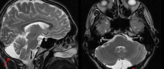
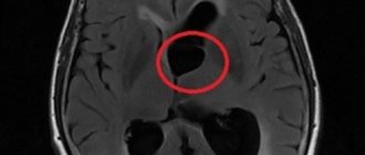
The term “retrocerebellar” is not a characteristic of the cyst itself, but an indication of its location behind the cerebellum (cerebellum), in the region of the posterior cranial fossa, as shown by MRI data, through which these same cysts are detected.
Thus, a retrocerebellar cyst is a cavity formation in the back of the skull, formed by the arachnoid mater, collagen fibers, containing cerebrospinal fluid and lying between the surface of the brain and its arachnoid membrane.
example of cystic expansion of the retrocerebellar arachnoid space
In most cases, a retrocerebellar cyst is discovered accidentally, in young adults who, for one reason or another, have had an MRI. As a rule, the reason for examination is neurological symptoms, which are not always associated with a cyst. A small-sized retrocerebellar cerebrospinal fluid cyst can be asymptomatic and is extremely rarely accompanied by any negative effects on the brain itself.
Indications for the study
The presence of a retrocerebellar cyst can be suspected by some characteristic symptoms:
- Severe headaches that do not go away even after taking painkillers;
- Unreasonable loss of consciousness or fainting state;
- Sensation of squeezing and pulsation in the head;
- Deterioration of hearing or vision;
- Arterial hypertension;
- Numbness of the limbs;
- Impaired coordination of movements;
- Epileptic seizures.
Magnetic resonance imaging is required if surgical intervention in the brain area is necessary.
Note that the clinical picture and severity of symptoms depend on the exact location and size of the retrocerebellar cyst.
How does the disease manifest itself?
The symptoms of the anomaly have a direct relationship with the size of the tumor. Localization and educational factors are important. If the cyst continues to increase in volume or the pressure of the substance in it increases, the patient may experience pronounced manifestations. At the same time, the formation of small cysts that stop their growth does not provoke symptoms. The main reasons for the progression of the disease are:
- neuroinfectious microorganisms;
- long infectious process;
- malfunctions in the functioning of the circulatory system of a chronic nature;
- autoimmune disorders.
The patient exhibits the following signs of an unstable condition:
- pain in the head area;
- discomfort from a feeling of pulsation inside the skull;
- temporary hearing problems;
- presence of noise in the ear canals;
- a feeling of fullness in the head and a surge in pressure;
- disturbances in visual abilities (formation of spots, blurriness, bifurcation);
- paralysis of the arms or legs, complete or partial;
- seizures;
- sudden loss of consciousness;
- numbness of body parts of a temporary or permanent nature.
Diagnostic measures
To prevent the development of an anomaly and establish a timely correct diagnosis, it will be necessary to rely on all the patient’s existing complaints along with the clinical picture. Medical staff use clinical tests such as magnetic resonance imaging.
Contraindications
Magnetic resonance imaging is safe for the body only if all contraindications are excluded.
Magnetic resonance imaging is not recommended in the following cases:
Very often, to obtain more informative images, doctors prescribe additional contrast. In this case, you must ensure that there are no following contraindications:
- Pregnancy of any stage.
- Breastfeeding period.
- Kidney failure.
- Chronic liver diseases.
- Allergic reaction to components included in contrast agents.
How to study a retrocerebellar cyst?
MRI testing with an additional enhancer in the form of a contrast agent helps to distinguish a neoplasm from a tumor. An anomaly of malignant origin can easily accumulate a contrast agent, while a benign cyst is not capable of such a reaction. Doctors identify the causes of the formations to further prevent the formation of new objects and the increase in size of old ones. To achieve this, various diagnostic techniques are used to help identify the sources of infections, problems with the functioning of the circulatory system and autoimmune disorders. These include:
- Doppler diagnostics of the vessels of the head and cervical region - the procedure is prescribed by doctors to identify narrowing of the vessels that serve the function of supplying the brain with arterial blood cells. Due to blood deficiency, gray matter may begin to die.
- Heart check - necessary to determine heart rhythm disturbances and look for insufficiency.
- Donating blood to analyze coagulation and cholesterol levels - an increase in cholesterol and coagulability can become a factor in blocking blood vessels, which will subsequently lead to the formation of a cyst.
- Controlling blood pressure - pressure surges often become a source of strokes and the appearance of post-stroke neoplasms.
- Testing blood cells for infections and autoimmune diseases - prescribed if there is a likelihood of developing a neuroinfection, multiple sclerosis, and other things.
You can make an appointment at any local clinic for diagnostics on the search portal piter-mrt.ru. Here you can apply for an appointment online, find out the current price lists of the centers, the number of vacancies for the coming days and read reviews from visitors.
Preparation rules
During a routine examination, doctors recommend following a few simple recommendations:
- If it is necessary to administer contrast, you must come to the clinic on an empty stomach, that is, do not eat food for 7-8 hours.
- If you are afraid of research or confined spaces, be sure to consult with a specialist. Your doctor may prescribe you sedatives to make the examination process more comfortable.
- Do not bring any metal products inside the tomograph. For this reason, doctors recommend removing metal accessories in advance.
- Prepare a change of clothes that do not have metal or metal-containing accessories.
- The duration of the diagnostic test is about 40 minutes. Empty your bladder in advance to avoid discomfort during the procedure.
Peculiarities
A retrocerebellar cyst is considered a benign formation that appears where the gray matter dies. This type of cyst is quite dangerous
, as it can cause more serious pathologies and diseases.
The peculiarity of this formation is that the retrocerebellar cyst of the brain increases in size over time and therefore, if not treated in a timely manner, the cyst can even destroy brain tissue.
But there are also cases of congenital retrocerebellar cyst, and it can lie dormant for a long time, but due to the influence of certain causes, the neoplasms begin to grow, disrupting the blood circulation of the brain and its functioning.
How is an MRI performed?
To ensure that the procedure is as informative as possible and that the images are clear, experts recommend performing magnetic resonance imaging in closed-type tomographs. Before starting the study, the patient must change into clothes without metal structures, take off his shoes and lie down on a retractable table. If the doctor has prescribed additional contrast enhancement, a special drug is injected into a vein, which is distributed throughout the circulatory system in just a few minutes. Then the table moves inside the tomograph, where the research process takes place.
Maintaining immobility is the main requirement for the patient! Remember that even minimal movements can lead to a decrease in the information content of the procedure.
It is also worth noting that when performing magnetic resonance imaging, a specific loud noise is observed. If this scares you, you can use earplugs.
Patients generally tolerate MRI well. Inside the tomograph there is good lighting and ventilation, there is a loudspeaker, as well as a panic button for emergency stop of the study.
These are relatively common benign and asymptomatic brain lesions. They are usually located in the subarachnoid space and contain cerebrospinal fluid. Richard Bright first described the arachnoid cyst in 1831.
Epidemiology
Arachnoid cysts account for about 1% of all intracranial formations. Although the vast majority of them are sporadic, they are most often observed in mucopolysaccharidoses. In a retrospective cohort study of 48,417 patients who underwent MRI, arachnoid cysts were identified in 661 patients (1.4%).
Clinical picture
Most arachnoid cysts are small and asymptomatic. Approximately 5% of patients have symptoms, and these are usually the result of a gradual increase in cyst size, leading to mass effect. And this leads to either direct neurological dysfunction or distortion of the normal cerebrospinal fluid pathways, which can lead to obstructive hydrocephalus. Arachnoid cysts are thought to arise from congenital clefting of the arachnoid layer with accumulation of cerebrospinal fluid in this space. The cyst wall consists of flattened arachnoid cells forming a thin translucent membrane. There is no solid component and no epithelial lining.
Radiological features
Arachnoid cysts can occur anywhere in the central nervous system, most often (50-60%) in the middle cranial fossa, where they invaginate and widen the Sylvian fissure. At this point they can be divided into three types depending on their size (Galassi Classification):
Classification
- type I
- small;
– limited to the anterior part of the middle cranial fossa, below the sphenoid crest;
– free communication with the subarachnoid space;
- type II
– length along the Sylvian gap;
– displacement of the temporal lobe;
– slow communication with the subarachnoid space;
- type III
– big
– fills the entire middle cranial fossa;
– displacement of not only the temporal lobe, but also the frontal and parietal lobes;
– often leads to a shift in the midline;
– weak connection with the subarachnoid space;
On CT: Arachnoid cysts are very well circumscribed, with an inconspicuous wall and displace adjacent structures. When large, they can have a remodeling effect on the bone over time. CT cisternography (injection of contrast into the subarachnoid space) demonstrates the communication of the cyst with the subarachnoid space. Because this communication occurs slowly, the cyst often fills later and the contrast can be seen to merge with it, highlighting its dependent part.
On MRI : Because arachnoid cysts are filled with cerebrospinal fluid, they have similar signal characteristics including FLAIR and DWI sequences. This makes it possible to distinguish them, for example, from epidermoid cysts. Because their wall is so thin, it can only be seen occasionally, and the displacement of surrounding structures suggests their presence. Phase contrast imaging can also be used to not only determine whether a cyst communicates with the subarachnoid space, but also to determine the location of this communication. Magnetic resonance cisternography: High-resolution sequences such as CISS and FIESTA help delineate the cyst wall and adjacent anatomical structures.
MRI with contrast
In most cases, magnetic resonance imaging of the brain is performed with contrast, especially if any neoplasm is suspected. This makes it possible to examine the tumor in detail and distinguish a benign tumor from a malignant one.
The contrast agent clearly highlights the vessels, which allows you to see any tissue pathologies. Don't worry, contrast agents are safe for the human body. Most patients tolerate this procedure well. You must first consult a doctor and exclude all possible contraindications.
Relationship between the symptoms of cystic formation and its size
The clinical course of the pathology is directly dependent on its increase (growth). Of course, there are many other factors - the location of the cyst, the source of the disease, but size plays a primary role
. An increase in cyst volume provokes an increase in fluid pressure in it, which entails an active course of the disease. The growth of cystic formation is influenced by:
- neuroinfection;
- chronic disorders of the blood supply to the heart;
- development of autoimmune processes (for example, multiple sclerosis).
Decoding the results
A retrocerebellar cyst is located in the brain behind the cerebellum, in the posterior cranial fossa. During magnetic resonance imaging, it is possible to clarify the exact location and size of the formation.
The report will contain all the information, but the MRI specialist cannot make a diagnosis - this must be done by your attending physician. If the diagnosis of retrocerebellar cyst is confirmed, the doctor will determine treatment tactics. For benign tumors, it is often possible to do without surgical interventions - they are required only in cases of large tumors or rapid growth. To avoid serious consequences, consult a doctor at the first alarming symptoms. Be healthy!
How is a retrocerebellar cyst treated?
For the recovery of a patient with such a diagnosis, an endoscopic session is considered an effective operation, since it provides a minimum number of injuries. During the operation, using surgical equipment, doctors puncture the skull, remove the cyst and suck out the substance. This method of therapy may not be carried out in every situation, since the location of the tumor significantly influences the choice of treatment. Shunt surgery may be indicated if hydrocephalus or persistent fluid influx develops. In this case, surgeons perform intracranial shunting, when during the intervention a clinical shunt is used, which helps drain the natural cavities of the department. When the cyst is localized in an accessible area for trepanation, surgery is prescribed to completely remove the anomaly. This procedure is quite traumatic for the patient.
Diagnostics
To detect a cyst, there is a special diagnosis:
- Magnetic resonance imaging is the most commonly used. It provides fairly accurate data and a complete description of the current state of the disease.
- Doppler ultrasound scanning is used as an additional diagnostic method . It allows you to identify more detailed information about the tumor.
- Diagnosis through blood pressure monitoring. To determine the nature of the formation (tumor or cyst), a contrast agent is injected, to which the brain tissue reacts.
In addition to the above diagnostic methods, ECG and computed tomography are used.
About prevention
There are no preventive measures to prevent the formation of a retrocerebellar cyst. In this case, the medical history is important: from birth or acquired during life.
If atheroma is acquired from birth and there is no growth trend in its volume, then no special measures are needed.
For prevention in this situation, a neck massage may be indicated or medications may be prescribed for good vascular tone.
When the formation of a cyst is due to some external reasons, then preventive measures are taken taking into account these reasons.