General information
Bruising is a fairly common occurrence and not always harmless.
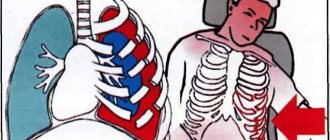
It occurs as a result of direct mechanical influence. The resulting blow leads to damage to soft tissue cells. Most often, with such an injury, the superficial tissues are not ruptured, only rupture of small vessels is noted. However, a very severe injury can lead to serious consequences and complications in the future. Chest injuries are especially dangerous in this context, since vital organs are located in this area, damage to which can occur as a result of a bruise. ICD-10 code for chest contusion is (S20-S29. Chest injuries). A chest injury, as a rule, does not go away without a trace, and if the blow is too strong, a life-threatening condition can develop. At the moment of a very severe bruise or after it, breathing and the heart may stop, since due to a strong blow to the chest area, the functions of two important systems - the respiratory and cardiovascular systems - are sometimes disrupted.
Often a chest bruise causes sharp pain, which intensifies during a deep breath. In the place where the damage occurred, damage and swelling develop. Severe bruising of the ribs can lead to their fracture . Therefore, injuries that cause pain and discomfort in the sternum are indications for hospitalization. The patient may need urgent care and even surgery, which can only be performed in a specialized surgery department. More details about what the consequences of such injuries can be and how to act correctly to help the victim will be discussed in this article.
Consequences
Possible consequences of a bruise in the thoracic area occur immediately or after some time. In this case, the risk of the following dangerous conditions increases:
- Fracture of the ribs, which entails damage to blood vessels and soft tissues.
- Violation of the integrity of the pleural tissue - pneumothorax, which leads to air getting between the lung and the pleural layer.
- Midline fracture of the sternum.
- Compression of the lung when blood enters the pleural cavity is hemothorax. The condition is caused by rupture of large vessels.
For women, a bruise is dangerous not only because of the above-mentioned consequences. The fair half risks damaging the mammary glands, as a result of which the risk of lumps increases. Fibrous hematomas can provoke the development of malignant tumors. Regardless of gender, bruises can cause stroke, myocardial infarction, and sudden cardiac arrest.
Pathogenesis
When the chest is contused, damage occurs in which the integrity of the tissue is not compromised. The mechanism of development of chest contusion varies. Such injuries occur during a fall, after being hit by a hard object, or when a person’s body is pinched on both sides. The severity of the injuries and the mechanism of their development depend on the characteristics of the injury and the type of tissue that was impacted (skin, muscles, bones, adipose tissue, internal organs). It is in the chest that vital organs are located - the lungs and the heart. Therefore, as a result of a blow to the sternum, not only external hematomas , but also very serious damage - rupture of the diaphragm or lung, heart contusion, etc. In such conditions, immediate medical attention is required.
Kinds
The classification of types of thoracic bruise consists of several elements. These include:
- external and open injuries;
- rib fractures;
- vascular injury;
- crushing of the sternum;
- injury to the heart muscle;
- dislocation, curvature of the capsular-ligamentous apparatus;
- damage to nerve endings, spinal cord;
- deformation of organs located next to the injury (abdominal wall trauma).
According to the degree of localization, contusion of the soft tissues of the chest can be of two types. These should include:
- Contusion of the chest on the right - can cause damage to the lung, resulting in a risk of organ rupture with internal bleeding.
- Left-sided contusion - in particularly difficult cases, can cause injury to the heart muscle, even leading to cardiac arrest with a fatal outcome.
Classification
Like other types of bruises, bruises of the sternum, depending on the damage received, can be divided into several degrees.
- First , minor damage is noted. There is practically no pain; abrasions and scratches may remain. After a few days, all unpleasant manifestations disappear without treatment.
- The second is pain, muscle tissue is damaged, resulting in swelling and hematomas.
- Third , in addition to external damage, internal damage to organs may also occur.
- The fourth is a serious injury, causing serious damage to internal organs and threatening health.
First aid for bruised sternum
In case of severe chest injury, you should call an ambulance. Before the team arrives, you need to do the following:
- arrange the victim in a semi-sitting position, ensure him peace;
- apply a cold compress to the damaged area, which will help reduce swelling and hematoma;
- if the pain is severe, give the patient an anesthetic and be sure to inform the arriving doctors about this.
Next, the patient should be treated by a traumatologist. An ambulance employee will decide on the spot whether hospitalization is necessary or recommend contacting a specialist for treatment.
The appointment is conducted by traumatologists
Denis Ivanovich Burmakin - Deputy Director for Medical Affairs, orthopedic traumatologist. Extensive experience in emergency traumatology, pediatric orthopedics, and rehabilitation after injuries.
Ask a question Read more...
Bryukhanov Anatoly Valentinovich - microsurgeon.
Ask a question Read more...
Bryukhanov Vladimir Innokentievich - traumatologist-orthopedist, highest qualification category, Honored Doctor of the Russian Federation, work experience of more than 35 years.
Ask a question Read more...
Zotov Vyacheslav Viktorovich - orthopedic traumatologist.
Ask a question Read more...
Pospelov Yuri Vladimirovich - orthopedic traumatologist.
Ask a question Read more...
Klimov Vladimir Aleksandrovich - orthopedic traumatologist. Operating traumatologist-orthopedist.
Ask a question Read more...
Dmitry Anatolyevich Shcherbovich - orthopedic traumatologist, chiropractor, physiotherapist, first medical category, 13 years of work experience. Head of the rehabilitation department.
Ask a question Read more...
Causes
A chest contusion occurs from a blow to a hard object, a blow from a fallen object, or compression between two hard objects.
Medical statistics show that chest contusions are a relatively rare occurrence. They make up no more than 15% of all cases of bruises treated by doctors. Most often, such injuries occur due to the following reasons:
- Car accidents - as a result of road accidents, drivers who hit their chest on the steering wheel most often suffer from such a bruise.
- Falling from height.
- Fight - bruise can occur as a result of a blow to the chest with a blunt object or fist.
- Household reasons.
- Sports activities.
- Industrial injuries.
Blunt trauma to the mammary gland
A mammary gland bruise (right or left) is a blunt injury or closed injury to the soft tissue of the chest. Such damage occurs during sports; girls involved in strength martial arts, artistic gymnastics, and cycling are especially often affected.
You can injure your chest:
- in case of an accident;
- during a domestic quarrel;
- when hitting a door frame as a result of dizziness;
- while playing with children;
- and even just when falling on the street.
Fortunately, mammologists report blunt breast injuries in women in only 1-2% of all requests for help. And this despite the open location of the organ. By the way, you need to go to a mammologist if you have suffered an injury in the mammary gland (right or left).
What happens with a closed injury?
Under the influence of a traumatic factor (impact, fall), the vessels of the mammary gland are destroyed and blood enters the tissue from the bloodstream, forming a hematoma (bruise).
If the bruise is minor, the vessels may remain intact or the bleeding will stop quickly and spontaneously with the formation of a small superficial hematoma. If there is a large area of damage or a strong blow, the bruise will be large and painful.
Considering that the mammary glands are well innervated, damage is accompanied by pain up to a state of shock. Shock often develops when the nipple and areola area are bruised, despite the fact that the tissues of the organ are very resistant to direct injury.
Symptoms of injury
For blunt breast trauma, you:
- feel severe pain;
- notice a change in breast volume, swelling, redness of the skin, thickening, and then bruising;
- You will find bloody discharge from the nipple (due to injury to the milk duct).
Injuries are accompanied by superficial and deep hematomas. If it seems to you that the blow did not rupture the blood vessels or cause a bruise, consult a doctor. Perhaps the hematoma is hidden deep in the tissues.
Consequences of a chest bruise
Small hematomas often resolve, like any superficial bruises, without consequences or medical intervention, but you should not expect that “everything will resolve.” If you do not contact a mammologist in a timely manner and there is no adequate treatment, a breast injury can be complicated by:
- abscess (when pathogenic bacteria penetrate into the contents of the hematoma, blood is an excellent environment for the proliferation of microorganisms);
- fat necrosis of tissue in the bruise area;
- deformation of the mammary glands due to the replacement of necrotic (dead) tissue with scar.
Please note that the hematomas themselves never become malignant, but most patients with breast cancer (breast cancer) have a history of bust injuries. Such damage is especially dangerous for patients with nodular mastopathy and breastfeeding.
Symptoms of chest contusion
The characteristic signs of any bruise are, first of all, hemorrhage in the tissue without damage to the skin. But the complex of symptoms of sternum injury depends on the severity of the injuries received. Therefore, the stronger the blow, the more pronounced and intense the symptoms will become.
Signs of a chest contusion may include the following:
- Pain in the place where the blow occurred - the pain can be aching, throbbing, or dull. If you experience throbbing pain, this may indicate damage to the nerve endings, as well as a heart injury. Pain can be bothersome both during movement and at rest. There is often an increase in pain when breathing, sneezing, coughing, or talking.
- Hematoma - a bruise forms in the place where the soft tissue of the sternum was damaged, since upon impact small capillaries are damaged and bleeding occurs into the subcutaneous tissue. The hematoma may be larger than the affected area. Minor lesions may resolve without bruising.
- Swelling – fluid accumulates at the site of injury and swelling forms.
However, such trauma is rarely isolated. Due to the anatomical proximity of the lungs, diaphragm, and heart, the risk of their damage is very high. Most often, a bruise of the sternum is accompanied by:
- lung contusion;
- bruised ribs, which may result in a rib crack or fracture (complete or partial);
- breast injury;
- heart bruise.
If such complications occur, then the symptoms after the blow will be more pronounced. In this case, the following symptoms are likely:
- Severe pain when breathing, touching, palpating.
- The appearance of breathing problems (they indicate damage to the lung or injury to the ribs). Sometimes the pain is so severe that it is difficult for a person to breathe air.
- If the injury causes damage to the lungs or pleura, the victim may develop pneumothorax or hemothorax . Pneumothorax is a condition in which a large amount of air collects in the chest cavity, causing compression of the lung. With pneumothorax, there are serious breathing problems, decreased pulse and increased blood pressure , and the development of emphysema under the skin. The man is in serious condition. Hemothorax - when large blood vessels located in the sternum, lungs or abdominal cavity are damaged, blood enters the pleural cavity and thickens there. In this condition, serious increasing breathing problems, severe weakness, headache and other general symptoms are observed. Both of these conditions are very dangerous to health and require urgent medical attention.
Symptoms of bruised ribs immediately manifest as sharp pain and redness of the skin. Later, swelling and hematoma develop. The victim feels a constant dull aching pain, and it increases with sudden movements, coughing, laughing, and deep breathing. The same are the signs of cracked ribs. If a person feels a crunch inside when moving, this may be evidence of a rib fracture. Pain from a chest injury may not go away for a long time.
Complications of rib fractures
Rib fractures, especially multiple ones, are often complicated by hemothorax, closed and valve pneumothorax, and subcutaneous emphysema.
Hemothorax
Hemothorax is the accumulation of blood in the pleural cavity, which leaked from damaged muscles or intercostal vessels, when wounded by fragments of the rib of the parietal pleura. There is less bleeding when the lung parenchyma is damaged, but then, as a rule, hemothorax is combined with pneumothorax, i.e. hemopneumothorax occurs. Depending on the degree of bleeding, hemothorax can be small - it occupies only the pleural sinus (100-200 ml of blood), medium, and does not reach the level of the lower angle of the scapula (300-500 ml). Total hemothorax (1-1.5 l) is extremely rare.
The level of hemothorax is determined by percussion and x-ray with the patient in an upright sitting position. During percussion, the upper limit of dullness of the percussion sound is especially clearly demarcated against the background of the box sound of pneumothorax. On the radiograph, the hemothorax area is darkened with a pronounced horizontal upper border. Under local anesthesia, the diagnosis is clarified by puncture of the pleural cavity. If the hemothorax is small, sometimes it is not possible to suction the blood from the sinus.
Symptoms . A small hemothorax has no special signs, and the clinical symptomatology is dominated only by signs characteristic of rib fractures. But the dynamics of hemothorax need to be monitored, since it can increase. Moderate, especially total, hemothorax compresses the lung, hypoxia, shortness of breath, sometimes hemodynamic disturbances, etc. appear. With hemothorax, body temperature predominantly increases (38-39 ° C).
Treatment . Considering that hemothorax is one of the complications of rib fractures, the patient is treated comprehensively. As for hemothorax, with minor hemorrhage into the pleural cavity, the blood gradually resolves, although puncture is done to minimize the amount of blood. Due to reactive inflammation of the pleura and blood residues, the pleural cavity becomes obliterated over time.
In case of significant hemothorax, blood from the pleural cavity is immediately sucked out with a puncture needle, since after some time it can settle into a clot, and then it is necessary to perform an operation.
If after the puncture blood appears again, which should be regarded as unstoppable bleeding from damaged vessels, the patient undergoes a thoracotomy - a surgical intervention to stop the bleeding. But before that, a puncture and a Ruvilois-Gregoire test are performed to determine whether the blood is fresh. Fresh blood obtained in a test tube in air quickly settles into a clot, but stale blood does not settle. Then you can limit yourself to repeated puncture.
There are cases when exudative pleurisy develops after hemothorax. Then the diagnosis is clarified by puncture and conservative treatment is carried out (repeated punctures, drug therapy, etc.).
Closed and valve pneumothorax
If the visceral pleura and parenchyma are damaged during inspiration, air from the lung enters the pleural cavity, where normally there is negative pressure (0.039-0.078 kPa, 4 8 mm water. in.).
The elastic lung tissue contracts and the lung collapses - a closed pneumothorax is formed. If, in addition to air, blood from damaged intercostal vessels or lung parenchyma enters the pleural cavity, hemopneumothorax is formed.
There are cases when the lung is injured so that pleural or lung tissue hangs over the rupture site. Then, when you inhale, air enters the pleural cavity, and when you exhale, this tissue, like a valve, closes the opening to the lungs and does not allow the air to escape - a valve pneumothorax is formed.
With each inhalation, the amount of air in the pleural cavity increases, its pressure rises sharply (tension pneumothorax), which leads to compression of the lung and displacement of the mediastinum. Disorders of gas exchange and hemodynamics appear quite quickly. The general condition of the patient becomes severe, severe shortness of breath, cyanosis of the skin and mucous membranes, and tachycardia occur. As a result of sudden suffocation, the patient develops fear and severe psychomotor agitation.
The presence of pneumothorax is determined by percussion by the characteristic box sound, comparing it with the healthy half of the chest. On auscultation, breathing is weakened, and with a collapsed lung, it cannot be heard. The radiograph shows a clear contour of the collapsed lung against the background of clearing of the pneumothorax area. Puncture of the pleural cavity clarifies the diagnosis; moreover, with valvular tension pneumothorax, air comes out through the needle under pressure.
Treatment . In case of closed pneumothorax, regardless of its degree, air is immediately sucked out from the pleural cavity. This, firstly, improves the general condition of the patient, and, secondly, with prolonged pneumothorax the lung becomes rigid, and then it is more difficult to straighten it.
If for hemothorax the chest is punctured in the lower section, then for pneumothorax it is punctured in the upper, mainly in the second intercostal space along the midclavicular line. The air is sucked out using a Janet syringe or a triampulle system. If the pressure in the pleural cavity becomes negative, then the triampulle system is excluded. Lung expansion is controlled by percussion and x-ray.
The general condition of a patient with a closed valve pneumothorax is so severe that he should immediately, directly at the scene of the accident, perforate the chest wall (with a thick injection needle) - convert the closed pneumothorax into an open one. After the puncture, air is immediately released from the pleural cavity under pressure. And then the pressure in the cavity is equalized with atmospheric pressure, and the patient’s general condition improves. Choking is significantly reduced. After a few hours, with collapsed lungs, the “valve” can be stuck with fibrin, and a regular closed pneumothorax is formed. In these cases, the air from the pleural cavity is sucked out using a triampulle system. If the lung has expanded, then the triampulle system is not excluded, but negative pressure is maintained in the cavity and monitored for a day or two. The system is turned off only when they are sure that the valve has closed and there is no air in the pleural cavity. This is confirmed by percussion, auscultation and x-ray.
If the amount of air sucked out exceeds the nominal volume of the pleural cavity, this indicates that air continues to flow from the damaged lung. In this case, the pleural cavity is drained using the Bulau method .
Execution technique . A finger of a surgical glove is hermetically secured to one end of a sterile rubber tube (diameter 5 mm and length 60-70 cm), the top of which is cut along the length by 1.5-2 cm. Thoracentesis is performed and the second end of the tube is inserted into the pleural cavity, fixed, sealed skin wound with a suture. The finger is dipped into a sterile jar filled with an aqueous solution of an antiseptic substance (furacilin (1: 500), etacridine lactate (1: 1000), etc.).
During inhalation, the tip of the finger collapses in the solution and closes the hole in it, preventing the solution from being sucked into the tube. When you exhale, the chest collapses and air escapes through the tube into the jar. This is how suction drainage works. After a day or two, when the valve in the lungs closes, negative pressure is created in the pleural cavity, and the lung expands, the drainage stops working, and after a day it is removed.
If the valve does not close after a few days, this indicates significant damage to the lung, and the patient is operated on. After elimination of pneumothorax, patients with rib fractures are treated according to general principles.
Subcutaneous emphysema
If there is pneumothorax and damage to the parietal pleura or mediastinum, then air from the pleural cavity through the wound enters the soft tissues of the chest or mediastinum, moves through the interfascial spaces into the subcutaneous tissue of the shoulder girdle, neck and face. Subcutaneous emphysema is especially pronounced with valvular pneumothorax.
Characteristic signs of subcutaneous emphysema: swelling in the area of air accumulation, and upon palpation - a specific crunch in the subcutaneous tissue (“walking in the snow”) due to the rupture of bubbles and movement of air. By percussion you can feel the difference in the percussion sound above the emphysema. Air in the soft tissues is also visible on a chest x-ray.
Subcutaneous emphysema gradually decreases, the air is absorbed and no special treatment is required. Only in case of excessive emphysema, when air accumulated under the skin of the neck compresses the veins or trachea, small fasciocutaneous openings with drainage are made above the collarbone, through which the air escapes.
Tests and diagnostics
A person who has suffered a chest injury needs to be examined by a traumatologist or surgeon. Confirming a bruise is not difficult. But at the same time, it is important to exclude the presence of injuries to the chest organs and ribs, and to promptly detect life-threatening conditions. The doctor uses the following diagnostic methods:
- Questioning, medical history - the doctor must find out how the injury was sustained.
- Inspection - allows you to get a general impression of the consequences of the injury. The doctor must pay attention to the presence of cyanosis , which is evidence of hypoxia caused by respiratory failure. The presence of subcutaneous emphysema is also determined as evidence of damage to the lung or bronchus, respiratory sounds, etc.
- Palpation - performed to determine whether a rib fracture has occurred. Sequential palpation of the ribs and sternum is carried out. The doctor pays attention to the symmetry of the sternum, the nature of breathing, the condition of the neck veins (non-pulsating swollen veins are a sign of cardiac tamponade), etc.
- Chest X-ray – allows you to evaluate the condition of the bones of the chest cavity.
- MRI - performed if there is suspicion of damage to internal organs. Allows you to assess the condition of blood vessels and soft tissues, and determine internal hematomas.
- CT scan allows you to assess the condition of bone structures in more detail.
- Electrocardiogram – to determine the condition of the heart. Blood pressure and pulse .
If necessary, other studies are also carried out. It is very important to conduct a complete and comprehensive examination so as not to miss serious internal injuries.
Diagnostics
To make a diagnosis, the doctor examines the patient, palpates the site of injury and prescribes a hardware examination of the breast:
- Ultrasound;
- mammography.
Radiation thermometry is performed less frequently. In doubtful cases, MRI or CT is recommended.
Treatment with folk remedies
You can use traditional methods aimed at rapid healing of injuries only if a slight bruise has occurred and the absence of serious injuries has been confirmed by a doctor. You can prepare folk remedies for a compress. The selected product should be used every day for 10-14 days.
- Herbal tincture for compress. Mix 20 g each of horsetail, bearberry, hernia, bean pods, horsetail, birch buds, cornflower flowers, knotweed and meadowsweet. Pour all the herbs into 0.5 liters of vodka and leave for three days. Use the product as a daily compress.
- Tincture of wild rosemary and nettle. Take 100 g of each herb and pour 100 ml of vodka. After 4 days, pour 200 ml of pomegranate juice into the product and leave for another day. Use for compresses.
- Tincture of birch buds. Take 100 g of birch buds and 50 g of barberry, mix with 100 ml of vodka and leave for several days. Make compresses every day.
- Tincture of St. John's wort and oak bark. Take 20 g of oak bark and marshmallow, 30 g of St. John's wort. Grind the herbs and pour in 0.5 liters of vodka. Leave for 4 days, strain. Use for daily compresses.
Treatment
Methods and methods of therapy depend on the severity of the injury. The recommendations are as follows:
- Mild bruise - treatment at home with limited motor function, using traditional and traditional medicine.
- Moderate degree - the patient can be treated at home with general normal condition. If there is a suspicion of the development of complications, therapy is carried out on an outpatient basis.
- Severe - immediate hospitalization for life-saving measures.
Treating a bruise at home
For 2 days after injury, cold compresses should be applied to relieve swelling and pain. The procedure is carried out for 15-20 minutes 5 times a day. On the third day, the compresses should be warm, which promotes the resorption of the hematoma. Apply a tight bandage all the time after an injury - it will allow the treatment process to proceed more efficiently and reduce pain.
Such injuries disturb the victim's sleep. To prevent pain from interfering with sleep, a pressure bandage is attached as you exhale, and a large pillow should be placed under the lower back. The mattress must be rigid, so the spine will not be deformed and the pain will become less pronounced. You should take a pain reliever at night. Physical exercise is limited, it is possible to do breathing exercises so that phlegm does not stagnate in the lungs. Magnetic therapy and electrophoresis procedures, which can be prescribed by a doctor, are useful.
Drug therapy
Treatment of chest contusion due to a fall, blow or accident is characterized by an integrated approach. The following forms and groups of drugs are effective:
- Painkillers (ointments, gels, tablets) – Baralgin, Tempalgin, Diclofenac, Spazmalgon.
- Anti-inflammatory (tablets, ointments) – Ibuprofen, Nise.
- Thrombolytic (ointments, gels) – Lyoton, Troxevasin. The drugs promote the resorption of the hematoma.
Drug treatment is often carried out using ointments. They can be classified as follows:
- Ointments with non-steroidal components (Voltaren, Diklak) - eliminate external and internal inflammation.
- Ointments based on badyagi (Comfrey, Larkspur) - resolve bruises and reduce pain.
- Heparin ointments (Lavenum, Trombless gel) – relieve pain and have an antithrombic effect.
Gels and ointments for chest bruises are used more often than other remedies. For this reason, it is necessary to take a closer look at effective drugs for treating bruises. One of them is Finalgon:
- Combined product for external use. It has a warming, vasodilating effect on bruises.
- It is recommended to apply a small amount of ointment 2-3 times a day for 10 days.
- The advantage of the product is its rapid action and wide range of uses.
- Cons: many side effects, use only from 12 years of age.
The next drug in the ranking is Troxevasin. Brief characteristics:
- Troxerutin-based gel, which strengthens blood vessels, is an antioxidant. The product has an anti-edematous, anti-inflammatory effect. It is used for pain and swelling after bruises, thrombophlebitis.
- The gel is applied to the damaged area in the morning and evening for 6-7 days.
- An undoubted advantage of the drug can be considered a small list of side effects.
- Disadvantage: cannot be used if the integrity of the skin is damaged.
For chest bruises, treatment is supplemented with systemic medications. Example of Flogenzyme:
- The tablets consist of enzymes of animal and plant origin. They prevent the formation of edema, blood clots, inflammation, and have immunostimulating and analgesic properties.
- The list of indications is huge. For bruises, take 3 tablets three times a day for 14 days.
- The advantages of the drug lie in its systemic effect on the body.
- The downside is the high cost of the tablets.
Article on the topic: Chronic viral hepatitis B - routes of transmission, symptoms, diagnosis and treatment
Heparin ointment is considered the most popular among people for bruises. Her description:
- The drug belongs to the class of anticoagulants and painkillers. Relieves pain, inflammation, prevents the formation of blood clots.
- The ointment is used for bruises and hemorrhoids. To speed up the resorption of the hematoma, a course of treatment is prescribed for 5 to 15 days. The product should be applied 2-3 times a day.
- The main advantage of the ointment, besides the price, is its rapid absorption and speed of onset of action.
- Negative aspects include the condition of controlling blood clotting during prolonged use of the ointment.
The final product in the review is the Badyaga cooling gel. Its characteristics:
- The gel is based on powder made from protozoa – river and lake sponges. The product accelerates the resorption of bruises, heals damaged vessels and capillaries, and relieves swelling.
- The scope of use of Badyagi is enormous, but for bruises, the gel is prescribed 4 times a day until complete recovery.
- Advantages: low cost, the gel can be used even by newborns.
- The drug has no disadvantages; individual intolerance to the components of the gel may occur.
Surgical intervention
Situations often arise that require resuscitation or surgical assistance in a hospital setting with subsequent rehabilitation. Such situations are possible in cases where complications arise after a bruise. These include:
- hemothorax;
- subcutaneous emphysema;
- heart failure;
- hematoma for more than 1 week;
- thromboembolism;
- pneumothorax.
If fluid, blood or air accumulates in the pleural cavity, the patient is given a drainage. It will help remove unnecessary components from the pleura. Then intensive care measures are carried out. If the hematoma lasts 7 days or more, the help of a surgeon is necessary. He makes a puncture in the damaged area, through which the stagnant blood comes out. Damage to the lungs, heart, and large vessels requires immediate extensive surgery under general anesthesia.
Traditional methods of treatment
For mild bruises, folk therapy in the form of compresses, wraps, ointments, and alcohol tinctures is effective. A herbal mixture of horsetail, hernia, knotweed, cornflower flowers, bearberry, birch buds and bean pods helps to eliminate pain. All components are taken in equal proportions - 20 g each. The resulting mixture must be poured with 250 ml of vodka, left for 3 days and compresses applied to the damaged part of the body.
There are many traditional medicines for treating bruises. Effective are:
- Soap ointment. Laundry soap should be finely grated and mixed with chicken yolk. The ointment should be applied to the damaged area 2-4 times a day.
- Table vinegar should be mixed in equal parts with honey. Soak a napkin with the resulting mixture and apply it to the hematoma. The procedure must be done for 3-5 days, 2 times a day.
- Cilantro for pain relief. 50 g of greens should be brewed with a liter of boiling water for 10 minutes. Take the strained infusion in a glass 2-3 times a day.
- Finely grate the horseradish root and apply it to the hematoma twice a day for a week.
First aid
To minimize the negative consequences of an injury, immediately after the incident you must act as follows:
- Apply cold to the affected area by unbuttoning your clothes. A water bottle, a cold heating pad, or a wet towel will do.
- If cracked ribs are suspected, a bandage may be applied to the chest. It will reduce breathing movements, which will relieve some of the pain.
- If you suspect serious damage, you should call an ambulance.
- If a person is unconscious, he needs to be given ammonia to sniff and brought to his senses. If vomiting occurs, tilt the patient's head to the side.
- If breathing stops, artificial respiration must be performed immediately.
- It is important to understand that even with minor injuries you need to consult a doctor. External signs are not always evidence of the true condition of the victim.
Symptoms
Signs of chest injury depend on the severity of the injury and the time it was received. Symptoms are divided into general and local. The latter include:
- Acute pain when pressing or sudden movements.
- Swelling of the tissues at the site of the bruise due to the accumulation of lymph in them.
- Constant pain syndrome is characterized by aching, pulsating sensations when coughing or talking.
- Formation of a hematoma due to damage to small vessels, subsequent hemorrhage into the soft tissue.
Symptoms of injury may increase. This happens when the lungs and pleura are damaged. The condition refers to general signs of complications of a bruise. Symptoms:
- slow heart rate;
- lowering blood pressure;
- increased temperature due to the inflammatory process;
- shortness of breath on exertion;
- pale skin;
- respiratory arrest;
- hemothorax, pneumothorax, traumatic shock.
Consequences and complications
The consequences of a chest injury can be very serious. As a result of the blow, such serious consequences as a ruptured lung, ruptured diaphragm, pulmonary hematoma (hemorrhage), and heart contusion are possible.
Lung rupture often occurs due to injury from a broken rib fragment. The causes of lung rupture can be associated with both a strong blow and tissue tension. The consequences of such an injury can be eliminated if you consult a doctor in time.
With a mild injury, the consequence is a hematoma and pain, which sometimes may not disappear for a long time.
Because sternum injuries cause severe pain, patients are sometimes forced to make limited breathing movements (rigidity). A complication of this phenomenon may be hypoxemia or pneumonia .
If hemothorax is poorly drained, a purulent intrathoracic infection is likely.
List of sources
- Bagnenko, S. F. Medical care for mechanical trauma of the chest and abdomen at the prehospital stage / S. F. Bagnenko, Yu. B. Shapot, A. N. Tuchunov // Bulletin of Surgery named after. I.I. Grekova - 2007. - No. 2. -S. 47-51.
- Tatyanchenko, V.K. Clinical and anatomical aspects in the surgical treatment of chest wall injuries. Educational and methodological manual / V. K. Tatyanchenko. — Rostov-n/D. : Publishing house of Rostov State Medical University, 2006. - 21 p.
- Seleznev S.A., Bagnenko S.F. Traumatic illness and its complications. St. Petersburg 2004.
Diagnosis of cardiac contusion in victims with chest trauma
A.A. Beloborodov, E.P. Danilina, M.A. Dektyarev, V.A. Beloborodov
State Educational Institution of Higher Professional Education “Krasnoyarsk State Medical University of the Ministry of Health and Social Development of the Russian Federation named after. V.F. Voino-Yasenetsky"
An analysis of public health shows that the problem of injury and its consequences is one of the most pressing in modern medicine. According to the World Health Organization, mortality from mechanical injuries ranks third after cardiovascular diseases and cancer, and among people under 40 years of age it ranks first.
Some of the most severe types of trauma include open and closed injuries to the chest, which are accompanied by high mortality, especially when the chest injury is combined with injuries to other parts of the body [5].
The most severe and life-threatening injuries to the chest are heart injuries. [1, 7]. According to various authors, the incidence of cardiac contusion in victims with closed chest trauma ranges from 5 to 75% [2, 6, 7]. In most cases, closed chest injuries complicated by cardiac contusion are combined.
The difficulty of early diagnosis of cardiac contusion lies both in the lack of uniform diagnostic criteria for cardiac contusion and in the polymorphism of its manifestations. According to SJ Rooney, JAJ Hyde, TR Graham., 2000 [8], cardiac contusion is the most commonly missed pathology in patients with closed chest trauma. Meanwhile, the presence of this pathology not only significantly worsens the prognosis, but also radically changes the tactics of intensive care in the early stages of treatment [4].
The purpose of the study was to study the objective diagnostic criteria for cardiac contusion in victims with closed chest trauma and the possibility of early diagnosis of this type of injury.
We observed 56 patients with closed chest trauma complicated by cardiac contusion aged from 17 to 78 years. The average age was 43.4 ± 2.2 years. There were 46 men (82.1%), 11 women (17.9%). Isolated chest trauma was noted in 7 (12.5%) patients, combined – in 49 (87.5%). The most common closed chest injury, complicated by cardiac contusion, was combined with traumatic brain injury (73.5% of cases) and abdominal injury (46.9% of cases). The main causes of injury were road traffic accidents (51.8%) and falls from height (33.9%).
36 patients died. Mortality rate was 64.2%. There were no deaths in the group of patients with isolated chest trauma. Among patients with concomitant chest trauma, 36 patients died (hospital mortality in this group was 73.5%).
Signs of myocardial contusion are quite characteristic: pale or cyanotic and moist skin, palpitations, a feeling of interruptions in the heart, shortness of breath, hypotension; upon auscultation, muffled heart sounds, systolic murmur at the apex, and pericardial friction murmur are heard [3, 4]. However, in case of severe combined trauma against the background of respiratory and circulatory depression, especially in the absence of consciousness in the patient, it is often difficult to identify and establish the exact nature and leading signs of cardiac contusion based only on a clinical examination. General symptoms of shock and massive blood loss come to the fore. Thus, we were able to diagnose cardiac contusion based only on the clinical picture in only 3 (5.4%) victims. In 53 (94.6%) patients, cardiac contusion was masked by intrathoracic and concomitant injuries.
Nowadays, ECG remains one of the main methods for diagnosing cardiac contusion. The ECG may show various arrhythmias, including atrial fibrillation, ventricular fibrillation, atrial and ventricular extrasystoles, disturbances of atrioventricular and intraventricular conduction of varying severity, and signs of myocardial ischemia [3, 4]. In cases of extensive myocardial necrosis, pathological Q waves are recorded on the ECG [4]. An electrocardiogram was recorded in 55 patients (98.2%). The ECG was not recorded in 1 (1.8%) patient with severe concomitant injury, who was urgently taken to the operating room and died on the operating table.
The most common ECG findings were rhythm and conduction disturbances. Rhythm disturbances were noted in 44 patients (80%), conduction disturbances – in 21 (38.2%). In 5 (8.9%) patients, no pathological changes were detected on the ECG, although the patients had pronounced signs of myocardial failure. These patients were diagnosed with cardiac contusion using additional diagnostic methods - echocardiography and Holter monitoring.
The myocardium in cardiac contusion histologically resembles that of an infarction, therefore one of the first non-invasive tests for diagnosing cardiac contusion was the determination of the level of enzymes (ALT, CPK, LDH) in the blood serum [4]. 48 (85.7%) victims with suspected cardiac contusion underwent determination of enzyme levels (ALT, CPK, LDH). Enzyme levels were elevated in 33 (68.8%) victims. In 23 (31.2%) patients, no increase in enzyme levels was observed.
Conclusions:
1. The most common causes of heart contusion are road traffic accidents (51.8%) and falls from a height (33.9%). The main group of victims are men – 82.1%.
2. Hospital mortality for cardiac contusion is 64.2% and is mainly due to severe concomitant injuries.
3. The conducted studies allow us to conclude that the diagnostic value of any currently known sign of cardiac contusion, if assessed without taking into account other diagnostic criteria, is low. From this point of view, for diagnosing cardiac contusion, especially with severe concomitant injuries, the optimal approach is based on a comprehensive assessment of the diagnostic signs of cardiac contusion.
Bibliography:
1. Wagner E. A. Surgery of injuries to the chest organs. M.: Medicine. 1981. – 316 p.
2. Gumanenko E. K., Gavrilin S. V., Boyarintsev V. V., Kuzmin A. Ya. Features of diagnosis, intensive care and surgical tactics for cardiac contusions // Vestn. hir. – 1998. – No. 4. – pp. 53-56.
3. Gumanenko E. K., Kochergaev O. V., Gavrilin S. V., Nemchenko N. S., Boyarintsev V. V. Diagnosis of cardiac contusion in victims with combined chest injuries // Vestn. hir. – 2000. – No. 6. – P. 36-40.
4. Stazhadze L.L., Spiridonova E.A., Lachaeva M.A. Questions of classification, pathogenesis, clinical picture and diagnosis of cardiac contusion // Medicine of critical conditions. – 2000. – No. 2. – P. 4-8.
5. Leidner, V., Adiels M., Aspelin P. et al. Standardized CT examination of the multitraumatized patient // Europ. Radiol. – 1998. – Vol. 8, No. 9. – P. 1630-1638.
6. Orliaguet, G., Jacquens Y., Riou B. et al. Combined severe myocardial and pulmonary contusion: early diagnosis with transesophageal echocardiography and management with high-frequency jet ventilation: case report // J. Trauma. – 1993. – Vol. 34, no. 3. – P. 455-457.
7. Paone, RF, Peacock JB, Smith DL Diagnosis of myocardial contusion // South. Med. J. – 1993. – Vol. 86, No. 8. – P. 867-870.
8. Rooney SJ, Hyde JAJ, Graham TR Chest injuries. In DriscollP, SkinnerD, EarlamReds. ABC of major trauma, 3rd edition. London: BMJ PublishingGroup. – 2000. – R. 16-27.
9. Suzuki I, Sato M, Hoshi N, Nanjo H. Coronary arterial laceration after blunt chest trauma // New Engl. J. Med. – 2000. – Vol. 343. – R. 742-743.
10. West J. An autopsy method for evaluating trauma // J. Trauma. – 1981. – Vol. 21, N 1. – R. 32-34.