Adhesions in the fallopian tubes and uterine cavity are the second most common cause of infertility (in first place are endocrine factors that disrupt the ovulation process). The insidiousness of this condition is that its occurrence does not lead to the appearance of specific symptoms. A woman often does not even realize that there is a problem in the pelvis. And only additional diagnostic methods help to identify internal and external synechiae.
The Center for Reproductive Health "SM-Clinic" has introduced advanced examination methods that make it possible to identify adhesions and obstruction of the fallopian tubes in the shortest possible time. Based on the data obtained, gynecologists create an individual treatment and rehabilitation program, helping the couple realize their long-standing dream of happy parenthood.
What are adhesions?
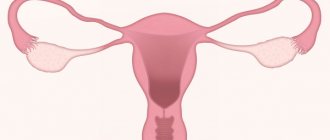
The organs of the abdominal cavity and small pelvis (uterus, fallopian tubes, ovaries, bladder, rectum) are externally covered with a thin shiny membrane - the peritoneum. The smoothness of the peritoneum, combined with a small amount of fluid in the abdominal cavity, ensures good displacement of the intestinal loops, uterus, and fallopian tubes. Therefore, normally, the functioning of the intestines does not interfere with the capture of the egg by the fallopian tube [1], and the growth of the uterus during pregnancy does not interfere with the normal functioning of the intestines and bladder.
Inflammation of the peritoneum - peritonitis - is a very dangerous disease. And the more dangerous it is, the more space it occupies in the abdominal cavity or small pelvis. But there is a mechanism in the body that limits the spread of peritonitis - the formation of adhesions.
With the development of the inflammatory process in the pelvis, the tissues at the site of inflammation become swollen, and the surface of the peritoneum is covered with a sticky coating containing fibrin (the protein that forms the basis of the blood clot). The fibrin film on the surface of the peritoneum at the site of inflammation glues adjacent surfaces to each other, resulting in a mechanical obstacle to the spread of the inflammatory process. After the end of the acute inflammatory process, adhesions in the form of transparent whitish films can form in places where internal organs stick together. These adhesions are called adhesions. The function of adhesions is to protect the body from the spread of a purulent-inflammatory process throughout the abdominal cavity.
The inflammatory process in the abdominal cavity does not always lead to the formation of adhesions. If treatment of adhesions
started on time and carried out correctly, the likelihood of adhesions is reduced. Adhesions form when an acute process becomes chronic and the healing process extends over time.
Adhesions can interfere with the normal functioning of internal organs. Impaired mobility of intestinal loops can lead to intestinal obstruction. Adhesions affecting the fallopian tubes, uterus, and ovaries disrupt the entry of the egg into the fallopian tube, the movement of sperm along the fallopian tube, the meeting of sperm and egg, and the advancement of the embryo after conception to the place of attachment in the uterine cavity. In gynecology, adhesions can cause infertility and pelvic pain.
Symptoms
Symptoms in the presence of adhesions in a woman are very different, however, several common signs can be identified:
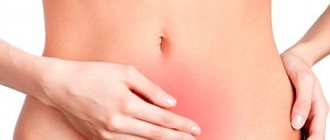
- Chronic pelvic pain syndrome is a constant, sometimes almost imperceptible, dull pain in the area of adhesion formation. It occurs due to tension and deformation of organs. In some cases, it can radiate to surrounding organs, the lower back and legs. The pain may intensify during sexual intercourse, physical exercise and prolonged exercise, hypothermia, and when the bladder is full.
- Impaired organ function - reduction or disappearance of menstruation, lack of ovulation, inability to become pregnant.
- In cases of formation of adhesions with intestinal loops - constipation, feeling of bloating, intestinal obstruction.
It is also worth noting that if a woman has adhesions, it is quite possible for her to experience increased pain during menstruation and ovulation. But it is worth considering that in half of the cases, adhesions do not cause any symptoms and are detected only during an ultrasound of the pelvic organs or a visual examination.
Adhesions formed as a result of inflammation
The fallopian tubes, uterus and ovaries can be involved in the adhesive process that occurs during inflammation of neighboring organs (appendicitis - inflammation of the appendix), as well as with lesions of the small and large intestines. In this case, the genital organs themselves suffer little: the adhesive process almost does not disturb their internal structure. If inflammation occurs inside the genital organs, not only the formation of adhesions occurs, but also damage to the genital organs themselves.
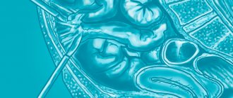
The most unprotected in this regard is the fallopian tube - one of the most delicate and finely structured smooth muscle organs. It plays a key role in facilitating conception and supporting early pregnancy.
Sperm entering the vagina are filtered through the mucus of the cervix, pass through the uterine cavity and enter the fallopian tube. Peristalsis - the movement of the fallopian tube - helps sperm enter the outer third (ampull) of the fallopian tube, where the process of conception occurs. During ovulation—the release of an egg from the ovary—the fallopian tube “sucks in” the mature egg. If at the time the egg enters the fallopian tube there are sperm there, fertilization occurs, and the resulting embryo moves to the uterine cavity within a few days, where it is immersed in the uterine mucosa (implantation). Delivery of the embryo into the uterine cavity is ensured by the movements of the fallopian tube and the active work of the microcilia of the fallopian tube.
Thus, the fallopian tube not only ensures the transportation of germ cells and the embryo, but also creates an environment for fertilization and development of the embryo during the first 5-6 days of intrauterine development. A change in the composition of the fluid that is produced in the fallopian tube can destroy the embryo. Local immunity inside the fallopian tube is minimal, that is, there are almost no mechanisms that ensure the rejection of foreign substances, because the embryo is half foreign, the fallopian tube does not reject it, and excessive activity of the immune system is unfavorable for the development of pregnancy. This is why the fallopian tubes so easily become victims of the so-called ascending infection (coming from the vagina and uterine cavity). Surgical and diagnostic interventions in the uterine cavity (abortion, diagnostic curettage, hysteroscopy[2], echohysterosalpingography[3]) facilitate the entry of infection into the fallopian tubes.
Once in the fallopian tubes, the infection first affects the mucous membrane of the fallopian tube (endosalpinx), then the muscular layer (myosalpinx), and only at the last stage the outermost layer of the fallopian tube (perisalpinx) is involved in the inflammatory process and conditions arise for the formation of adhesions. If treatment of adhesions
belated or not effective enough, after recovery not only adhesions remain, but also irreversible damage to the mucous membrane of the tube and its muscle layer. The cilia disappear, and scar tissue forms in place of the smooth muscle fibers. The fallopian tube can turn into a connective tissue sac (sactosalpinx), i.e. it loses the ability to promote a fertilized egg. With such disorders, elimination of adhesions cannot restore the function of the fallopian tube, and the presence of a focus of the inflammatory process leads to a decrease in the likelihood of pregnancy even in the tube from the opposite side or with the help of in vitro fertilization. In such cases, to increase the chances of pregnancy using IVF, which can be performed after recovery, the entire tube has to be removed. As a result of inflammation, gluing and fusion of the walls of the fallopian tube can occur, which leads to obstruction of the tube for the egg and is also an indication for separation of adhesions or removal of the tube.
How are adhesions on the female genital organs and infertility related?
The most well-known consequence of the appearance of synechiae is partial or complete obstruction of the fallopian tubes. Most often, the presence of adhesions is detected when a woman consults a gynecologist with a complaint of prolonged inability to become pregnant.
The fallopian tubes are the place where, during ovulation, the egg prepares for fertilization.
After colliding with a sperm, the egg moves through the tube to exit into the uterus and attach there. If synechiae are present in the fallopian tube, the path of the egg becomes obstructed, and pregnancy ultimately does not occur. In addition, adhesions can change the shape of the fallopian tube, which also prevents the egg from attaching to the wall of the uterus. Sometimes, especially with chronic forms of tubal obstruction, adhesions can provoke an ectopic pregnancy.
Thus, adhesions are one of the main causes of infertility. Fortunately, modern medicine can effectively get rid of them, increasing the likelihood of a successful pregnancy.
Postoperative adhesions
During surgical interventions, adhesions are formed due to:
- tissue hypoxia or ischemia - insufficient supply of tissues with blood and oxygen;
- drying tissue during surgery;
- rough manipulation of fabric;
- presence of foreign bodies;
- presence of blood;
- separation of former adhesions.
Foreign bodies that cause adhesions often include talc particles from doctor's gloves, small cotton fibers from gauze or tampons, and suture material. Adhesions also form with endometriosis. During menstruation, a small amount of menstrual blood containing living cells from the lining of the uterus (endometrium) may enter the abdominal cavity through the fallopian tubes. Normally, these cells are removed by the body's own immune system, but if there are any problems, they take root and form functioning endometrial islands that menstruate into the abdominal cavity. Adhesions form around these foci.
Diagnostics
The presence of adhesions in the abdominal cavity can be suspected in patients who have previously undergone pelvic inflammatory diseases, surgical operations on the pelvic and abdominal organs, and in women suffering from endometriosis. However, only in half of the patients with more than two risk factors for the development of adhesions in the anamnesis, adhesions are detected during laparoscopy (an operation during which small holes are made in the anterior abdominal wall through which an optical device is inserted to examine the cavity and special surgical instruments) .
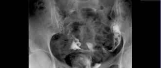
A gynecological examination suggests the presence of adhesions in the abdominal cavity with a probability of 75%. Obstruction of the fallopian tubes according to hysterosalpingography (a contrast agent is injected into the uterus, X-ray images are taken) and ultrasound examination with a high degree of certainty indicate the presence of adhesions, but the patency of the fallopian tubes does not exclude the presence of adhesions that seriously impede pregnancy. Conventional ultrasound does not reliably detect the presence of pelvic adhesions. Today, the method of nuclear magnetic resonance (NMR, or magnetic resonance imaging, MRI) seems to be very promising in diagnosing the adhesive process. Using this method, images are obtained that reflect the “state of affairs” at different levels.
The main method for diagnosing adhesions is the laparoscopy method. It allows not only to detect the presence of adhesions and assess the severity of the adhesions, but also to treat adhesions
.
There are 3 stages of the adhesive process according to laparoscopy: Stage I: adhesions are located around the fallopian tube, ovary or in another area, but do not interfere with the capture of the egg; Stage II: adhesions are located between the fallopian tube and the ovary or between these organs and other structures and can interfere with the capture of the egg; Stage III: either torsion of the fallopian tube occurs, or it is blocked by adhesions, or a complete blockade of egg capture.
general information
All human internal organs easily move relative to each other thanks to the slippery membrane covering them. The mobility of an organ is an important criterion for its good condition and ability to perform its functions. Conversely, any violation of mobility, fixation, attachment to another structure means pathology, which inevitably leads first to malfunctions in the functioning of the organ (functional disorders), and over time to changes in its tissues (structural disorders).
error
The formation of adhesions on the ovaries, tubes (uterine appendages) is a connection (soldering) of the internal organs of the small pelvis that is not provided for by nature, disrupting their normal mobility.
Causes
The most common causes of the formation of adhesions of organs in the pelvis in women are as follows:
- abdominal surgeries,
- surgical abortions,
- long-term “hidden” infections,
- untreated or insufficiently treated inflammation of the uterine appendages,
- appendectomy,
- some venous diseases (gonorrhea, chlamydia),
- consequences of ovarian apoplexy,
- internal endometriosis.
The number of adhesions after delivery by cesarean section is two to three times greater than after laparoscopic operations on the pelvic organs. They can attach not only to the uterus, but also to the ovaries, rectum, and bladder, which leads to the appearance of certain symptoms. More than 50% of cases of abdominal pain and ovario-menstrual cycle disorders in girls are a consequence of the formation of adhesions in the pelvis.
How adhesions form in the pelvic organs
Let's consider the mechanism of formation of adhesions in women - regardless of the cause, it is approximately the same.
The space between the organs in our abdomen is filled with abdominal fluid. It is secreted by the peritoneum - a thin layer lining the abdominal cavity from the inside. With inflammation of internal organs, especially with genital infections, after surgery, the peritoneum becomes irritated, the secretion of fluid increases sharply, while the latter becomes more viscous and sticky (this is how nature helped our ancestors to localize, “seal up” deep damage from the inside).
In addition, when getting to the diseased organ during surgery, the surgeon cuts the multilayer films that ensure the free sliding of the organs. After the operation to remove an ovarian cyst, apoplexy, or appendicitis, sutures remain that tie this entire complex structure into knots, and the sticky liquid continues to be intensely released into the abdominal cavity for some time and begins to “glue” neighboring organs and tissues together.
As a result of the formation of the first point adhesions near the ovary or uterus, the axes of movement of these adherent organs change: they begin to move around the point of connection. Since movement near the commissure is limited, its area increases, increasingly reducing the mobility of the organ. Over time, a dense scar forms at the junction, which “tightly” holds the organs and tissues together.
ICD-10 code N73.6 Pelvic peritoneal adhesions in women.
What do adhesions look like in the pelvis (photo)
Photo 1. Adhesions around the ovary (stage of removal surgery).
Photo 2. Adhesions in the pelvis behind the uterus (chronic adnexitis)
Photo 3. Adhesion and adhesive process in the area of the appendages and uterus.
Due to the restriction of natural mobility, inadequate position and trajectory of movement of the organ, the work of the communications going to it is disrupted, which are stretched, pinched, twisted, etc. In this case, normal blood circulation and lymph flow may be disrupted, and spasms of the supporting ligaments and muscles may occur. To avoid consequences, after gynecological manipulations and operations in the pelvic area, it is necessary to undergo preventive treatment (read more about methods of treating adhesions in gynecology below).
Why is the adhesive process dangerous?
It would seem, what danger is fraught with removing the appendix? However, in this case, intestinal loops, primarily the cecum, are often involved in adhesive processes in the pelvis. Typical consequences are constipation, dysbacteriosis, biliary dyskinesia, etc. Girls often develop adhesions in the appendages, which can subsequently lead to painful periods, infertility, and ectopic pregnancy.
Recently, laparoscopy through a small hole has become popular in gynecology. With it, for a better view, the abdominal cavity is filled with gas. But if before the intervention the organs were perfectly ground in and easily slid relative to each other, then after surgery on the appendages or uterus, when the gas pressure subsides, their relative position differs from the original one. As a result, their mobility also decreases. In addition, during laparoscopy, the peritoneum is still noticeably irritated and secretes a sticky fluid, which leads, as during abdominal surgery, to the formation of adhesions in the pelvic organs, only on a smaller scale.
The most common complications of untreated adhesions on the ovaries in women are:
• infertility, • pain in the lower abdomen and during sexual intercourse, • varicose veins of the uterus and appendages; • backward bending of the uterus, • obstruction of the fallopian tubes, • ectopic pregnancy, • various menstrual cycle disorders.
Doctors often have to deal with the consequences of an episiotomy (an incision in the vagina during childbirth). A small incision to facilitate the passage of the fetus is a common occurrence in obstetric practice. However, over time, scarring of the vaginal tissue can lead to disruption of the position of the internal genital organs, dilated veins and varicose veins, and prolapse of the bladder and uterus. What can we say about possible adhesions in the pelvis after a cesarean section.
Symptoms of adhesions in the pelvis
So, in women, the adhesive process from the ovaries can spread in different directions, ultimately forming long chains of rigidly connected organs, tissues, and ligaments. Further damage occurs according to the principle “where it is thin, it breaks,” that is, the disease affects the most weakened organ located anywhere in such a chain (ovary, fallopian tube, ligaments).
The range of symptoms of adhesions in the appendage area is very wide. For example, restriction of lymph flow in the reproductive organs leads to a decrease in local immune defense, provoking inflammatory diseases. In turn, the obstructed inflow and outflow of blood causes symptoms such as congestion in the pelvis, varicose veins of the uterus and periuterine space, menstrual cycle and ovulation disorders, pain during intimacy.
Symptoms of pelvic adhesions are especially dangerous in girls and adolescents (after removal of appendicitis, surgery, chronic inflammation of the appendages, etc.). The organs and bones in this case have not yet formed. And the occurrence of adhesions in children at an early age affects the entire growing organism - in its development it begins to adapt to the axis or point of fixation. As a result, the diaphragm and other organs do not move as they should (one-sidedly), and the skeleton does not develop as expected—the child sits and moves askew.
If an adhesive process occurs in the female organs during adolescence, there is a risk of disruption of ovarian function, the menstrual cycle, and the appearance of pain in the lower abdomen. In this regard, not only treatment, but prevention of these complications is important, so be sure to consult a good pediatric or adolescent gynecologist.
What pain occurs with adhesions in the pelvis?
Most people suffering from this problem have approximately the same ovarian pain. Women who have adhesions on the ovaries leave approximately the same reviews of their sensations. What common signs do all such patients have:
- Causal connection with adnexitis, cystitis, surgical abortion, appendicitis, curettage after miscarriage;
- There has been a history of gynecological surgery or complicated appendicitis in the past;
- Prolonged ignoring of the symptoms of inflammation of the appendages and their careful treatment;
- The ovaries hurt, the adhesions of which were caused by the surgery, almost constantly, with a tendency for the pain to increase with exercise;
- During sexual intercourse, there is a pulling sensation, dull pain in the vagina, closer to the top, which intensifies with aggressive and overly active movement, in a knee-elbow position and with deep penetration of the penis;
- Pain in the iliac regions on the left or right, which occurs regularly and intensifies during sudden, intense physical activity, as well as before and during menstruation.
conclusions
From the above it follows that medical care is needed not only for established pathology. Any woman who has undergone an abortion, abdominal surgery, cauterization of the cervix, inflammation of the appendages, hysteroscopy and laparoscopy, it is advisable to consult a gynecologist to prevent the development of the disease. To prevent the development of adhesions after appendicitis in children (girls, adolescents), it is necessary to visit a specialist in pediatric gynecology (accepted in our center).
In addition, it is advisable to visit a gynecologist before pregnancy, as well as after childbirth. This will help avoid many serious problems in the future. Our gynecology doctors in Moscow treat adhesions without surgery using proven techniques, which in the vast majority of cases avoids possible complications.
Treatment of adhesions
The main method of treating adhesions is laparoscopy . Using special micromanipulators, adhesiolysis - cutting and removing adhesions. Techniques for separating adhesions include laser therapy (cutting adhesions with a laser), aquadissection (cutting adhesions with pressurized water), and electrosurgery (cutting adhesions with an electric knife).
To prevent the formation of new postoperative adhesions during laparoscopy, the following methods can be used:
- introduction into the spaces between anatomical structures of various barrier liquids (dextran, povidin, mineral oils, etc.);
- wrapping the fallopian tubes and ovaries with special polymer absorbable films.
In addition, after laparoscopy, control diagnostic laparoscopy several months after the first laparoscopy has become increasingly common in recent years.
Is it possible to completely get rid of adhesions?
Most often, obstetricians-gynecologists give a positive answer to this question. Modern methods of treating synechiae make it possible to effectively and accurately remove adhesions, preventing their further occurrence. According to statistics, after removal of adhesions, pregnancy occurs in almost 85% of cases, which indicates a favorable prognosis for treatment of adhesions.
We will talk in more detail about ways to deal with adhesive threads later.
Prevention
The cellular and molecular mechanisms of adhesion formation are now quite well understood. Therefore, the process of adhesions after surgical operations, including after laparoscopy, can be sharply slowed down using so-called adjuvant (auxiliary) therapy. This therapy should begin as early as possible after the operation (in the first days and hours) and continue for several weeks. Treatment is aimed at suppressing the inflammatory reaction, suppressing fibrin deposition in the abdominal cavity, and activating fibrin dissolution.
Adjuvant therapy includes the use of the following drugs:
- Fibrinolytic agents are substances that dissolve fibrin, around which adhesions are formed: FIBRINOLYSIN, STREPTOKINASE, UROCINASE, HYALURONIDASE, CHEMOTRYPSIN, TRYPSIN, TISSUE ACTIVATORS OF PLASMINOGEN.
- Anticoagulants - drugs that prevent blood clotting: drugs HEPARIN, OXALATES, CITRATES.
- Antibiotics : TETRACYCLINES, CEPHALOSPORINS, SULPHANAMIDES.
- Anti-inflammatory drugs : corticosteroids, antihistamines, non-steroidal anti-inflammatory drugs, progesterone, calcium channel blockers.
The choice of drugs and treatment regimens depends on each specific case and can only be made by the attending physician.
How to prevent adhesions from occurring?
For the prevention and treatment of adhesions, it is possible to use enzyme preparations based on hyaluronidase. This enzyme acts on the “skeleton” of connective tissue fibers. Unfortunately, natural hyaluronidase, introduced into the body, is quickly inactivated by enzymes and blood plasma inhibitors, without having time to exert its therapeutic effect.^(3)
Longidase is a new generation enzyme preparation based on hyaluronidase—Unlike the previous generation enzyme preparations, hyaluronidase in its composition is stabilized by a high-molecular carrier, which allows it to remain resistant to the action of enzymes and unimpededly exert its therapeutic effect. Longidase facilitates the movement of fluid in the intercellular space, which leads to a reduction in edema, resorption of hematomas and increases the availability of antibacterial drugs to the site of infection. In addition, the decrease in the viscosity of the connective tissue base under the influence of the drug increases the elasticity of adhesions, which reduces pain (instructions for the drug Longidaza).
If treatment for adhesions does not help
Unfortunately, laparoscopy cannot solve all problems associated with adhesions. It is possible to free the internal genital organs from adhesions, but it is impossible to restore the structure and function of the fallopian tubes. Therefore, if pregnancy does not occur within several months after laparoscopy, you should consider switching to more radical methods of treating infertility. In vitro fertilization[4]
(IVF) is one of the most famous methods of in vitro fertilization.
To increase the effectiveness of in vitro fertilization (IVF), the following is taken into account: 1. The presence of sactosalpinxes (limited accumulation of fluid in the fallopian tube) sharply reduces the effectiveness of the IVF method. Sactosalpinxes are removed using laparoscopy. 2. Before the in vitro fertilization (IVF) procedure, it is advisable to undergo a special immunological examination to increase the chances of embryo engraftment.
[1]
The fallopian tube is a thin, hollow tube that extends from the uterus, connecting the uterine cavity to the abdominal cavity. After the egg is released from the ovary into the abdominal cavity, it travels through the fallopian tube into the uterine cavity. [2] Hysteroscopy is the insertion of a special optical device into the uterus, which allows you to examine the uterus from the inside. [3] Hysterosalpingography – injection of an X-ray contrast agent into the uterus and taking a series of X-rays. [4] In vitro fertilization (IVF) is a relatively new method of treating infertility. It was first used in England in 1978. The essence of in vitro fertilization is that the egg is fertilized and develops outside the uterus, and an embryo (sometimes consisting of only a few cells) is transferred into it to bear a child. Tags: infertility
How to reduce the risk of formation of adhesions on the fallopian tubes?
Unfortunately, it is impossible to completely prevent the formation of adhesive threads. However, it is quite possible to reduce the likelihood of their occurrence or minimize harm at the initial stage of education.
Gynecologists talk about adhesions in the fallopian tubes. Source - Live Healthy!
First of all, it is important to maintain hygiene in intimate areas. Regular washing with one of the Ginocomfort washing gels with antiseptic and softening components will help cleanse the vulnerable vaginal environment and prevent pathogens from entering. Of course, it is important, if possible, to avoid constantly wearing synthetic underwear, giving preference to natural materials: poorly breathable underwear can provoke the growth of bacteria.
In addition, you need to visit a gynecologist at least once every 6 months; if you are planning a pregnancy or have recently had surgery, the frequency of visits to the doctor should be increased.
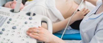
It is recommended to do an ultrasound of the pelvic organs at least once a year.
Don't forget about contraception. Frequent surgical abortions can cause infertility. For this reason, intrauterine devices are not highly recommended. From this point of view, the most preferable methods of protection against unwanted pregnancy are oral and chemical contraceptives and condoms. If emergency protection is necessary, you can use post-coital means.
Of course, it is important to move more, while trying not to overcool your legs and pelvic area. Moderate physical activity and exercise will ensure normal blood circulation.
These measures significantly reduce the likelihood of synechia formation, and in addition, make your reproductive system healthier.
Fallopian tube adhesions are no longer a death sentence and can be effectively treated.
A variety of techniques for removing connective tissue threads allows many patients to cope with this disease, getting the chance to become a mother. Remember that no less important than timely consultation with a doctor is the prevention of diseases and proper care of intimate areas. The Ginocomfort line of intimate hygiene products allows you to maintain the sensitive microflora of the vagina after childbirth, surgical interventions or infectious diseases. Herbal ingredients and the latest pharmaceutical developments in combination provide effective assistance in eliminating ailments of the reproductive system.
Take care of yourself and watch your health!
Sources:
- Minimally invasive surgery in gynecological practice. Adamyan L.V. // Obstetrics and gynecology. — 2006, application. — P. 11–17.
- The evolution of reproductive surgery in gynecology. Kira E.F. // Obstetrics and gynecology. - 2007. - No. 5. — pp. 59–62.
- Rational approaches to the treatment of tubo-peritoneal infertility (literature review). Adamyan L.V., Mynbaev O.A., Uskova M.A., Kuzmichev L.N. // Problem reproductions. - 2009. - No. 4. — pp. 24–27.
- https://cyberleninka.ru/article/n/tazovye-peritonelnye-spayki-endoskopicheskaya-harakteristika
- https://cyberleninka.ru/article/n/rekanalizatsiya-matochnyh-trub-pri-besplodii
- https://cyberleninka.ru/article/n/modelirovanie-neprohodimosti-matochnoy-truby
- https://www.kp.ru/guide/prokhodimost-matochnykh-trub.html