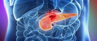
Pancreatic cancer is a malignant neoplasm that develops from glandular tissue or ducts. Pancreatic cancer affects the organ and very quickly invades nearby tissues. The symptoms of pancreatic cancer are often invisible until the final stages occur. The first signs of pancreatic disease begin to appear when the formation increases in volume and begins to spread in the body, metastasizing to the lymph nodes, liver, bones, lungs and other organs and systems.
The Scientific and Practical Surgery Center has ultra-modern equipment to combat cancer. Diagnosis and treatment are carried out by highly qualified specialists who have modern methods of treatment, who have undergone practical and theoretical training in advanced cancer treatment technologies in recognized global cancer centers.
Stages of pancreatic cancer:
Stage 1 – tumor up to 2 cm. Does not spread beyond the organ.
Stage 2 – cancer cells penetrate into neighboring organs and the lymph system. There are no metastases.
Stage 3 – the tumor grows into the spleen, nerves, duodenum 12, renal artery and aorta.
Stage 4 – the tumor metastasizes. appears , and diabetes mellitus develops.
Symptoms
In the initial period, the disease does not manifest itself. When the tumor grows and grows into nerve fibers, pain appears in the right hypochondrium. Then the bile duct is blocked and obstructive jaundice develops. General pancreatic head cancer clinic:
- pain;
- yellowing of the skin, sclera of the eyes, mucous membrane;
- weight loss;
- colorless feces, dark brown urine;
- signs of intoxication (suppression of appetite, nausea, vomiting, weakness, arrhythmias, bradycardia, drop in blood pressure, dizziness, loss of consciousness);
- bruises and subcutaneous hemorrhages, deep vein thrombosis;
- possible fever;
- skin itching.
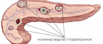
The pancreas is conventionally divided into three parts - head, body and tail. They all perform the same function, but the head is in close contact with the common bile duct (common bile duct), upper intestines, liver, and stomach. It is the damage to neighboring organs that gives such symptoms and worsens the patient’s condition. With neoplasms in the body and tail of the pancreas, the clinical manifestations will be different.
Pancreatic tumor treatment:
Surgical methods:
Pancreaticoduodenal resection – removal of the head and body of the pancreas, duodenum 12, part of the common bile duct and stomach.
Tail pancreatic resection is the removal of the distal part of the pancreas along with the spleen and lymph nodes.
Palliative surgery is performed when the tumor is unresectable, its purpose is to make the person’s life easier.
Chemotherapy is the introduction into the patient’s body of chemotherapy drugs that destroy cancer cells.
Radiation therapy (radiotherapy ) is the irradiation of cancer cells with a small dose of x-rays, during which they are destroyed.
OTHER ISLE CELL TUMORS
Lipomas can cause diarrhea, “pancreatic cholera”, WDHA syndrome (water diarrhea, hypokalemia, achlorhydria), 50% of them are malignant. Glkzagonoma is characterized by skin lesions (necrolytic erythema migrans), diabetes, glossitis, anemia, weight loss, depression and venous thrombosis; 75% of glucagon are classified as malignant tumors. The best treatment is resection. Clinical manifestations of somatostatin include diabetes, diarrhea, steatorrhea, achlorhydria, gallstones, malabsorption and abdominal pain. All of these symptoms are attributed to excess somatostatin levels. Streptozocin, dacarbazine, and doxorubicin are most often used in treatment.
Consultation on paid services
Show phone numbers
Forecast
Pancreatic cancer is one of the most unfavorable in terms of prognosis. This is due to the fact that this pathology progresses very quickly, without showing any signs. The consequences of surgical intervention on the pancreas cannot be accurately predicted. Surgical intervention can be prescribed only in 30-40%, and the five-year survival rate after surgery is only 50-60%. Without surgery, with palliative treatment, it does not exceed 1-2 years. And only 3-4% of patients who did not undergo surgery can overcome the 5-year survival threshold.
GASTRINOMA
Clinical manifestations. The disease is characterized by the original Zollinger-Ellison triad: peptic ulcers with atypical localization and a fulminant, aggressive course, extreme hypersecretion of gastric contents and the presence of a non-P islet cell tumor of the pancreas. It can begin as a simple disease and end with the appearance of severe complications (perforation, obstruction, bleeding, refractoriness to therapy). Standard medical and surgical treatment is ineffective. There are cases accompanied by significant diarrhea and steatorrhea.
Diagnostics. Manifestations typical of gastrinoma include high basal secretion rates of VAO/MAO (= or >0.6), hypertrophied gastric mucosal folds, fasting hypergastrinemia (>200 pg/ml) or paroxysmal increase in gastrin levels in response to secretin administration, imaging pancreatic tumor using CT.
Pathology and pathophysiology. Gastrinomas are non-β islet cell tumors of the pancreas that range in size from 2 to 10 mm. There is varying evidence in the literature regarding the malignant potential of gastrinomas, although rates equal to or greater than 90% have been reported. The presence of gastrinomas in the wall of the duodenum is observed. Neoplasms grow slowly and metastasize late; The cause of death is often peptic ulcer disease. Cases of gastrinomas have been described as manifestations of MEA-1 syndrome (Wermer's triad: pituitary gland, parathyroid glands, pancreas), in which the tumors are distinguished by the fact that they are multiple and benign.
Treatment. The original treatment method is gastrectomy. However, the use of histamine H2 receptor blockers and omeprazole with or without proximal selective vagotomy can control the condition of patients with disease that cannot be resected.
Causes
The causes of the development of diseases in this group are very diverse. Among the main ones are the following:
- Smoking.
- Alcohol abuse.
- Metabolic diseases (obesity, type 2 diabetes).
- Poor nutrition (consumption of fast food, fatty and fried foods, lack of plant fiber in the diet).
- History of chronic inflammatory diseases of the pancreas (pancreatitis).
- Living in an unfavorable environmental environment, in conditions of high levels of asbestos, cadmium, benzene, petroleum products, soot, phenolic resins in the atmosphere; harmful working conditions (heavy dust, high temperature).
How is jaundice treated?
A special case of the disease is concomitant jaundice due to compression of the common bile duct by a cancerous tumor, therefore, at the first stage, the outflow of bile is surgically restored, and after some time the issue of radical surgery is resolved.
Restoring the patency of the bile duct is possible in several ways, which are selected individually depending on the patient’s condition, the location of the lesion and its size: an expander is installed in the duct - a stent or catheter; a bypass route for the excretion of bile into the intestine is formed.
Research methods
Based on the patient's complaints and initial examination data, the doctor decides how to identify pancreatic cancer and prescribes the necessary examination. If the results obtained do not provide the necessary information, additional research is carried out.
To diagnose pancreatic cancer, the following are used:
- Laboratory diagnostic methods - analysis of the level of tumor marker CA-19-9 in the blood and pancreatic biopsy.
- Imaging methods for the lesion are CT, MRI, PET, ultrasound, endoscopic ultrasonography (EUS) and retrograde cholangiopancreatography (ERCP). Considering that scanning by any method does not always provide an accurate and complete picture, to clarify the nature and stage of the process, patients diagnosed with pancreatic cancer may require examination of the abdominal organs using a laparoscope.
Pancreatic biopsy - how they do it and what they want to know
During EUS, ERCP or laparoscopy, the doctor may perform a biopsy - remove a small piece of tissue from the problem area for examination under a microscope.
Material for analysis can also be obtained using a puncture biopsy. To do this, the specialist pierces the patient’s abdominal wall with a special thick hollow needle and, under ultrasound or CT control, brings it to the desired location.
A pancreatic biopsy makes it possible to differentiate between cancerous and benign tumors, as well as to determine the degree of malignancy of the tumor.
Analysis of specific tumor markers
The main tumor marker for pancreatic cancer is CA19-9. Determining its level is part of a comprehensive examination when making a diagnosis, is used to assess the severity of the disease, and is used to monitor the effectiveness of treatment and prognosis.
In addition, the content of CA242, TPS and other specific markers can be analyzed. As a rule, a conclusion is made based on the results of several tests.
When undergoing any examination, it is important not to forget: diagnostic results depend not only on how pancreatic cancer is determined, but also on proper preparation for the examination. Careful adherence to the doctor’s recommendations facilitates the tasks of specialists, speeds up the process of making a diagnosis, allows you to prescribe adequate treatment and adjust it if necessary.
Transabdominal ultrasound
This classic method is included in the standard complex of primary diagnostics. The study allows the specialist to see abnormal changes in the organ. The information content of traditional ultrasound is not high enough, so its results cannot be used as the only or main criterion for making a diagnosis.
CT scan
This type of scan allows you to obtain a detailed image of the internal organs. The study is carried out if it is necessary to clarify the size and location of the tumor, as well as to determine the presence of secondary foci (metastases).
Magnetic resonance imaging
The principle of obtaining images with MRI differs from the principle of scanning internal organs using a computed tomograph. In some cases, magnetic resonance imaging is more informative than CT. Compared to CT scans and other types of scans, MRI takes longer to complete.
Positron emission tomography
PET is a modern imaging method, the capabilities of which are invaluable when studying tumors with unclear boundaries or malignant neoplasms, the tissues of which differ little from the healthy tissues of surrounding organs.
The method is based on the different rates and intensity of uptake of radiopharmaceuticals by cancer and normal cells. When performing positron emission tomography, radioactive glucose is usually used as such a drug. In terms of its effect on the human body, PET is no more dangerous than ordinary x-rays.
PET/CT
Simultaneous visualization of the tumor focus by both methods using a special installation increases the accuracy of diagnosing pancreatic cancer by an order of magnitude.
Endoscopic ultrasonography
EUS is usually ordered when only a slight shadow is visible on CT or MRI images. The study is carried out using a special sensor attached to an endoscopic probe. The endoscope is inserted into the patient's stomach through the mouth, exactly as is done during fibrogastroduodenoscopy (FGDS).
Endoscopic retrograde cholangiopancreatography
ERCP is also performed using an endoscope. With its help, a contrast agent is delivered to the duodenum. A series of x-rays are then taken. The study is indicated in the presence of jaundice and allows you to identify tumors blocking the bile ducts and pancreatic ducts, in which contrast accumulates. To ensure complete relaxation of the patient before EUS and ERCP, sedatives are usually prescribed. As an alternative to ERCP, if appropriate equipment is available, magnetic resonance cholangiopancreatography (MRCP) can be performed without the use of contrast agents, which is especially valuable for patients with allergies to iodine-containing drugs.
Laparoscopy
This method refers to the so-called “minor surgery”. The procedure is performed under general anesthesia using a special instrument - a laparoscope, which is inserted into the abdominal cavity through small incisions.
If you need a second opinion to clarify your diagnosis or treatment plan, send us an application and documents for consultation, or schedule an in-person consultation by phone.
Expert opinion