Malfunctions of the liver and gallbladder are studied by hepatology. Hepatologists at our clinic treat hepatitis, cirrhosis, cholecystitis, tumor neoplasms, diseases of the biliary tract and much more. The clinic performs a thorough examination to establish an accurate diagnosis, as well as identify the causes of the disease. Next, our qualified doctors select the appropriate treatment method. We use the latest equipment. If you are concerned about hepatological problems, we are guaranteed to help you get rid of them.
Cholelithiasis (GSD)
- a disease caused by impaired metabolism of cholesterol and bilirubin, characterized by the formation of stones in the gall bladder and bile ducts with the possible development of life-threatening complications.
Factors contributing to the formation of cholesterol and mixed gallstones:
- obesity,
- starvation,
- dyshormonal disorders in women associated with pregnancy, taking contraceptive medications,
- diseases of the ileum (Crohn's disease, etc.),
- irrational and unbalanced diet (food rich in fats, insufficient consumption of vegetables and fruits, etc.),
- gallbladder dyskinesia,
- age.
Factors contributing to the formation of pigment stones - black stones consisting of bilirubin and calcium and making up 70% of all radiopaque stones:
- chronic hemolytic anemia,
- alcoholism,
- alcoholic cirrhosis of the liver,
- chronic infections,
- helminth infections of the liver,
- age.
In 10-15% of cases, gallstones are diagnosed during a preventive ultrasound examination of the abdominal organs. The development of cholelithiasis (GSD) goes through several stages.
- first stage - there are no clinical symptoms of the disease, the diagnosis is based on the results of a study of gallbladder bile (the tendency to form stones is determined),
- the second stage is asymptomatic “stone carriage” in which most of the stones located at the bottom of the gallbladder remain asymptomatic,
- the third stage – with a pronounced complicated clinical picture (acute or chronic calculous cholecystitis, etc.).
The manifestations of cholelithiasis depend on the size of the stones, their location and the activity of the inflammatory process, the patency of the biliary system, as well as on the condition of other organs of the gastrointestinal tract.
Diagnostics
THE INSTITUTE OF ALLERGOLOGY AND CLINICAL IMMUNOLOGY has a fundamental base that allows it to carry out a full range of diagnostic and therapeutic measures that meet the level of international standards. Many of the diagnostic and treatment methods were developed by the staff of our Institute.
Instrumental and radionuclide methods are informative methods of research. At the initial stages, laboratory tests of blood, urine, and feces will be mandatory methods.
- Ultrasound of the abdominal organs gives a picture of obstructions in the patency of the biliary tract, allows one to determine the size, structure and location of the liver, structural anomalies;
- fibrogastroduodenoscopy (FGDS) allows you to assess the condition of the esophageal veins and determine the risks of bleeding;
- MRI reveals narrowing of the bile ducts, the presence of stones, neoplasms;
- computed tomography (CT) – detects small, from 1 cm, neoplasms. Allows you to diagnose changes in the structure of the liver, cirrhosis, fatty degeneration;
- computed tomography with contrast determines the patency of the hepatic ducts and vessels;
- Liver scintigraphy (scan) is a radiological procedure to detect liver cancer, hepatitis, cirrhosis, tumors and abscess. It can also be used to assess the body's response to therapy.
Options for housing and communal services
| Biliary (hepatic, biliary) colic. A stone lodged in the neck of the gallbladder closes the exit and causes biliary colic. A stone up to 0.5 cm in size will most likely pass into the lumen of the duodenum; a larger stone may lodge in the common bile duct, which will lead to the development of complete or intermittent obstruction (“valve stone”) with a typical clinical picture of biliary colic. The presence of stones in bile is always accompanied by inflammation (cholangitis). Colic is accompanied by intense short, “minute-long” pain with an interval of up to an hour, long-term recurrent pain in the right hypochondrium is characteristic of chronic cholecystitis. If the pain lasts more than 72 hours and is accompanied by fever, this is most often a sign of acute cholecystitis. Constant pain that continues continuously throughout the day is not typical for biliary colic. The pain is accompanied by flatulence and nausea. Acute cholecystitis. The main cause of the development of the disease is strangulation of a stone in the cystic duct. Characterized by fever and constant pain in the right hypochondrium. Typical pain in acute cholecystitis occurs in less than 50% of patients. Most often, pain occurs soon after eating and increases in intensity over an hour or more. Fever usually appears 12 hours after the onset of the attack and is associated with the development of inflammation. The pain becomes constant. Acalculous acute cholecystitis is extremely rare and can occur secondary to salmonellosis, sepsis and trauma. Complications of acute cholecystitis.
Chronic calculous cholecystitis. Usually characterized by recurrent attacks of biliary colic, less often by constant pain. The development of an attack is provoked by fatty foods, spices, smoked foods, hot seasonings, severe physical stress, working in an inclined position, as well as infection. Less commonly, colic occurs “for no reason.” Usually accompanied by fever, nausea, and sometimes vomiting. The pain intensifies with movement and deep breathing. Sometimes the pain radiates to the heart area, simulating an attack of angina. The pain is varied - from intense cutting to relatively weak aching. Vomiting with cholecystitis does not bring relief. In women, colic sometimes coincides with menstruation or occurs after childbirth. Choledocholithiasis (common bile duct stones) occurs when a stone from the gallbladder enters the common bile duct. Symptoms are characterized by pain in the right hypochondrium similar to biliary colic, fever, chills, jaundice and characteristic changes in blood biochemistry. Cholangitis. A variant of the course of gallstone disease is accompanied by pain in the upper abdomen, jaundice, fever, and often chills. Bacterial cholangitis is one of the most dangerous complications of cholelithiasis; it is usually associated with cholestasis, which occurs when a stone blocks the common bile duct. The severity of cholangitis depends, first of all, on the duration of cholestasis and the level of cholemia. Gangrene and empyema of the gallbladder are manifested by sepsis, a serious condition of the patient with symptoms of peritonitis. Requires immediate medical attention. Empyema is an acute purulent inflammation of the gallbladder, characterized by intoxication and a high risk of perforation. Open perforation into the abdominal cavity is accompanied by peritonitis, causing high mortality (up to 25%). Bile stasis in the gallbladder is hypomotor dyskinesia of the gallbladder. As a rule, it accompanies cholelithiasis. Stasis of bile in the gallbladder is usually manifested by constant dull aching pain in the right hypochondrium, aggravated by shaking, walking quickly, carrying a heavy object in the right hand, or bending forward. It is also observed during pregnancy, long-term use of antispasmodics, after vagotomy, diabetes mellitus, obesity, i.e. for conditions and diseases that are risk factors for the development of cholelithiasis. |
Treatment: surgical. Absolute indications for surgery:
- acute cholecystitis,
- chronic cholecystitis with recurrent biliary colic,
- non-functioning gallbladder,
- common bile duct stones,
- gangrene of the gallbladder.
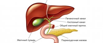
Structure of the gallbladder and bile ducts
Liver and gallbladder
The gallbladder is pear-shaped and has a capacity of about 40 cm2. This is a pouch 8–12 cm long and 4–5 cm wide. The widened end of the gallbladder is called the bottom, the narrowed end is called the neck. Between them is the body of the bubble. The neck of the bladder continues into the cystic duct, about 3.5 cm long. The gallbladder is covered with peritoneum only from the lower surface; its upper part, as a rule, is in contact with the liver. The bottom of the bladder is adjacent to the abdominal wall in the place where the right costal arch crosses the rectus abdominis muscle.
The wall of the gallbladder is formed by a layer of involuntary muscle fibers, covered on the outside with loose connective tissue, and on the inside with a mucous membrane that forms folds and contains many mucous glands. The mucous membrane of the gallbladder is capable of intensively absorbing water, so the bile in the gallbladder thickens 3–5 times compared to that coming from the liver. In the cystic duct, the folds of the mucous membrane are arranged in a spiral, which allows bile to move along the duct in both directions: into and out of the bladder.
The cystic duct, connecting with the common hepatic duct, forms a common bile duct with a length of about 7 cm. It goes down, pierces the wall of the duodenum and opens together with the pancreatic duct into an extension called the hepatopancreatic ampulla (see Fig. 3). The ampulla is located inside the large duodenal papilla, which is clearly visible on the mucous membrane of its descending part. Circular bundles of muscle fibers in the thickness of the major papilla form the sphincter of the ampulla (sphincter of Oddi), which regulates the flow of bile into the duodenum and prevents the flow of intestinal contents into the common bile duct and pancreatic duct. Another sphincter, the sphincter of the common bile duct, is located in the wall of the common bile duct slightly above the ampulla and directly regulates the flow of bile into the intestine.
In the absence of digestion, the sphincters are closed and bile accumulates and concentrates in the gallbladder. When food enters the stomach, the wall of the gallbladder contracts, the sphincters open and bile enters the duodenum. The structure of the bile ducts explains why either more liquid and lighter bile or darker and thicker bile can enter the duodenum. In the first case, it is hepatic bile, which directly from the liver through the common hepatic and common bile ducts enters the intestine. In the second case, it is cystic bile, which from the liver first enters the gallbladder, where it accumulates and concentrates, and when food arrives, it is released from the bladder and enters the duodenum through the cystic and common bile ducts. The secretion of bile is regulated by the nervous system. Fatty substances and some hormones (for example, secretin) stimulate contraction of the gallbladder and the secretion of bile.
CHRONIC AALCTIC CHOLECYSTITIS (CAC)
It occurs much less frequently than is diagnosed. If there is a clinical picture of chronic cholecystitis, it is necessary to exclude, first of all, cholelithiasis and parasitic infestations. CBC can be caused by microbial flora. The infection can enter the gallbladder from the gastrointestinal tract through the common bile and cystic ducts (ascending infection). Downward spread of infection from the intrahepatic bile ducts is also possible. The development of the inflammatory process in the gallbladder is promoted by stagnation of bile. CBC is characterized by a long progressive course with periods of remissions and exacerbations, aching pain in the right hypochondrium, less often in the epigastric region, lasting for many hours, days, sometimes weeks. The occurrence or intensification of pain is associated with the consumption of fatty and fried foods, eggs, cold and carbonated drinks, wine, beer, and spicy snacks. The pain may be combined with nausea, belching, bloating, and fever. There is a constant feeling of heaviness in the upper abdomen. Treatment. Diet, fractional meals, pain relief, anti-inflammatory and antiparasitic therapy, taking enzymes.
Symptoms
In the early stages, symptoms of liver disease may be mild. Over time, signs of liver and biliary tract disease become more distinct.
- dyspepsia: nausea, vomiting, diarrhea, constipation, flatulence, belching;
- yellowing of the skin, mucous membranes;
- skin itching;
- fatigue, weakness;
- dark urine and at the same time light feces;
- symmetrical red spots on the palms;
- sometimes - an increase in temperature;
- heaviness in the epigastric region, right hypochondrium;
- swelling of the legs;
- increase in abdominal size (ascites);
- drowsiness during the day, insomnia at night;
- the appearance of spider veins;
- weight loss, loss of appetite.
If these symptoms occur, you should consult a gastroenterologist-hepatologist!
CHOLESTATIC (subhepatic) JAUNDICE
Cholestatic jaundice develops when there is an obstruction to the flow of bile at any level of the biliary system. Obstructive or subhepatic (obstructive) jaundice develops when bile flow is obstructed at the level of the extrahepatic bile ducts (choledochal stones, pancreatic cancer, chronic pancreatitis, post-traumatic narrowing of the common bile duct, pancreatic pseudocysts, etc.). 70% of cases are associated with stones in the common bile duct and cancer of the head of the pancreas. Intrahepatic cholestasis is most often observed in acute drug-induced hepatitis and primary cirrhosis, less often in viral, alcoholic hepatitis, cholangitis, liver cirrhosis, metastases, etc. Jaundice develops slowly, often preceded by itchy skin. It is typical for both extrahepatic and intrahepatic cholestasis. Diagnostic criteria:
- light-colored (non-pigmented) stools
- dark brown urine
- traces of scratching on the skin,
- yellowish gloss of nails,
- xanthelasma (cholesterol deposits on the eyelids),
- xanthoma (cholesterol deposits on the palmar folds and tendons),
- hepatomegaly – enlargement of the liver.
When blocked by a stone, jaundice develops within 24 hours after the onset of pain, and fever occurs due to developing cholangitis. Itchy skin does not occur in all patients. Unlike a tumor, long-term stone blockage is rarely complete. Diagnostics: Laboratory tests of blood, feces, urine. Instrumental diagnostics: Ultrasound, CT, MRI diagnostics with contrast, Endoscopic diagnostics.
Major liver diseases: pathology and distribution
- Hepatitis is liver damage, the appearance of which is triggered by viruses and infections. Experts distinguish several forms of the disease:
- Hepatitis A - occurs due to improper preparation of food or exposure to unsanitary conditions.
- Hepatitis B and C - infection occurs after contact of the body with untreated needles and other medical instruments, as well as during unprotected sex with an infected person.
- Hepatitis D - spreads in the same way as the previous type of disease.
- Hepatitis E occurs after contact with contaminated food or liquid.
- Cirrhosis is a disease whose main cause is considered to be alcoholism. Due to severe poisoning and intoxication, scars form on the liver. Organ tissues begin to harden, and basic functions slow down. The disease is fatal, the damage is irreversible and leads to liver failure.
- Fatty liver disease – cholesterol and triglycerides accumulate in the body. This disrupts the functioning of the organ and causes irreversible pathological processes.
- Autoimmune liver diseases are diseases in which the body independently destroys healthy cells. The immune system protects our body from viruses, bacteria, infections and other malignant microorganisms. In autoimmune diseases, the body begins to attack itself. Damage to healthy liver tissue leads to inflammation and tissue scarring.
Autoimmune organ diseases include:
- Autoimmune hepatitis
- Primary sclerosing cholangitis
- Primary biliary cholangitis
- Genetic lesions of the organ
Some people are predisposed to liver disease. Experts include such ailments as:
- Wilson's disease is an excessive accumulation of copper in the body.
- Hereditary hemochromatosis is excessive accumulation of iron in the body.
- Alpha-antitrypsin deficiency is a disease in which our body is unable to produce sufficient quantities of substances necessary for life.
- Infections that cause liver disorders
Such ailments include:
- Epstein-Barr virus
- Adenovirus
- Toxoplasmosis
- Cytomegalovirus
POSTCHOLECYSTECTOMIC SYNDROME
In 15% , pain and dyspeptic disorders remain or recur These symptoms are associated with impaired motility of the sphincter of Oddi, dyskinesia of the extrahepatic bile ducts and duodenum . The main features are:
- recurrent colicky pain in the right hypochondrium,
- fat intolerance,
- diarrhea,
- bloating.
Patients often have duodenogastric reflux, antral gastritis, duodenitis, causing a feeling of heaviness and pain in the epigastrium, nausea, bitterness in the mouth, flatulence, unstable stool, etc. Inflammation of the gastroduodenal mucosa is usually associated with a microbial factor and the damaging effect of bile acids
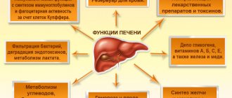
Liver bile
Hepatocytes produce bile constantly, 0.5–1 liters per day. 95–98% of liver bile consists of water and has an alkaline reaction. It contains bile salts, bilirubin, cholesterol, fatty acids, lecithin, ions Na+, K+, Ca2+, Cl-, HCO3-, etc. The color of bile is due to bile pigments (bilirubin, etc.), which are formed from the breakdown products of red blood cells. It is bile pigments that color the intestinal contents brown. The role of bile in digestion is reduced to emulsification and breakdown of fats, which facilitates their digestion and absorption. Bile enhances intestinal motility.