Mark Anatolyevich, what could be the reason for liver enlargement?
- In general, liver enlargement itself is just a symptom of the disease. It is necessary to first find out the cause of the disease, and then eliminate it.
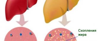
Diseases that contribute to liver enlargement are cirrhosis and fatty hepatosis (fatty liver).
These diseases are chronic and are caused by various factors, including malnutrition, alcohol abuse, or infectious viral hepatitis.
What is the treatment?
— One of the most common causes of liver disease is viral hepatitis. In this case, antiviral therapy is prescribed.
If it is toxic, alcoholic hepatitis, then you need abstinence from alcohol, medication support and diet.
In case of fatty hepatosis associated with excess body weight, measures aimed at reducing excess body weight, diet, drug treatment, and surgical intervention if necessary are prescribed.
In the case of cirrhosis, that is, more severe liver damage, treatment is more difficult, but it is possible. First of all, therapy is aimed at eliminating the causes of cirrhosis.
The next step will be treatment with various medications that allow the liver to function more optimally. Well, the last resort treatment for cirrhosis is liver transplantation.
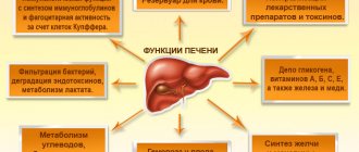
CAUSES OF HEPATOMEGALY, RISK FACTORS
The cause of the development (etiotropic factor, impetus for the occurrence) of hepatomegaly can be a variety of diseases. In clinical practice, hepatologists (the so-called specialists whose profile is focused on the management of patients with a diseased liver), as a rule, note hepatomegaly caused by the following pathologies:
- Oncological diseases of the liver - both benign and malignant. Moreover, the enlargement of the liver will most likely be uneven;
- Hepatitis of a very different nature - viral, toxic, autoimmune, alcoholic;
- Polycystic disease;
- Parasitic diseases, for example - echinococcosis (echinococcus is a worm that, when it enters the liver, forms a large cavity filled with a toxic liquid);
- Chronic infectious diseases accompanied by liver damage;
- Severe intoxication, in which the effect of toxic substances extends to the entire body;
- Liver cirrhosis (this disease in the early stages of development leads to hepatomegaly, and in the terminal stage of cirrhosis, on the contrary, the liver decreases in size).
Metabolic disorders in the body and various cardiovascular diseases can also provoke the occurrence of hepatomegaly (for example, with chronic cardiovascular failure of the 2nd stage, stagnation in the pulmonary circulation is formed, the outflow of blood from the liver is disrupted, as a result of which its size increases).
Are there any measures to prevent liver disease?
— Quitting the harmful use of alcohol is one of the most important and effective measures in the fight against liver diseases.
Secondly, this is a diet that should not contain too much fat and easily digestible carbohydrates.
You need to control your body weight and prevent it from growing beyond the norm.
Prevention will also include protection with a condom during sexual intercourse, because hepatitis B and C are also transmitted through sexual contact.
It is necessary to stop using drugs, which are associated with a very high risk of contracting viral hepatitis, since blood is another route of transmission.
In addition, there is a vaccine against hepatitis B, which I recommend for everyone.
DIAGNOSIS OF HEPATOMEGALY
The presence of hepatomegaly is determined by the doctor after examination, questioning and a thorough physical (without the use of instruments) examination of the patient. A significant diagnostic method in this case is palpation, during which the density and degree of enlargement of the liver is determined.
However, the mere statement of liver enlargement does not give anything - it is necessary to establish the cause, identify the disease, the development of which led to the occurrence of this syndrome. For this purpose, a number of additional examination methods are carried out - instrumental and laboratory:
- General and biochemical blood test with determination of the renal-hepatic complex;
- Serological blood test with determination of antibodies to the hepatitis virus and;
- Survey radiography of the abdominal organs;
- Ultrasound of the abdominal organs;
- CT and MRI of the abdominal organs;
- Coagulogram - determination of blood clotting.
This is what an enlarged liver looks like on computed tomography images in different projectionsIf these studies do not help determine the cause of hepatomegaly, doctors decide to perform diagnostic laparoscopy - surgery to identify the primary disease. But this happens extremely rarely.
What cases from practice related to liver disease do you remember?
— Severe liver damage occurs with viral hepatitis, when a person for a long time did not know that he had a virus, for example, hepatitis C. This hepatitis can be asymptomatic for many years, ultimately leading to cirrhosis of the liver.
That is, a person lived a normal life and suddenly he began to develop symptoms of cirrhosis. Upon further examination, it turned out that the patient’s liver functions were impaired, the disease was already at a serious stage, and the person was on his way to the operating table for a transplant. We have to fight such cirrhosis, and we are talking about survival, not about cure.
Hepatomegaly
Symptoms of liver enlargement are determined by the underlying disease. Moderate hepatomegaly, which develops during acute viral infections and malnutrition in children, may not manifest itself in any way. When the liver reaches a significant size, there may be discomfort in the right hypochondrium, painful sensations that increase with movement. Skin itching, rashes, dyspeptic complaints (nausea, stool problems, flatulence), and bad breath are also typical.
In the case of hepatomegaly against the background of viral hepatitis, compaction of the liver parenchyma is determined, which is easily detected even by palpation. An enlarged liver is accompanied by yellowness of the sclera and skin, and symptoms of intoxication. With timely effective treatment, the syndrome can regress. Hepatomegaly in liver cirrhosis occurs due to damage to hepatocytes and the formation of connective tissue in their place. Characterized by significant compaction of the organ, constant pain in the right hypochondrium, sallow skin tone, and a tendency to bleed.
Liver enlargement due to primary neoplastic lesions occurs quite rarely, with the leading symptoms being: hepatosplenomegaly, pain, dyspeptic disorders, jaundice, edema and ascites. With secondary (metastatic) lesions, the symptoms of hepatomegaly are usually less pronounced than the signs of primary tumor growth. In the case of benign liver tumors, organ enlargement is usually the first and leading sign. When the formation reaches a significant size, an asymmetrical enlargement of the abdomen and signs of compression of neighboring organs are possible.
A feature of hepatomegaly with degenerative changes (fatty liver disease) is scanty symptoms and the rare development of severe damage. Typically, this disease is a diagnostic finding when a patient presents for other reasons. With amyloidosis, the liver can reach significant sizes, its structure is dense, the edge is smooth, and there is no pain on palpation.
Hepatomegaly in heart disease develops in the case of right ventricular failure; the syndrome progresses rapidly, which leads to stretching of the organ capsule and severe pain. The size of the liver is variable and decreases with successful treatment of the underlying disease.
In the case of toxic damage to hepatocytes, liver enlargement may be the only sign; less often it is combined with itching, jaundice of the sclera and skin, and moderate changes in laboratory parameters. In case of traumatic damage to the liver tissue, hepatomegaly against the background of the patient’s severe general condition is accompanied by signs of intra-abdominal bleeding and hemorrhagic shock. Arterial hypotension and tachycardia, hypoxia progress; On palpation the liver is sharply painful.
Symptoms
Signs of moderate hepatomegaly due to minor changes in the size of the organ are weakly expressed. Symptoms may not appear at all for a long time. Many patients do not even suspect that they have a pathological liver condition. The disease is most often diagnosed accidentally when a person undergoes an examination for a completely different reason. So, during an ultrasound of the pelvic or abdominal organs, the doctor identifies echo signs that indicate an increase in liver volume.
In some cases, signs of moderate hepatomegaly still appear.
A patient with this diagnosis will feel:
– heaviness and feeling of constriction in the lower part of the right hypochondrium;
– pain in the above area upon palpation;
– mild nausea;
– the skin may acquire a jaundiced tint;
– general health will worsen.
Due to the fact that the signs of moderate hepatomegaly are nonspecific, many people attribute the discomfort to malaise, fatigue, or banal poisoning. However, doctors advise not to delay visiting the clinic. This will allow the disease to be diagnosed in time and treatment of moderate hepatomegaly to be started in order to avoid more dangerous consequences. Ignoring contact with a qualified specialist increases the likelihood of scarring of healthy liver parenchyma tissue. This will lead to the formation of cysts and other pathological formations.
Stages of liver cancer
The basis of clinical classification is the TNM system. It is based on the pathological features of the tumor, and its prognosis depends on the stage of liver cancer. Each classification criterion will indicate the presence or absence of certain changes.
- T is the growth and spread of the tumor, the presence of several tumor nodes, the size of the largest of them, as well as the growth of the tumor focus into neighboring areas.
- N - reflects tumor metastasis to nearby (regional) or distant lymph nodes;
- M - indicator reflects metastasis of liver cancer to other organs.
In addition to these criteria, the course of the disease, the features of its treatment and the prognosis of liver cancer are influenced by the presence of fibrosis and the degree of differentiation of tumor cells. The lower the degree of differentiation, the more the cancer cells differ from healthy ones and the more aggressive the cancer. These indicators have an extremely negative impact on survival, regardless of TNM stage.
One of the classification options is based on blood parameters for liver cancer. They determine the functional activity of the liver and the severity of cirrhosis. The assessment is carried out based on the levels of total bilirubin, serum creatinine, and blood coagulation parameters.
How do you know if you have fatty liver disease?
STEP 1. Calculate your body mass index.
In the table below, you can find out your body mass index by comparing height and weight.
Interpretation of the result:
BMI up to 22 – underweight
22-25 – normal weight
25-29 – excess body weight
Over 30 – obesity
In the table below you can see the body mass index and its interpretation in color:
If the body mass index is more than 30, this is grade 1 obesity, in which there is always fatty hepatosis
STEP 2: Ultrasound-diffuse changes in the liver parenchyma. FIBROSCAN?
In its normal form, liver tissue is a weakly echogenic homogeneous structure. When examined in the liver tissue, you can see blood vessels with bile ducts, which have increased echogenicity. Signs of diffuse changes in the liver parenchyma indicate that the liver tissue has been completely changed. Such signs are typical for both minor changes and very severe lesions. Unfortunately, the ultrasound method does not allow determining the stage of hepatosis, as well as changes in the liver (fibrosis) that lead to cirrhosis.
Therefore, INSTEAD of an ultrasound of the abdominal cavity, you can immediately perform a FIBROSCAN OF THE LIVER TO DETERMINE THE STAGE OF FIBROSIS AND STEATOSIS (THE AMOUNT OF FAT IN THE LIVER TISSUE).
This is an absolutely painless and non-invasive technique that is close to the accuracy of a liver biopsy. Further, there is no need to conduct a laboratory examination to determine how affected the diseased organ is, since the degree of liver damage will already be clear.
STEP 3. TREATMENT OF FATTY HEPATOSIS
Treatment of fatty liver disease begins with lifestyle modifications. This includes an individually tailored diet and physical activity, treatment of other target organs affected by obesity. Treatment programs are mandatory for persons with a body mass index of more than 30 and include hepatoprotective drugs that relieve inflammation in the liver and promote the elimination of fat. Medical has all the above capabilities. At the Center for Weight Correction and Diabetes Treatment
Modern diagnostics and free treatment programs for fatty hepatosis are also possible.
To diagnose and treat fatty hepatosis, you need to make an appointment with a hepatologist at the center for a treatment program.
Treatment of hepatomegaly
With an enlarged liver, first of all, lifestyle correction is necessary: therapeutic diet Table No. 5, giving up bad habits.
The basis of therapy is the treatment of the primary disease that caused the development of hepatomegaly. Drug treatment involves the prescription of drugs from the following groups:
Treatment
It is possible to restore the normal size of the liver in a moderate form of the disease through diet therapy and taking hepatoprotective medications aimed at protecting the liver, as well as vitamin complexes and enzymes.
It is recommended to strictly follow the diet.
The following should be excluded from the diet:
- alcohol;
– fried, spicy, fatty, salty and spicy dishes;
– canned food and marinades;
– carbonated sweet drinks;
– dairy products with a high fat content – butter, cream, sour cream over 20%, ice cream, cheese.
You need to eat small portions 5 times a day. The main menu should include boiled vegetables and fruits, water porridge, lean meats and fish.
BENIGN TUMORS
Most benign liver tumors are clinically asymptomatic formations originating from both epithelial tissue (hepatocellular adenoma) and stromal (nodular hyperplasia) and vascular (hemangioma) elements.
adenomas are extremely rare, and their occurrence may be associated with the use of contraceptives. If an adenoma is detected, resection is indicated, since with its rapid growth, “tumor rupture”, damage to adjacent vessels and bleeding are possible.
Nodular hyperplasia of the liver is a benign tumor, the central part of which is represented by scar connective tissue, and the peripheral part by modified tissue against the general background of healthy liver tissue. Foci of necrosis and hemorrhage are often observed in the tumor; sometimes these tumors are called “focal cirrhosis.”
Hemangiomas , originating from vascular, predominantly venous elements of the liver, are usually found accidentally during ultrasound or CT. Sometimes they can be mistaken for metastases, the difference lies in the varying degrees of ultrasound density. Treatment is usually not required.
Liver cancer treatment methods
If the diagnosis of liver cancer is confirmed, treatment must be started immediately. It will be long and includes several stages. In addition, depending on the type of tumor, its size and location, several methods are combined. The main method is surgical removal of cancer
– resection of part of the liver or its complete removal with transplantation of a donor organ.
If resection is not possible and there are no suitable donors for transplantation, doctors can use non-resection therapy
- percutaneous ethanol injections, radiofrequency ablation therapy, arterial embolization or chemoembolization of tumor vessels. Traditional treatment of primary liver cancer is also possible - chemotherapy, the use of radiotherapy, targeted drugs or a combination of several techniques.
In the initial stage of liver cancer, surgery will be the main method of treatment. The tumor is resected within healthy tissue. One-stage resection and two-stage surgery with preliminary ligation or embolization of the portal vein are possible.
Contraindications to such an intervention include damage to distant lymph nodes and cancer metastasis. However, if the remaining portion of the liver is not enough for its full functioning, if the functions of the organ are significantly impaired, there are signs of liver failure, the tumor has grown into the hepatic vein, or affects the portal system, doctors resort to alternative methods of therapy.
If the tumor develops against the background of cirrhosis, the only radical treatment method is organ transplantation
. However, the criteria for transplantation are very strict, so such an operation is performed infrequently.
Chemotherapy
for liver cancer it is used infrequently due to its low activity against cancer cells. It is carried out using cytostatic drugs, but the effectiveness does not exceed 20%. When chemotherapy is combined with immunotherapy for liver cancer, toxic side effects may develop (decreased platelet and leukocyte levels). But the tumor can be reduced to a size where it can be removed. Research into the use of new immunotherapy drugs is still ongoing, but they have shown some success.
Radiation therapy
for liver cancer, it is used to a limited extent, since the development of radiation hepatitis and organ failure is possible. It is used for inoperable tumors in the area of the portal vein and the porta hepatis. Modern equipment is used to minimize damage to surrounding healthy tissue.
One of the promising areas is targeted therapy
in oncology for liver cancer. This is a targeted effect on cancer cells without affecting healthy tissue. It is used in stages when surgery is dangerous or impossible; the drugs help reduce the size of the formation.
Prognosis and prevention
The prognosis for hepatomegaly depends on the underlying disease. For example, with viral hepatitis A it is most often favorable, but the outcome of chronic hepatitis B can be cirrhosis or liver cancer.
A similar situation occurs with storage diseases. Some of them can be completely controlled with specific therapy, while others are practically untreatable.
There is no cure for hepatomegaly, but a balanced diet, moderate physical activity and weight control help maintain liver and cardiovascular health.
Sources:
1 I.V. Kornelyuk [and others]. Practical skills in internal medicine. Differential diagnosis of hepatosplenomegaly: educational method. manual/ – Minsk: BSMU, 2015
LIVER METASTASES
In Europe, 90% of all liver tumors are metastatic. The most common primary location of a tumor metastasizing to the liver is:
- colon and rectum,
- breast,
- stomach,
- pancreas.
Characterized by predominant damage to the lymph nodes rather than the liver parenchyma. Clinic: fever, pain in the right hypochondrium, hepatomegaly, jaundice. Early diagnosis of liver metastases increases the patient's life expectancy.
Types of primary liver cancer
There are several classification options for primary liver cancer. There are various types of tumors, which differ in course and prognosis. Experts identify several of the most typical types of primary liver cancer (the tumor is formed from the cells of the organ).
Hepatocellular cancer
is the most common tumor (carcinoma) that occurs in the liver area.
As the name suggests, these tumors form in hepatocytes, which form the basis of liver tissue. The course of hepatocellular cancer is possible in two clinical forms - diffuse
(when a large part of the liver is affected) or
nodular
(one focus is formed - a node or several areas).
A more favorable variant of this cancer is possible - fibrolamellar carcinoma
, with it there are possibilities for more active treatment.
Cholangiocarcinoma
is a malignant tumor that originates in the area of the bile ducts.
Cholangiohepatoma
is a mixed type of tumor in which cancer cells originate both in the area of hepatocytes and in the area of epithelial cells in the bile ducts.
Variants of tumors such as angiosarcomas, mesodermal formations
are lesions that grow from lymphatic capillaries, blood vessels or connective tissue cells.
Possible hepatoblastoma
cystadenocarcinoma
is also identified . It occurs as a result of the degeneration of a benign tumor.
Primary liver cancer: diagnosis
All people who are at high risk for developing liver cancer are regularly screened. First of all, doctors look for ultrasound signs
Liver cancer in people suffering from chronic inflammatory processes of the organ have different degrees of cirrhosis. They are advised to perform an ultrasound examination every six months, even in the absence of complaints.
In addition, they are advised to perform laboratory blood tests to determine the tumor marker of primary liver cancer - AFP
(stands for alpha-fetoprotein). With this type of observation, it is possible to identify tumor lesions at an early stage (there are no symptoms or complaints yet), when the cancer can be radically cured. Changes in tests for liver cancer with an increase in AFP typically occur in up to 50-90% of patients. Levels greater than 400 ng/mL may occur in individuals with large tumors or rapidly growing tumors. But a temporary increase in the indicator is possible against the background of cirrhosis or inflammation of hepatocytes. However, high AFP in combination with ultrasound data and MRI of the liver confirms the diagnosis in almost 100% of patients.
Performing a CT
or
MRI with contrast
helps to identify a tumor and clarify its size and location, which is important for drawing up a treatment plan for liver oncology.
If all this data is not enough to make a diagnosis, a percutaneous biopsy
(puncture or aspiration) under ultrasound control. The risk of complications with such diagnostic methods is minimal.