Hematuria is blood in the urine. This disorder is characterized by increased production of red blood cells, which can provoke the presence of various serious diseases.
There are two types of hematuria:
- microhematuria, such a disorder can only be determined in laboratory conditions using a microscope. In patients with microhematuria, the urine may appear normal;
- macrohematuria, in this case the disorder can be determined even with the naked eye: there will be streaks of blood in the urine, it will become a pronounced red hue.
A healthy person has 1-2 red blood cells in the urine.
Disease or symptom?
Urinalysis is one of the main laboratory tests that is carried out at the stage of diagnosis. If the test results indicate hematuria, the doctor will definitely refer the patient for further examination. Why? Hematuria is the presence of blood in the urine. Obviously, normally there should be no blood in the analysis.
Since this is not an independent disease, but a symptom that may indicate serious health problems, the doctor will prescribe an additional examination to establish an accurate diagnosis.
Cost of services
| Code | Service | Price |
| 02.00 | Initial appointment with an obstetrician-gynecologist-endocrinologist | 1700.00 rub. |
| 02.01 | Initial appointment with an obstetrician-gynecologist-endocrinologist, PhD, Honored Doctor of the Russian Federation (Ivanova N.V.) | 3500.00 rub. |
| 02.02 | Repeated appointment with an obstetrician-gynecologist-endocrinologist | 1200.00 rub. |
| 02.03 | Repeated appointment with an obstetrician-gynecologist-endocrinologist, PhD, Honored Doctor of the Russian Federation (Ivanova N.V.) | 2000.00 rub. |
| 02.04 | Consultation with an obstetrician-gynecologist-endocrinologist based on test results without prescribing treatment | 500.00 rub. |
| 03.00 | Initial appointment with a urologist | 1500.00 rub. |
| 03.02 | Repeated appointment with a urologist | 1200.00 rub. |
| 03.03 | Initial appointment with a urologist (candidate of medical sciences) | 2500.00 rub. |
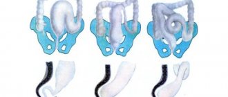
Types of hematuria
There are micro- and macrohematuria. Macrohematuria can be seen with the naked eye - the urine turns red or brown. Microscopic hematuria is determined laboratory.
Despite the fact that with the latter the amount of blood in the urine is not large, both conditions can be dangerous.
It is worth noting that patients sometimes see blood in the urine when there is none. Urine changes color when consuming certain foods (beets, black currants and beans, blackberries, rhubarb) or medications (metronidazole, rifampicin, etc.).
About hematuria
Hematuria is the appearance of red blood cells (red blood cells) in the urine.
At the same time, the urine changes color to a reddish-brown, dark, comparable to the color of tea, coffee, or classically defined as the “color of meat slop.” Hematuria is a symptom of many dangerous diseases, so this sign is a reason to immediately consult a doctor. If the color of urine differs significantly from normal, and small blood clots can be detected by eye, we speak of gross hematuria. If the number of red blood cells in the urine exceeds the norm slightly and is detected during examination, this is called microhematuria. We must remember that urine does not always change color solely due to blood getting into it. Eating foods rich in food dyes, beets, and taking certain medications can cause such coloring. A woman may mistake the mixing of urine with menstrual blood from the vagina for hematuria , which, of course, cannot speak of pathology.
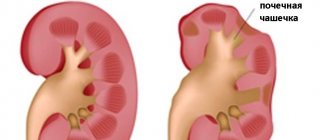
Causes
“Kidney hematuria” is a diagnosis made by patients who find blood in their urine. We recommend not to engage in self-diagnosis, but to consult a urologist or nephrologist in time, because this phenomenon has many causes. Let's look at the main ones.
- Infections of the kidneys and urinary tract (cystitis, urethritis), prostate gland, genital organs
- Urolithiasis (sand and stones in the kidneys, bladder, ureter)
- Neoplasms (tumors of the kidneys, bladder, benign prostatic hyperplasia, etc.)
- Glomerulonephritis
- Kidney and urinary tract injuries
- Kidney infarction
- Hereditary pathologies
- Drug therapy (taking certain antibiotics, anticoagulants, drugs used in oncology).
M.M. Batyushin, D.G. Pasechnik State Educational Institution of Higher Professional Education Rostov State Medical University of the Russian Health Service, Rostov-on-Don
The concept of Hematuria is the leading symptom in the clinic of pathology of the kidneys and urinary tract. Hematuria refers to the appearance of blood cells (namely red blood cells) or their components (hemoglobin) in the urine. In this regard, the concept of hematuria includes erythrocyturia, hemoglobinuria and hemoglobin cylindruria (excretion of hemoglobin casts in the urine). Normally, during a general analysis, there are either no red blood cells in the urine, or their number when counted in Goryaev’s chamber does not exceed 1–2 elements in the field of view. Sometimes a higher number of red blood cells may be normal. This may be due, for example, to intense physical activity. Such episodes are short-lived. The appearance of 3 or more red blood cells in the field of view is considered erythrocyturia. There is no clear definition of persistent erythrocyturia, however, an increase in the number of red blood cells in the urine and persistence according to the results of three or more tests can be conditionally considered persistent erythrocyturia. In the case of erythrocyte adhesion and hemoglobin precipitation in the lumen of the renal tubules, the formation of erythrocyte and hemoglobin casts, which have a characteristic appearance and are stained red with hematoxylin, is possible. As part of the definition of hematuria, micro- and macrohematuria are distinguished. Microhematuria (more correctly, microerythrocyturia) refers to erythrocyturia that does not lead to a change in the color of urine, assessed by eye. With gross hematuria, the urine becomes red (from light pink to cherry). It may be diffusely colored or have blood clots. Thus, the concept of micro- or macrogematria is a qualitative symptom assessed visually. Previously, the phenomenon of “leached erythrocytes” was widely used in the differential diagnosis of erythrocyturia. It is now generally accepted that red blood cell leaching is related to the physicochemical properties of urine. Often, with undoubted renal origin of hematuria, fresh red blood cells are found; on the other hand, gradual leaching of red blood cells can occur when urine is stored for several hours before examination. In this regard, the phenomenon of “leached erythrocytes” should not be leading in differential diagnosis and retains a certain significance only when using phase-contrast microscopy. Hemoglobinuria develops due to the excretion of free extra-erythrocyte hemoglobin in the urine through glomerular filtration. Due to the oxidation of hemoglobin and its conversion into hemosiderin, which is a black pigment, urine with hemoglobinuria and hemosiderinuria may acquire a dark cherry color.
Prevalence The prevalence of erythrocyturia in the population ranges from 0.18 to 16.1%. According to our data obtained during a one-time study of 1446 patients in somatic hospitals in Rostov-on-Don (the study did not include patients from the departments of nephrology and urology), erythrocyturia was detected in 3.7% of cases. When examining young soldiers, P. Froom et al. found erythrocyturia in 39% of cases. However, it was transient in nature, since during subsequent examinations it occurred with a frequency not exceeding 16%. In postmenopausal women, erythrocyturia occurs with a frequency of 13%. In screening studies in pediatric populations, hematuria occurs with a frequency of 1–4% and increases with age, reaching 12–18% in adolescence.
Causes of hematuria and pseudohematuria Pseudogematuria Not every phenomenon of red urine is hematuria. Redness of urine not accompanied by erythrocyturia and/or hemoglobinuria is called pseudohematuria. Pseudohematuria can be caused by taking a number of medications, food nutrients, and chemicals that color it red (Table 1).
Table 1. Causes of pseudohematuria
| Urine color | Cause |
| Red | Antipyrine, amidopyrine, santonin |
| Pink | Acetylsalicylic acid in large doses, carrots, beets |
| Brown | Phenol, cresol, bears ears, activated carbon (carbolene), urates, porphyrins |
| Dark brown | Salol, naphthol |
In addition, it is possible to verify hematuria that does not actually exist due to a laboratory error or deliberate distortion of the result of a urine test, but the latter is extremely rare. This phenomenon should also be interpreted as pseudohematuria.
Hematuria of extraurinal origin If pseudohematuria is excluded, the next step in the differential diagnosis should be the exclusion of hematuria of extraurinal origin. In this case, we are talking about blood getting into the urine not from the kidney and urinary tract, but from other organs, as well as from the outside. The following types of hematuria of extraurinal origin include:
simulated hematuria:
- blood is added to the urine after urination from a wound inflicted by the simulator on a finger, lip, scrotum, etc., as well as the blood of another person;
- blood is introduced into the bladder through a catheter before urination;
- a foreign object injures the mucous membrane of the urethra before urination;
- the malingerer’s urine is mixed with the urine of a patient suffering from kidney pathology, manifested by hematuria.
hematuria of genital origin:
- taking a urine test during menstruation, as well as the day before or within 3-4 days after its end;
- bleeding from tumors of the uterus, vagina, atrophic colpitis;
- formation of vesico-uterine anastomosis (tumor, traumatic);
- preservation of menstruation with desquamation of the functional layer of the mucous membrane of the cervix during supravaginal amputation of the uterus;
- hematuria in pregnant women;
- postcoital hematuria.
hematuria of rectal origin:
- bleeding from a hemorrhoid;
- bleeding from anal fissure;
- rectal cancer;
- chronic proctosigmoiditis with the opening of a fistula in the perianal region, vesico-rectal anastomosis.
hematuria of perineal origin:
- boil, perineal carbuncle;
- perineal injuries.
During screening examinations of pregnant women, hematuria is observed in 20% of cases. Moreover, in 53% of cases it is detected before the 32nd week of pregnancy. Its appearance is a risk factor for the development of complications during childbirth and is usually due to obstetric reasons (threat of miscarriage, premature birth, premature abruption of the low-lying placenta, etc.). Postcoital hematuria is characterized by the appearance of erythrocyturia in a portion of urine given immediately after sexual intercourse. We observed 10 patients who had erythrocyturia after coitus. In 4 cases, erythrocyturia appeared after the introduction of foreign bodies into the urethra, and in one case gross hematuria was observed. It can be assumed that this cause of hematuria is not rare, but due to its delicacy it is rarely considered when carrying out differential diagnosis. In 6 cases, the cause of erythrocyturia was prostate disease and the patients were referred for examination and treatment to a urologist. As a rule, postcoital hematuria is combined with hemospermia. Isolated hemospermia and isolated postcoital hematuria are possible. The latter is an extremely rare option. Variants of isolated postcoital hematuria caused by a urethral polyp are described. Combined postcoital hematuria and hemospermia in young men in most cases is caused by the presence of benign papillary adenoma, in older men - prostate cancer. Isolated hemospermia in 40% of cases is caused by prostate infection; a malignant tumor also occurs; in some cases, the cause cannot be determined. Simulating pseudohematuria is not widespread due to the lack of difficulties in most cases in differentiating it from true hematuria. Their appearance is due to the motivation of malingerers and aggravators to enhance or direct the clinical manifestations of the disease in order to achieve some social benefits. Understanding the motive for malingering or aggravation is key to a successful differential diagnosis. Most often, elements of aggravation or simulation are observed in expert practice when conducting military medical, medical, social or forensic examinations.
True erythrocyturia There are many reasons for the development of erythrocyturia. There are no less attempts to classify this phenomenon. The appearance of red blood cells in the urine may indicate the development of pathology of the kidneys and urinary organs, as well as the genital organs. In 65% of patients, erythrocyturia is of extrarenal origin. The pathogenesis of renal anemia is poorly understood. A possible cause is the formation of damage in the basement membrane of the glomeruli, the so-called breaks or gaps. When the size of the defects exceeds 0.25 microns, red blood cells move through the membrane into the urinary space through them under the influence of intracapillary pressure and retraction forces. In this case, severe deformation and damage to the membranes of erythrocytes occurs, which explains the dysmorphism of these cells in the glomerular type of hematuria. Among the causes of hematuria, infectious, traumatic, autoimmune, toxic, tumor and mixed are distinguished. The classification of R.Cohen and R.Brown (2003) seems to us to be the most acceptable. When hematuria is detected, the diagnostic search is aimed at identifying these diseases. Noteworthy is the fact that hematuria is only one of the symptoms of the disease. The appearance of other symptoms often accompanies hematuria and forces the doctor to analyze their relationship and diagnostic significance. The classification of hematuria, distinguishing symptomatic and asymptomatic forms, is very conditional and is of an exclusively applied nature. Asymptomatic hematuria occurs in the adult population with a frequency of 0.19–21%. In approximately 10–12.1% of cases, it is caused by cancer of the kidneys and urinary tract. In this case, macrohematuria occurs in 3/4 of cases. As literature data show, in 39–90% of cases, the detection of asymptomatic hematuria is not subsequently accompanied by a diagnostic search. According to our data, only in 8% of cases the detection of asymptomatic hematuria led to further examination of patients (n=1446). In childhood, asymptomatic hematuria is most often caused by thin membrane disease (27.5% of all cases of asymptomatic hematuria) and IgA nephropathy (26.2% of all cases of asymptomatic hematuria). Of the glomerulopathies, the most common causes of gross hematuria in children are IgA nephropathy (54.2%) and Alport syndrome (25%). Among non-glomerular causes of hematuria, the most common are hypercalciuria (16%), urethrorrhagia (14.3%), and hemorrhagic cystitis (12.5%). An unidentified cause appears in 46% of cases.
Let's look at some of the causes of hematuria. 1. Urolithiasis Urolithiasis is the cause of 20% of all cases of hematuria. Hematuria with it is intermittent or persistent in nature and can be represented by both micro- and macrohematuria. In case of urolithiasis, Pasternatsky syndrome is described in the form of the appearance of erythrocyturia after walking or tapping the lumbar region. More often, hematuria is combined with pain in the lumbar region or flanks. The cause of hematuria is trauma to the mucous membrane of the urinary tract.
2. Tumors of the kidney and urinary tract Kidney tumors account for about 3% of all human tumors. 85–90% of all cases of oncological processes in the kidney are renal cell carcinoma, which develops from the epithelium of the proximal tubule. Of the benign neoplasms, angiomyolipoma, adenoma and oncocytoma predominate (6–8%). In 40–70% of cases, the disease is asymptomatic. In 5% of cases, the cause of microhematuria is cancer of the kidney and urinary tract. The risk of the latter increases with the patient's age, as well as in the presence of risk factors such as smoking, use of phenacetin, herbs containing aristolochic acid, and cyclophosphamide in high doses. Hematuria is a typical symptom of a tumor. Both micro- and macrohematuria, isolated or less often in combination with low proteinuria, can be observed. With gross hematuria, blood clots are often observed in the urine. Hematuria may be accompanied by pain (pain in the flank, hypochondrium, lumbar region, along the ureter), and may be asymptomatic. With kidney cancer in men, a varicocele may be observed, caused by compression of the testicular vein by the tumor. Paraneoplastic syndrome may develop, observed in 35% of patients. With prostate stromal tumors, hematuria occurs in approximately 14% of cases. The disease develops mainly in older men. The clinical picture of prostate cancer is caused by tumor growth and bladder outlet obstruction and is manifested by micro- and macrohematuria, chronic ischuria, stranguria, pain in the perineum and suprapubic region. Tumors of other locations are also possible: bladder, ureter, urethra.
3. Glomerulonephritis Hematuria is one of the clinical manifestations of acute, chronic and rapidly progressive glomerulonephritis. It is part of the nephritic syndrome. Acute post-streptococcal glomerulonephritis always manifests itself as isolated hematuria or hematuria in combination with proteinuria; urinary syndrome in chronic and rapidly progressive glomerulonephritis can occur without hematuria (proteinuric variant, nephrotic syndrome). Hematuria occurs in various morphological variants of nephritis, less often in minimal change disease and membranous glomerulonephritis. Hematuria with glomerulonephritis is usually preserved if other causes of hematuria are excluded, as well as in the presence of criteria for nephritic syndrome (hypertension, overhydration). Hematuria is observed both in idiopathic variants of glomerulonephritis and in its development as part of systemic connective tissue diseases (collagenosis, systemic vasculitis, arthropathy).
4. Connective tissue diseases Kidney damage in systemic vasculitis is varied and is represented by renal angiitis, interstitial nephritis and fibrosis with or without areas of necrosis, and various types of glomerulonephritis. The appearance of erythrocyturia in systemic vasculitis is evidence of the involvement of the kidneys or urinary tract in the pathological process. Differential diagnosis is usually aimed at excluding vasculitis, guided by criteria developed and widely used in rheumatological practice. In addition to systemic vasculitis, systemic lupus erythematosus, arthropathy such as gout, rheumatoid arthritis, and ankylosing spondylitis are often the causes of hematuria (Fig. 6).
5. Polycystic kidney lesions Polycystic kidney lesions may be accompanied by hematuria. It is usually asymptomatic. Clinical manifestations appear in the case of the development of infectious complications, as well as chronic renal failure.
6. Tubulointerstitial nephritis Interstitial nephritis of toxic origin Develops due to the effects of exo- and endotoxins on renal tissue. This group of kidney damage is diverse and includes exposure to industrial substances and household chemicals, medicinal and other substances.
Drug-induced interstitial nephritis The problem of drug-induced kidney damage is one of the pressing problems of modern nephrology. Approximately 6–60% of all cases of acute renal failure (ARF) are due to interstitial nephritis, as determined by nephrobiopsy. In half of the cases, the etiology of acute interstitial nephritis is drugs. Interstitial nephritis most often develops in response to antibiotics and nonsteroidal anti-inflammatory drugs (NSAIDs). NSAIDs are the cause of 44% of cases of acute interstitial nephritis, antibiotics – 33% of cases. Cases of interstitial nephritis have also been described during therapy with warfarin, thiazide diuretics, indapamide, mesalazine, ranitidine, and cimetidine. Erythrocyturia is present in 87–100% of cases when examining urinary sediment in acute drug damage and in 43–56% of cases in chronic cases.
Other drug-induced damage to the kidneys and urinary tract, accompanied by erythrocyturia
- Papillary necrosis - can be caused by NSAIDs, aspirin, analgin.
- Hemorrhagic cystitis - caused by cyclophosphamide, ifosfamide, mitotane.
- Urolithiasis - may occur when taking carbonic anhydrase inhibitors, dichlorphenamide, indinavir, mirtazapine, ritonavir, triamterene.
- Tumors of the urinary tract - can occur during therapy with cyclophosphamide, analgesics (phenacetin).
- Drug-induced vasculitis.
Interstitial nephritis due to herbal medicine Nephropathy due to the intake of Chinese herbs is known under the term “chinese herb nephropathy”. It is characterized by rapid progression of chronic renal failure (CRF) and is manifested morphologically by extensive interstitial fibrosis without glomerular damage. Occurs in women taking Chinese herbal supplements. Nephrotoxicity is determined by the presence of aristolochic acid in herbs. A cumulative dose of Aristolochia fangchi extract from the site of Stephania tetrandra has been shown to result in the development of CRF in 30.8% of cases. Erythrocyturia is one of the manifestations of the disease.
7. Congenital anomalies of the urinary tract Most often, hematuria is observed with nephroptosis, ureteral stricture, compression of the ureteropelvic segment or group of calyces by an aberrant renal artery, as well as with complete and incomplete duplication of the kidney, renal venous hypertension. The main cause of hematuria in patients with congenital anomalies is an increase in intrapelvic urine pressure due to compression and impaired urine outflow from the pelvis. As a result, microtraumatization of the mucous membrane of the pelvis develops with the development of microhematuria. With venous hypertension, venule ruptures with the development of microbleeding are observed. Typically, hematuria is recurrent and combined with renal colic or dull and weak flank and lumbar pain.
8. Infectious diseases Pyelonephritis, cystitis, prostatitis In some cases, erythrocyturia can be observed with pyelonephritis. It always occurs against the background of leukocyturia and is often caused by an unfavorable background in the form of a congenital anomaly of the urinary tract, urolithiasis, etc. With cystitis, erythrocyturia may be the only laboratory phenomenon. Prostatitis often leads to a combination of erythrocyturia and leukocyturia. In 10% of cases, isolated microhematuria is possible.
Septic nephropathy With sepsis, according to various sources, septic nephropathy develops in 10–45% of cases. It can be represented by septic acute glomerulonephritis, chronic glomerulonephritis, acute interstitial nephritis, tubular or cortical necrosis, renal carbuncle, apostematous nephritis and pyelonephritis, thrombosis of the renal arteries and veins. A wide range of possible injuries dictates the need for detailed differential diagnosis.
Tuberculosis With tuberculosis of the genitourinary system, epididymitis (58.1%) and kidney damage (16.3%) most often develop, less often than other organs: orchitis (9.3%), cystitis (7%), urethritis (4.7%) , damage to the prostate (2.3%), testicular membranes (2.3%). Tuberculosis of the kidney and urinary tract is manifested by erythrocyturia. Moreover, ultrasound or urographic imaging often reveals gross structural changes in these organs. Often a combination of tuberculosis of the kidneys and lungs.
Schistosomiasis An infection caused by a schistosome, most often Schistosoma haematobium. Distributed in the countries of Western (Benin, Burkina Faso, Gambia, Mali, Nigeria, Chad), Southern (Kenya, Madagascar, Mozambique, Tanzania, Zambia) and Northern (Egypt, Ethiopia, Sudan) Africa, also found among tourists who visited these countries . Less commonly, infection is caused by S. mansoni, S. japonicum, S. intercalatum and S. mekangi. Fever (44%), diarrhea (40%), itching (25%), chills (21%), and hematuria (20%) develop. Hypereosinophilia is observed in 82% of cases, increased liver enzymes – in 82%. Schistosomes are found in urine and stool in 60% of cases. With schistosomiasis, damage to the bladder is observed. Ultrasound examination reveals a change in the contours of the bladder wall, wall thickening of more than 5 mm, and visualization of a shadow in the form of a pseudopolyp. In this case, hematuria is a mandatory phenomenon.
Leptospirosis A zoonotic infection caused by Leptospira icterohaemorrhagia (50% of cases), L. canicola (41.4%), L. pomona (2.3%), L. grippotyphosa (5.5%), L. tarassovi (0.8% ). The most common sources of infection are rodents and dogs. Kidney damage in the anicteric form of leptospirosis is observed in 36.2% of cases and is represented by lower back pain (17.1%), a slight decrease in diuresis (21.3%), an increase in creatinine (12.8%), changes in urine tests (erythrocyturia , leukocyturia, low proteinuria). Morphological examination reveals interstitial nephritis. Jaundice forms are more severe and in 66.7% of cases are accompanied by the development of acute renal failure. Kidney damage in the icteric form is observed in 92.6% of cases. In this case, the clinical manifestations of kidney damage are most pronounced on the 5th–6th day of the disease.
Virus-associated nephropathies Kidney damage has been described in infections caused by hepatitis A, B, C viruses, HIV, parvovirus B 19, cytomegalovirus, Coxsackie virus B, Epstein-Barr virus, polyoma virus, hantavirus, adenovirus. Parvovirus infection is associated with focal segmental glomerulosclerosis, Coxsackie virus with IgA nephropathy, polyoma and hantaviruses with interstitial nephritis, hepatitis C virus with glomerulonephritis as part of cryoglobulinemic vasculitis.
9. Hematuria due to venous hypertension Nutcracker syndrome Nutcracker syndrome develops due to compression of the left renal vein between the aorta and the superior mesenteric artery [29]. The clinical manifestation of Nutcracker syndrome is hematuria, which develops as a result of venous intrarenal hypertension. According to WHO, varicocele occurs in 36% of men. Moreover, in 43% of cases of varicocele, hypertension is detected in the left renal vein [8]. The causes of varicocele development are aortomesenteric, less often retroaortic compression of the left renal vein, stenosis of the left renal vein. Changes in the kidney with venous hypertension are referred to as phleborenohypertensive nephropathy.
Montenbaker's hematuria ( Mountainbiker ), or physical effort hematuria Physical effort hematuria can be of renal and cystic origin. Most often develops after running. Cases of its development with minimal physical activity have been described.
10. Kidney infarction The main cause of kidney infarction is atherosclerosis of the aorta. The rupture of an unstable plaque is accompanied by the release of nuclear fragments rich in cholesterol into the blood, with the development of cholesterol embolism of the renal vessels. The absence of signs of cardiac pathology during renal infarction is confirmed by detailed examination in approximately 59% of cases. In this case, we are talking about idiopathic renal infarction. However, aortic atherosclerosis can occur without clinically manifested coronary atherosclerosis. Risk factors for cholesterol embolism are acute and chronic forms of coronary heart disease, including myocardial infarction, as well as atrial fibrillation of various origins, old age, diabetes mellitus, and a history of cerebral stroke.
11. Injuries to the kidney and urinary tract Traumatic injuries to the kidney and urinary tract are observed with impacts and compression of the lumbar region and pelvis. Micro- or macrohematuria is observed. Bleeding from the upper urinary tract can lead to the development of bladder tamponade. The appearance of blood clots is characteristic. Traumatic hematuria also develops during catheterization of the bladder, cystoscopy, catheterization of the ureter, urological operations, and kidney biopsy. In the latter case, microhematuria is observed the next day in 70% of patients, macrohematuria – in 3–5% of patients.
12. Coagulopathy Acquired coagulopathy is caused by the use of warfarin and direct anticoagulants. This occurs in approximately 3–15% of cases and usually resolves after reducing the drug dose. Renal bleeding is less common (0.5–3% of cases). The appearance of hematuria in DIC syndrome in the hypocoagulation phase is explained by the development of microinfarctions in the renal parenchyma and the capillary type of bleeding. Among congenital coagulopathies, hemophilia (A, B, C) and von Willebrandt's disease lead to micro- and macrohematuria. Administration of cryoprecipitate or concentrate of factor 8, 9 or 11 or fresh frozen plasma stops renal bleeding in hemophilia.
13. Rare causes of hematuria Tuberous sclerosis Tuberous sclerosis is a dominantly inherited disease with multifocal damage to the body. Hamartiasis (hamartomas) develops, affecting the skin, heart, kidneys, eyes, and brain. In approximately 80% of cases, a mutation of the TSC1 or TSC2 gene is observed. Kidney damage in tuberous sclerosis occurs in the form of angiomyolipoma, cysts or renal cell carcinoma). Renal manifestations are asymptomatic hematuria, less often - pain in the lumbar region, renal bleeding.
Sarcoidosis and kidney damage Most often occurs between the ages of 20 and 40 years. Frequency: 1 case per 2,500–10,000 people. Kidney damage is observed in 1% of cases, but during autopsy it increases to 20%. Three types of kidney damage have been described: nephrocalcinosis and nephrolithiasis, glomerulonephritis and granulomatous lesions (interstitial nephritis, destruction of renal parenchyma).
Psoriasis Kidney damage in psoriasis is rare. It is manifested by tubular dysfunction (47.5%), oxaluria (45%), uraturia (6%), leukocyturia (12%), according to some data, 50% of patients have a decrease in nitrogen excretory function of the kidneys (N.N. Panasyuk, 1988 ). Erythrocyturia is a common manifestation of psoriatic nephropathy. In the case of nephritis, IgA nephropathy is most often recorded.
14. Hemoglobinuria Hemoglobinuria is observed in hemolytic anemia in the case of the development of intravascular hemolysis. Acute intravascular hemolysis develops with the toxic effects of hemolytic poisons (viper venom, acetic acid, etc.), infusions of hypertonic or hypotonic solutions, sepsis, trauma (crash syndrome). In the first hours, the urine is red or pink, and later becomes brownish or black (hemosiderinuria). Under microscopy, erythrocyturia sediment is usually not observed. There may be cases of paroxysmal hemoglobinuria - paroxysmal cold hemoglobinuria and paroxysmal nocturnal hemoglobinuria (Marchiafava-Micheli disease). Thus, a wide range of pathological conditions that can cause hematuria requires a detailed diagnostic search with the involvement of urological and nephrological specialists. The most common mistakes when examining hematuria are the following.
- Lack of search for the causes of hematuria (in 92% of cases - own data);
- Incorrect interpretation of the cause of hematuria (chronic pyelonephritis, lack of interpretation);
- Refusal of endoscopic examination (cystoscopy, ureteroscopy);
- Refusal to perform renal nephrobiopsy with a probable diagnosis of chronic glomerulonephritis (12% of refusals – own data).
Conducting a narrow screening search If it is impossible to further examine a patient with hematuria in the medical institution where you work, the patient must be referred to a specialist with a wide range of diagnostic capabilities.
Conclusion In the process of managing a patient with the phenomenon of hematuria, it is necessary to use a wide range of studies, but their choice must be clearly related to the diagnostic hypothesis. Despite the great diagnostic capabilities of modern medicine, in approximately 5–9% of cases the cause of hematuria remains unclear. Often, long-term observation of such patients is not accompanied by verification of the diagnosis. The variety of diseases manifested by hematuria and the clinical features of the course and development of these pathological processes dictate the need for an integrated approach based on solid knowledge in the field of therapy, urology, oncology, gynecology, infectious pathology and toxicology. The wide occurrence of hematuria in outpatient therapeutic practice determines the relevance of the presented topic.
Literature 1. Alekseeva E.A., Antonova T.V. Kidney damage in anicteric and icteric forms of leptospirosis. Nephrology. 2002; 4:74–8. 2. Alyaev Yu., Krapivin A. Choice of diagnostic and therapeutic tactics for kidney tumors. Tver: Triad. 2005. 3. Berman R.E., Vaughan V.K. Pediatrics (director), book. 5. 1993; 314. 4. Eliseeva L.I., Varennikova E.I., Kurinnaya V.P., Shcherbinina I.G. Acute tubulointerstitial nephritis: diagnostic problems. Nephrol. and dialysis. 2002; 4 (2). 5. Mukhin N.A. (ed.) National Guidelines for Nephrology. M.: GEOTAR-Media, 2009. 6. Panasyuk N.N. Kidney damage in psoriasis. Ter. arch. 1988; 6:130–4. 7. Strakhov S.N., Spiridonov A.A., Prodeus P.P. and others. Changes in the renal and testicular veins in left-sided varicocele and the choice of surgical method in children and adolescents. Urol. and nephrol. 1998; 4:13–8. 8. Strakhov S.N., Burkov I.V., Spiridonov A.A. Nephropathy of phlebohypertensive origin and the choice of treatment method for varicocele in children and adolescents. Nephrol. and dialysis. 2001; 3 (4). 9. Tareeva I.E., Nikolaev A.Yu., Androsova S.O. Nephrology. 1995; 3. 10. Tareeva I.E., Androsova S.O. The effect of non-narcotic analgesics and NSAIDs on the kidneys. Ter. arch. 1999; 4: 17–22. 11. Abdel-Wahab MF, Ramzy I, Esmat G et al. Schistosoma haematobium infection in Egyptian schoolchildren: demonstration of both hepatic and urinary tract morbidity by ultrasonography. J Urol 1992; 148: 346–50. 12. Agbessi CA, Bourvis N, Fromentin M et al. La bilharziose d'importation chez les voyageurs: enquête en France métropolitaine. Rev Med Interne 2006; Jun. 13. 13. Albersen M, Mortelmans LJ, Baert JA. Mountainbiker's hematuria: a case report. Eur J Emerg Med 2006; 13: 236–7. 14. Aliabadi H, Cass AS, Gleich P. Utricular papilloma. Urology 1987; 29: 317–8. 15. Anagnostopoulos GK, Doriforou O, Sakorafas G, Missas S. Tuberous sclerosis associated with giant bilateral bleeding angiomyolipomas. Postgraduate Med J 2004; 80: 580. 16. Belfer MH, Stevens RW. Sarcoidosis: a primary care review. Am Fam Phys 1998. 17. Bolderman R, Oyen R, Verrijcken A et al. Idiopathic renal infarction. Am J Med 2006; 119 (4): 356. 18. Brown MA, Holt JL, Mangos GJ et al. 'Microscopic hematuria in pregnancy: Relevance to pregnancy outcome. Am J Kidney Dis 2005; 45 (4): 667–73. 19. Buysen JG, Houthoff HJ, Krediet RT, Arisz L. Acute Interstitial Nephritis: A Clinical and Morphological Study in 27 Patients. Nephr Dial Transpl 1990; 5:94–9. 20. Cameron JJ. Lupus nephritis. Am Soc Nephr 1999; 10: 413–34. 21. Cohen RA, Brown RS. Microscopic Hematuria. New Engl J Med 2003; 348:2330–8. 22. Collar JE, Ladva S, Cairns ThDH, Cattell V. Red cell traverse through thin glomerular basement membranes. Kidney Internat 2001; 59:2069–72. 23. Davison AM, Jones CH. Acute interstitial nephritis in the elderly: a report from the UK MRC glomerulonephritis register and a review of the literature. Nephr Dial Transpl 1998; 13(Suppl. 7): 12–6. 24. Edwards TJ, Dickinson AJ, Natale S et al. A prospective analysis of the diagnostic yield resulting from the attendance of 4020 patients at a protocol-driven haematuria clinic. BJU Int 2006; 97(2):301–5. 25. Enriquez R, Cabezuelo JB, Gonzalez C et al. Granulomatous interstitial nephritis associated with Hydroclorotyazide/Amiloride. Am J Nephrol 1995; 15:270–3. 26. Farrington K, Levison DA, Greenwood RN et al. Renal biopsy in patients with unexplained renal impairment and njrmal kidney size. QJ Med 1989; 70: 221–33. 27. Froom P, Ribak J, Benbassat J. Significance of microhaematuria in young adults. Br Med J (Clin. Res. Ed.) 1984; 288:20–2. 28. Gaughan WJ, Sheth VR, Francos GC et al. Ranitidine-induced acute interstitial nephritis with epithelial cell foot process fusion. Am J Kidney Dis 1993; 22: 337–40. 29. Gorospe EC, Aigbe MO. Macroscopic Haematuria in a 15-year old Male: A Case of Nutcracker Syndrome Managed by Endovascular Stenting. Scient World J 2006; 6:745–6. 30. Herawi M, Epstein JI. Specialized stromal tumors of the prostate: a clinicopathologic study of 50 cases. Am J Surg Pathol 2006; 30(6):694–704.
When the kidneys have nothing to do with it
Hematuria and kidney (genitourinary) diseases are not always related. Sometimes the cause of blood in the urine is heavy physical activity. For example, this condition can occur in long-distance runners.
Blood is also detected in the tests of women who neglected the rules for collecting urine for testing during menstruation.
Occasionally, the “culprit” of the problem is sexual intercourse preceding the clinical analysis.
The presence of single erythrocytes (red blood cells, not hemoglobin, as some patients incorrectly believe) in morning urine, which is collected according to all the rules after hygiene measures, is considered the norm.
Diagnostic methods
In order not to make a mistake in the diagnosis, the attending physician - a nephrologist or urologist - will prescribe a number of clarifying tests, including a general blood test, sediment microscopy according to Nicheporenko, creatinine level, concentration of sodium, calcium and potassium in the blood serum.
Additionally, the patient is sent for studies that visualize the kidneys and urinary tract: urography, CT, cystoscopy. Men may be prescribed an ultrasound of the prostate, and women may be prescribed an ultrasound of the pelvic organs.
An important role is played by collecting anamnesis: clarifying symptoms (where the pain is located and how severe it is), lifestyle and hereditary factors.
Hematuria in children: difficulties in making a diagnosis
Determining the causes of blood in a child’s urine has its own characteristics. In a conversation with the parents, the doctor finds out when hematuria was first noted in the tests. Early appearance of blood indicates a congenital pathology. Suspicions are aggravated if the child has chronic hematuria.
An important diagnostic sign that helps the doctor make a diagnosis is the combination of hematuria and proteinuria (protein in the urine) in the analysis. It confirms that the source of the blood is a kidney disorder.
How is hematuria diagnosed?
A healthcare professional diagnoses hematuria or the cause of hematuria by:
- disease history
- physical examination
- Analysis of urine
- additional testing
Disease history
Taking a medical history can help a healthcare professional diagnose the cause of hematuria. He will ask the patient to provide a medical history, an overview of symptoms, and a list of prescription and over-the-counter medications. The healthcare professional will also ask about current and past medical conditions.
Physical examination
During a physical examination, the health care provider will often tap your abdomen and back to check for pain or tenderness in the bladder and kidney areas. A healthcare professional can perform a digital rectal exam on a man to identify any prostate problems. A doctor may perform a pelvic examination on a woman to look for the source of possible red blood cells in the urine.
Digital rectal examination . A digital rectal exam is a physical examination of a person's prostate and rectum. To perform the exam, the health care professional has the person bend over a table or lie on their side with their knees close to their chest. The health care professional places a lubricated gloved finger into the patient's rectum and feels the part of the prostate gland that lies in front of the rectum. A digital rectal exam is used to check for prostate inflammation, an enlarged prostate, or prostate cancer.
Gynecological examination . A pelvic examination is a visual and physical examination of a woman's pelvic organs. The healthcare worker has the woman lie on her back on the examination table and place her feet on the corners of the table or in supports. The health care professional looks at the pelvic organs and places a lubricated gloved finger in the vagina to check for problems that may be causing blood in the urine.
Analysis of urine
A healthcare professional can test your urine in the office with a dipstick or send it to a laboratory for analysis. Sometimes urine dipstick tests can be positive even if the patient does not have blood in the urine, resulting in a "false positive" test. A healthcare professional may look for red blood cells by examining the urine under a microscope before ordering further tests.
Before obtaining a urine sample, the health care provider may ask the woman when she last menstruated. Sometimes blood from a woman's menstrual cycle can get into her urine sample and cause a false positive test for hematuria. The test should be repeated after the woman stops menstruating.
Additional testing
Sometimes the health care professional will test the patient's urine again. If urine samples reveal too many red blood cells, your healthcare provider may order additional tests:
- Blood analysis . A blood test involves drawing blood at a health care provider's office or commercial establishment and sending the sample to a laboratory for analysis. A blood test may reveal high levels of creatinine, a waste product of normal muscle breakdown, which may indicate kidney disease. Other blood tests can detect signs of autoimmune diseases, such as lupus, or other diseases, such as prostate cancer, that can cause hematuria.
- Computed tomography (CT). CT scans use a combination of X-rays and computer technology to create images of the urinary tract, especially the kidneys. The healthcare provider may give the patient a solution to drink and an injection of contrast material. CT scans require the patient to lie on a table that slides into a tunnel-shaped device that receives X-rays. A radiologist performs the procedure in an outpatient center or hospital, and a radiologist interprets the images. The patient does not require anesthesia. A CT scan can help your doctor diagnose urinary tract stones, obstructions, infections, cysts, tumors, and traumatic injuries.
- Cystoscopy . Cystoscopy is a procedure performed by a urologist, a doctor who specializes in urinary problems, to view a patient's bladder and urethra using a cystoscope, a tube-like instrument. A health care professional performs a cystoscopy in his or her office, outpatient center, or hospital. The patient may need pain medications. Cystoscopy can detect cancer in a patient's bladder.
- Kidney biopsy . A kidney biopsy is a procedure that involves removing a small piece of tissue from the kidney. A health care professional performs the biopsy at an outpatient center or hospital. The healthcare professional will give the patient light sedation and local anesthesia. In some cases, the patient will require general anesthesia. A pathologist—a doctor who specializes in diagnosing disease—examines tissue in a laboratory. A biopsy can help diagnose if hematuria is caused by kidney disease.
- Magnetic resonance imaging (MRI). An MRI is a test that takes pictures of a patient's internal organs and soft tissue without the use of X-rays. A specially trained technician performs the procedure at an outpatient center or hospital, and a radiologist interprets the images. The patient does not require anesthesia, although patients with fear of confined spaces may receive light sedation. An MRI may include an injection of contrast material. In most MRI machines, the patient will lie on a table that slides into a tunnel-shaped device that may be open or closed at one end. Some machines allow the patient to lie in a more open space. During an MRI, the patient must remain completely still while the technician takes the images. During the procedure, the patient hears a loud mechanical knock and a buzzing sound coming from the device.
We are treated correctly
It is important to know: if you have hematuria, medication is not the first thing you should worry about. The priority is to consult a doctor, since blood in the urine is not a disease, but a symptom.
Infectious diseases that cause blood to appear when urinating are treated with medication. Surgery cannot be avoided if advanced urolithiasis, tumors and some types of congenital pathologies are detected.
If the cause of unpleasant symptoms is a kidney infection, the patient must adhere to a special diet for some, possibly a long time.
Hematuria: medications that may be required during treatment
Most cases of blood in the urine are not associated with dire diagnoses, but with common kidney and urinary tract infections. This means that the treatment will be simple and effective. Patients will be prescribed antibiotics, anti-inflammatory drugs, and decongestants.
Severe hematuria requires additional drug treatment: these are drugs that stop the blood - dicynon, transekam. During the recovery period, iron supplements, for example, heferol, are prescribed. Vitamin complexes have also proven themselves well.
List of used literature
- Nikolaev A. Yu., Shcherbin A. A. et al. The mechanism of hematuria in hematuric nephritis // Ter. archive, 1988, No. 6, p. 34–37.
- Burtsev V.I., Turchina L.P. Hematuria // Clinical Medicine, 1997, No. 6, p. 66–69.
- Pediatric nephrology [Text]: practical work. hands / ed. E. Loyman, A. N. Tsygin, A. A. Sarkisyan. - Moscow: Litterra, 2010. - 400 p.
- Urolithiasis disease. Modern methods of diagnosis and treatment [Text] / Yu. G. Alyaev [etc.]; edited by Yu. G. Alyaeva. - Moscow: GEOTAR-Media, 2010. - 224 p. : ill. - (B-specialist doctor).
FAQ
How is hematuria diagnosed?
Blood in the urine is detected during a general urine test. Since it does not represent a ready-made diagnosis, the causes of hematuria need to be further clarified.
Is hematuria dangerous?
Blood in the urine may be a sign of life-threatening diseases, such as acute glomerulonephritis or kidney cancer. Therefore, the doctor will definitely prescribe additional tests that will help in making a diagnosis.
How does pediatric hematuria differ from adult hematuria?
It is not the phenomenon itself that is different, but its possible causes. Chronic hematuria that occurs in a young child leads the doctor to believe that the cause may be a congenital pathology. However, its risk is minimal: in most cases, blood in the urine in both children and adults is associated with infectious diseases of the kidneys, bladder and urethra.
What is the urinary tract?
The urinary tract is the body's drainage system for removing waste and excess fluid. The urinary tract includes:
- two kidneys
- two ureters
- bladder
- urethra
The kidneys are two bean-shaped organs, each about the size of a fist. They are located just below the rib cage, one on each side of the spine. Each day, the kidneys filter 120 to 150 liters of blood to produce 1 to 2 liters of urine, which consists of waste and excess fluid. Children produce less urine than adults. Urine flows from the kidneys to the bladder through tubes called ureters. The bladder stores urine until it is released through urination. When the bladder empties, urine flows out of the body through a tube called the urethra at the bottom of the bladder.