Take the first step
make an appointment with a doctor!
Ovarian cyst is a common disease of the female reproductive system, occurring in 30-50% of women of reproductive age (15-39 years). This pathology, depending on the severity, can cause consequences ranging from minor discomfort and pain to infertility and potentially even death. Removing an ovarian cyst is one of the most effective ways to treat it. However, it has both undoubted advantages and obvious disadvantages. They need to be known so that surgical treatment is adequate and does not bring even more problems than the disease itself.
What is an ovarian cyst?
This pathology consists of the appearance in the ovary of a benign neoplasm, consisting of a clearly defined membrane filled with liquid or porridge-like contents. Depending on the reasons for their appearance, the following types of cysts are distinguished:
- Functional. Such a cyst is formed in women of childbearing age (less commonly, menopause) from ovarian follicles when the ovulation process is disrupted (for example, due to hormonal imbalance). In this case, the egg sac does not dissolve, giving rise to a mature egg, but is preserved and increases in volume. A functional cyst in most cases only causes discomfort and does not threaten a woman’s health. Over the course of about 2-3 menstrual cycles, it independently decreases in size, resolves and disappears.
- Organic. A cyst of this type no longer goes away on its own and is a full-fledged pathology. It differs from the functional one by a denser wall and different types of contents. The only way to treat it is a careful operation to remove the ovarian cyst while preserving intact organ tissue (as far as possible).
The mechanism of appearance of an organic cyst can be different, which also determines the difference in the structure of its subtypes - for example:
- dermoid variety (teratoma) is formed due to a disruption in the process of embryogenesis in the ovary and is a capsule containing tissues atypical for this organ - hair, skin, teeth, bones, sometimes even the rudiments of full-fledged organs;
- an endometrioid cyst is formed due to the introduction of uterine endometrial cells into the ovaries; its cavity contains a characteristic dark brown thick liquid consisting of old menstrual blood;
- a parovarian cyst is formed from the tissues of the ligamentous apparatus of the ovaries; it can be located near the ovaries themselves or closer to the fallopian tubes.
The causes of ovarian cysts can be:
- accidental or violent injuries, careless surgery, etc.;
- hormonal imbalances caused by taking medical hormones, contraceptives, eating disorders, various diseases, stress;
- inflammatory diseases of the pelvic organs caused by infectious pathogens, exposure to negative environmental factors (for example, cold);
- various metabolic disorders - in particular diabetes mellitus.
In the vast majority of cases, the cyst is a benign formation that does not threaten the woman’s life. However, if left untreated, it can cause the following complications:
- Cyst rupture. This is a violation of the integrity of the membrane, due to which its contents enter the abdominal cavity. Depending on the type of tumor, its size and other factors, rupture can be accompanied by either minor pain or severe shock.
- Twisting of the cyst. In this case, the tumor does not burst, but the ligament that connects the affected ovary to the uterus is twisted. Because of this, the blood supply to the organ is disrupted, which leads to its atrophy and, as a consequence, impaired reproductive function.
- Malignization. This is the transformation of a cyst from benign to malignant. Malignization occurs quite rarely and depends on the type of tumor, its size and other factors. Most often, a mucinous cyst, characterized by a high growth rate, degenerates into a malignant tumor.
- Impact on surrounding organs. Increasing in size, the ovarian cyst affects the fallopian tubes and uterus, bladder, and intestines. This often leads to discomfort, pain, disturbances in urination, defecation, and reproductive function.
Take the first step
make an appointment with a doctor!
Obviously, pathology that can potentially lead to such complications must be treated. Curing a functional cyst is quite simple: it either goes away on its own or with the help of medications. With an organic cyst, the situation is more complicated - as a rule, drug therapy in this case is ineffective. The only way to remove it is surgical treatment.
Types of operations
Larascopy can be of several types. Diagnostic – carried out to identify a diagnosis and prescribe treatment. Planned – suitable in cases where one prepares for it and chooses a suitable clinic and specialist. In gynecology, many diseases have been identified that can be cured using laparoscopy.
- With endometriosis, a disease where endometrial tissue extends beyond the area of the uterus. As a result, inflammation occurs in the area of the disease, leading to the formation of adhesions and infertility.
- With an ovarian cyst - a benign tumor formed on the ovaries. They come in different types, which depend on the type of cells - epithelial, dermoid, endometriotic.
- With uterine fibroids - a benign formation on the walls of the uterus. Nodes can appear on different layers - in the middle (intramural), on the surface (subserous), on the inside (submucosal). Laparoscopy of the uterus is painless.
- For infertility, it is prescribed when drug treatment has not given a positive result.
Methods for removing ovarian cysts
To remove an ovarian cyst, it is necessary to carefully separate the tumor from healthy tissue and remove it from the abdominal cavity. The methods used can be divided into 2 categories:
- Classic cystectomy. This operation is performed through an incision in the wall of the abdominal cavity under the direct visual control of the doctor over the tumor and surrounding tissues. Classic cystectomy does not require the use of special instruments and is generally easier to perform. However, the open incision can become infected and cause irritation, and the operation leaves a fairly large and noticeable scar on the body.
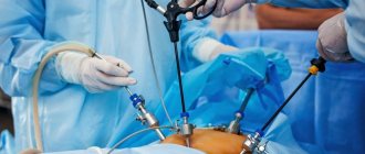
- Laparoscopic removal of ovarian cyst . This minimally invasive method involves excision of the tumor using an endoscopic device (laparoscope) inserted through a small puncture in the abdominal wall. This somewhat complicates the operation itself and increases the requirements for the qualifications of the specialist performing it. But after it there is no open incision, which reduces the risk of infection, and there are practically no unaesthetic scars left.
Today, laparoscopic cystectomy is the preferred method for removing ovarian cysts. Classic surgery through an incision is performed only in extreme cases or in the absence of appropriate specialists and instruments. Therefore, laparoscopic cystectomy will be discussed in detail below.
Contraindications before laparoscopy
Not all patients at the clinic can undergo surgery. Laparoscopy is contraindicated for patients in the following cases:
- detection of inflammatory processes in the pelvic organs;
- state of shock;
- presence of bronchial asthma;
- anorexia;
- disruption of the respiratory system;
- hiatal hernia;
- pathologies of the nervous system, etc.
Indications and contraindications for removal of an ovarian cyst
Resection of an ovarian cyst using a laparoscope is prescribed if it does not disappear on its own or as a result of drug (hormonal) therapy within 3 menstrual cycles. Indications for this operation are also:
- The appearance of a cyst after menopause - in this case it is not a violation of the natural process of ovulation and, therefore, will not go away on its own;
- Cysts and neoplasms are relatively small in size (up to 10 cm).
There is a list of situations in which laparoscopic removal of an ovarian cyst is not recommended or is strictly excluded - these include:
- Obesity of 3-4 degrees due to an increased fat layer of the abdominal wall, which interferes with the use of a laparoscope;
- High risk of stroke or myocardial infarction, decompensation of chronic pathologies;
- Infectious process, massive bleeding, peritonitis (inflammation) in the abdominal cavity;
- Malignancy of a cyst is its transformation from a benign tumor to a malignant one, in this case laparoscopy can only be used to biopsy its sample;
- Excessive (over 10 cm) size of the cyst, due to which it cannot be removed without an incision on the abdominal wall;
- A bleeding disorder that, if treated surgically, can cause extensive blood loss and death;
- Extensive adhesive process, making it difficult for the laparoscope camera to view the problem area.
At the request of the patient, the operation can be performed without any clear indications. For example, if she does not have the opportunity or desire to wait 3 months until the cyst goes away on its own (which may not happen). However, doctors still recommend using drug therapy first as the most gentle way to get rid of the problem.
Features of recovery after laparoscopy
After the operation has been performed, the patient is left in the hospital to regain consciousness. The most important recommendation from experts is movement no later than 4 hours after surgery. Physical activity in small quantities is useful for normalizing blood circulation.
In the normal course of the operation, the average recovery time is 3 days. After this, the patient returns home. But heavy physical activity is contraindicated for 2-3 weeks. It is recommended to carry out special gymnastics to strengthen the abdominal muscles.
Preparing to remove an ovarian cyst
It begins with diagnostics, which is necessary to accurately determine the presence of a neoplasm, its size, location, indications and contraindications for surgery. A comprehensive medical examination before removal of an ovarian cyst includes:
- Initial consultation and medical history , thanks to which the doctor learns about other problems with the reproductive system and the body in general, possible injuries, hypothermia, metabolic disorders and other factors contributing to the development of cystic pathology;
- Manual examination , which consists of palpating the suspected cyst through the abdominal wall and allowing one to determine the size, shape, position, density and other characteristics of the neoplasm;
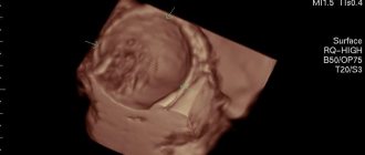
- Pelvic ultrasound is a non-invasive study that provides a clear visual representation of the cyst using ultrasound “candling” of the abdominal organs;
- Colposcopy is a visual examination of the vagina and cervix using an optical colposcope, which allows one to assess possible changes in the genital tract due to cyst growth;
- Fluorography - with its help, the doctor can identify neoplasms in a woman’s lungs and mammary glands, thereby confirming the possible oncological nature of the ovarian cyst;
- Blood tests - general, biochemical, clinical, histological, coagulability, blood group and Rh factor, infectious diseases.
Take the first step
make an appointment with a doctor!
Also, in order to clarify the diagnosis, magnetic resonance imaging of the pelvic organs, taking a smear from the vagina or cervical canal and other tests may be prescribed.
Laparoscopy has certain restrictions on the thickness of the abdominal fat layer under which it can be performed. If the patient is overweight, the doctor may prescribe a special diet and exercise to reduce body weight. It is recommended to start taking activated charcoal approximately 1 week before surgery.
Immediately the day before laparoscopy, the patient must:
- take a bath and epilate the abdomen and intimate area so that hair does not interfere with the operation;
- eat no later than 19:00 and drink before 22:00 the evening before;
- immediately before the procedure, cleanse the intestines with an enema.
Such preparation for surgery to remove an ovarian cyst is necessary for the most convenient access of the laparoscope to the abdominal cavity and the maximum possible view. This significantly reduces the risk of damage to healthy tissues and organs, internal bleeding and other complications.
Stages of laparoscopy
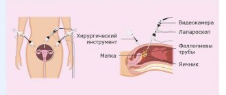
After completing the preparatory procedures, a small incision is made in the umbilical area. Next, doctors lift the abdominal wall and insert a Veress needle into the pelvic area. Carbon dioxide is injected through it in a volume of 3-4 liters. In this case, it is necessary to monitor the pressure (not higher than 14 mm) so that vasoconstriction does not occur.
The final stage of the operation in gynecology is the insertion of tubes, before which the needle is removed. After the puncture, the trocar is removed and the laparoscope is inserted through the tube. Due to the fact that it is equipped with a light indicator and a video camera, specialists can immediately see the condition of the internal pelvic area and prescribe treatment. Additionally, manipulation tools can be installed. The course of further treatment depends on the individual clinical picture.
How is surgical removal of an ovarian cyst performed?
Although laparoscopy is a minimally invasive procedure, it is performed only by a suitably qualified specialist in a hospital setting. The operation is performed under general anesthesia (anesthesia) with tracheal intubation or the use of a mask. This method of pain relief allows for better control over the quality of anesthesia.
The patient is positioned on the operating table, the head of which is tilted down at an angle of 30 degrees. Thanks to this, the intestines move towards the diaphragm and free access to the ovaries. To further improve visibility and provide the instruments with the necessary freedom of action, carbon dioxide is released into the abdominal cavity through a puncture in the navel through a tube, which inflates the abdomen and increases its internal volume. The operation then proceeds as follows:
- A laparoscope, a tubular instrument with a miniature camera and a light source, is inserted into a puncture made in the navel. With its help, the doctor establishes visual control over the operated organ and the surrounding area, assesses the size and shape of the cyst, and its condition. Based on this examination, a conclusion is made about the need for further use of laparoscopy or classical cystectomy.
- Through lateral punctures in the abdomen, the doctor inserts additional instruments into the abdominal cavity. With their help, under visual observation through a laparoscope, he peels out the inner surface of the cavity and sucks out its contents. In some cases, the doctor performs partial or complete removal of the ovary with a cyst in women with a strong spread of the pathological process in its tissue.
- At the final stage, the doctor examines the operated organ and surrounding tissues for bleeding or other injuries. If there are none, the instruments are removed from the patient’s body, the gas is sucked out from the abdominal cavity, the punctures are sutured and covered with bandages. The anesthesia tube is removed from the patient's trachea, she is brought to consciousness and transferred to a regular ward.
The postoperative period after laparoscopy lasts an average of 14 days. The patient is discharged from the ward after 3-5 days, the stitches from the punctures are removed after 7-10 days. If complications arise during the operation, the rehabilitation period can be increased depending on their severity.
Preoperative preparation
| Stages | Name | Process |
| 1st | Premedication | 1 hour before surgery, the anesthesiologist administers medications to speed up the recovery process and improve the state of anesthesia. |
| 2nd | Catheter placement | Occurs in the operating room when prescribed drugs are administered intravenously to a patient. A specialist is monitoring his condition. All data on blood and the working condition of the heart is displayed on the monitor. |
| 3rd | Introduction of anesthesia | The first medications are administered to quickly fall asleep, and subsequent medications simplify the process of inserting the tube into the tracheal tract. As a result, the procedure is painless and effective. |
| 4th | Connection of tube and anesthesia machine | After they are completed, the operation begins. |
The manipulation takes on average 10 minutes.
Efficiency and safety of ovarian cyst removal
The invasive and minimally invasive methods of surgical removal of ovarian cysts used today show fairly high efficiency and a low level of risk associated with complications. However, they do happen sometimes and you need to be aware of them:
- Pain syndrome. It occurs most often due to the accumulation of carbon dioxide residues in the abdominal cavity. They act on large clusters of nerves, causing them to become irritated. This does not pose a threat to life, but it causes discomfort that passes over time.
- Adhesive process. Occurs due to scarring of tissue in places where surgical instruments impact them. Adhesions after removal of an ovarian cyst occur quite rarely, but can lead to disruption of the anatomical structure of the reproductive organs and, as a result, to infertility.
- Subcutaneous emphysema. Also a non-life-threatening complication caused by the accumulation of carbon dioxide in the upper layer of subcutaneous fat. It occurs when the surgical technique is violated, but goes away on its own.
- Bleeding. This complication already poses a real threat to the patient’s life. It occurs due to a violation of the surgical technique, poor diagnostics, or for other reasons. Internal bleeding leads to the accumulation of blood in the abdominal cavity, causing acute peritonitis to develop in it.
Take the first step
make an appointment with a doctor!
The cost of surgery to remove an ovarian cyst using a classical or laparoscopic technique is today affordable for the vast majority of patients of reproductive age. The price may vary depending on the chosen method of surgical intervention, size, type, position of the cyst, and other complicating factors. In most cases, this procedure provides a high chance of complete recovery from this pathology.
Postoperative period
Sometimes after laparoscopy, a woman feels slight pain at the puncture sites, which is considered a completely normal manifestation, since this is the body’s reaction to damage to soft tissues and surgical intervention. This phenomenon will not affect the healing process in any way. If the pain is unbearable, then you should immediately seek advice from a specialist to assess the condition of the body and the wound surface and prescribe appropriate treatment.
After laparoscopy of the ovaries, a disruption occurs in the menstrual cycle, and vaginal discharge is not dangerous and is considered normal. They are small in volume and mucous in nature, lasting up to several weeks. An alarming symptom may be a change in the color of the discharge to a dark green or brown tint, in parallel with signs of general weakness, drowsiness, fever, and severe lower back pain. This situation indicates the addition of pathogenic microflora and the development of an infectious process.
What are the complications after laparoscopy?
Often, complications after this procedure rarely occur, since defects can be identified in the first stages of the examination. Complications may occur after insertion of the tube and trocars:
- sudden bleeding;
- damage to the outer lining of the intestine;
- emphysema.
Also, sometimes adhesions can form, which lead to infertility and disruption of the gastrointestinal tract. This is due to minor damage to blood vessels. If doctors are inexperienced, hernias and hematomas can form.
Diagnosis of ovarian cysts
At the initial appointment, the doctor collects anamnesis and conducts a gynecological examination, which allows you to assess the size of the ovaries, as well as identify space-occupying formations in the pelvis.
In addition, to diagnose ovarian cysts, studies such as:
- blood tests for tumor markers CA 125, CA 19-9, CA 15-3, CA 72-4;
- Ultrasound examination (ultrasound) of the abdominal organs;
- MRI of the abdominal cavity and pelvis;
- Laparoscopy is a minimally invasive surgical intervention that allows you to visually assess the condition of the reproductive organs, the size and macrostructure of the formation.