Urate stones
Violation of purine metabolism, for example with gout, provokes the appearance of urate stones in the kidneys. Basically, these stones dissolve well. To check for stones, patients are advised to check the amount of uric acid in their blood. Urate stones are clearly visible on ultrasound, but due to their low density, they are not detected on x-rays. For a more accurate diagnosis, the patient undergoes a computed tomography scan.
Open cavity stone removal
Open cavity removal of stones (nephrolithotomy) is used in only 3% of patients. The operation is forced and is performed when other methods have not brought benefit. Open manipulations are carried out when the patient has abnormalities of the pyelocaliceal system that require elimination. At the same time, the procedure removes stones.
- Urate urolithiasis is the most common type of all types of urolithiasis. Its treatment with conservative and surgical methods is carried out in all countries of the world, so the principles of treatment of the disease have been carefully developed.
- Citrate treatment is an innovation. It shows high efficiency with urates, which do not exceed 1.5 cm in size.
Modern minimally invasive procedures are used for stones from 1.5 to 2.5 cm, when conservative methods do not bring a positive result.
In any situation, the choice of treatment tactics for the disease should be made by a qualified specialist. Only he will be able to diagnose the pathology and select the optimal treatment tactics.
Dissolving urate stones
- Before dissolving the stones, it is necessary for the urine to change from acidic to slightly acidic or even alkaline. For treatment, the patient is prescribed citrate preparations and an alkaline liquid. You will have to drink from 2 to 6 months. It depends on the size of the stones. When the stone size is more than 2 cm in diameter, endoscopic crushing is first required. Only then begin to dissolve small fragments. It is important to combine the dissolution of stones with plenty of drinking.
- Unfortunately, patients come to the doctor late, when the stone has already moved from the kidney to the ureter. Then it’s too late to dissolve them. The patient suffers from renal colic and may develop pyelonephritis. The doctor has to urgently perform an operation to remove stones from the kidneys. The remaining small stones can be dissolved using citrate preparations.
Modern diagnostic methods
Detecting urate stones is not difficult for modern medicine. If urolithiasis is suspected or after renal colic has occurred, the doctor will prescribe the following examination:
- blood test (inflammatory changes are possible: increased leukocytes, accelerated ESR);
- urine analysis (examination of urinary sediment), pH measurement using special methods;
- Ultrasound of the kidneys and bladder (allows you to clearly visualize stones, their location, size, etc.);
- survey and intravenous renal urography;
- MRI or CT of the urinary system is the most modern and reliable diagnostic method.
Computed tomography and magnetic resonance imaging can very accurately detect urate stones
Advice for patients
- Drinking plenty of fluids reduces the acidity of urine. Therefore, small pebbles can dissolve. If the water treatment was successful, then you should continue taking citrate medications. But such drugs have negative consequences, so they will have to be used only as prescribed by a doctor.
- Some patients hope to use lemon or cranberry juice to dissolve and remove kidney stones. This is strictly forbidden. Drinking this may cause gastric perforation or internal bleeding. It is necessary to break and dissolve kidney stones only under the supervision of a doctor. You don’t need to listen to your neighbor about how he got rid of stones in his organ. It may have helped him, but such treatment could be harmful to another person.
- Many people expect that using fir oil along with diuretic herbs can dissolve stones. While the stone is in the organ, you just need to watch it and follow the right diet. Taking diuretic herbs can dislodge the stone and cause renal colic. The patient will have to be urgently hospitalized and have the stones surgically removed.
- To prevent the formation of oxalates, you should consume more buckwheat, carrots, rose hips, broken wheat, beans, and nuts. Rhubarb, lettuce, sorrel and chocolate should be excluded.
- If small grains of sand are found on an ultrasound, then these may be compacted papillae or dense tissue. It is better not to take diuretics, but to perform an ultrasound scan once every six months. This is how they know if the stone is growing.
Urolithiasis (kidney stones) - symptoms and treatment
General principles of therapy
Treatment of urolithiasis depends on the size and location of the stone (kidney, ureter or bladder), the condition and characteristics of the urinary tract (for example, narrowings or fixed bends that make it difficult to pass the stone), and the presence of complications.
In mild cases, if the stones are small (usually up to 5 mm), drug stone therapy can be used with the prescription of diuretics, antispasmodics and painkillers.[2]
Conservative treatment
Diet therapy. Diet is very important to prevent the recurrence of kidney stones. To normalize metabolic processes, all patients with urolithiasis are recommended to limit table salt to 5-6 grams per day (food is prepared without salt and add salt already on the plate), limiting animal and vegetable protein (up to 1 gram per kg of body weight). For urate stones (that is, consisting of uric acid salts), in addition to the above-mentioned dietary restrictions, dark beers, red wine, pickles, smoked meats, offal, coffee, cocoa and chocolate are not recommended.
To speed up the spontaneous passage of stones, it is recommended to drink plenty of fluids in combination with physical activity.
Phytotherapy. For the prevention and treatment of urolithiasis, herbal products are widely used. “Cyston” and “Canephron” are used more often. The drugs have a diuretic, anti-inflammatory and litholytic effect, but large kidney stones will still require more serious treatment; self-prescription of such drugs without consulting a doctor can only lead to a waste of time.
Among the traditional methods of treatment, woolly erva, or half-fell (lat. Aerva lanata), is effective. The plant is especially useful for oxalate stones.
Citrate mixtures . Some types of urinary stones (for example, urates) can be easily dissolved using so-called citrate mixtures (“Blemaren” or “Uralit-U”). This method is based on increasing the solubility of urate stones by shifting the acidity of urine (pH) to the alkaline side. The dissolution process is quite lengthy and labor-intensive, requiring regular monitoring of the pH (indicator strips are included in the package), but with the right approach it allows you to completely get rid of stones without additional intervention.[10]
Lithotripsy
Remote lithotripsy (or non-contact crushing of stones) is a unique method of getting rid of kidney and ureteral stones, when the stones are destroyed directly in the body without the introduction of instruments. Crushing is carried out using a special apparatus - a lithotripter. Lithotripsy is used in the treatment of urolithiasis in the presence of small kidney stones.
Previously, due to their high cost, such complexes were installed only in large research centers and hospitals, but today the method is more accessible, including in commercial clinics. A modern device for remote lithotripsy is a fairly compact shock wave generator combined with a device for targeting the stone.
Structurally, ultrasonic or x-ray guidance is possible. At the same time, ultrasonic guidance is advantageous in the absence of ionizing radiation (radiation exposure) and the possibility of continuous monitoring of stone destruction in real time. In addition, ultrasound can be used to target stones that are X-ray negative (that is, invisible to X-rays). The crushing procedure usually takes no more than an hour and does not require serious pain relief. Recently, external lithotripsy has been performed on an outpatient basis, that is, without hospitalization.
During crushing, the stone is destroyed by shock waves into small fragments, which then independently pass through the natural urinary tract. To facilitate and speed up this process, antispasmodic and diuretic drugs are often prescribed. Using extracorporeal lithotripsy, you can effectively destroy kidney stones of relatively low density up to 2 cm in size.[2]
Surgery
Transurethral contact lithotripsy. When a stone gets stuck in the ureter and blocks the outflow of urine, which is manifested by recurrent attacks of renal colic, which are difficult to relieve with conventional medications, endoscopic intervention is used to quickly remove the stone and restore the outflow of urine - transurethral contact lithotripsy. As the name suggests, in this operation, performed through the urethra (urethra), the instrument, under visual control, is brought directly to the stone and the latter is destroyed by contact - laser, ultrasound or a pneumatic probe.
The advantage of contact lithotripsy is the complete destruction and removal of the stone immediately during surgery, restoration of urine outflow and the absence of the stage of passage of fragments. In some cases, for additional drainage of the upper urinary tract, a plastic catheter (internal stent) is installed in the ureter after surgery.
Contact lithotripsy is usually performed under spinal anesthesia and requires short-term hospitalization. An additional advantage of transurethral lithotripsy is the ability to simultaneously eliminate narrowing or fixed kinks of the ureter below the stone, which can be an insurmountable obstacle to the passage of stones (or even fragments after remote crushing).
Percutaneous nephrolithotripsy . Large and dense kidney stones, which cannot be destroyed using extracorporeal lithotripsy, are today removed through a small puncture in the lower back. This operation is called percutaneous nephrolithotripsy. Under ultrasound and X-ray guidance, an instrument is inserted into the kidney through a puncture, with the help of which, under visual control, the stone is destroyed and fragments are removed. As with transurethral contact lithotripsy, destruction is achieved using a laser, ultrasound or pneumatic probe. This method can destroy stones of any size and density. True, in some cases it is necessary to make additional punctures for this. The operation often ends with the installation of a thin drainage tube (nephrostomy) into the kidney through an existing puncture, which is removed after a few days. Percutaneous nephrolithotripsy is usually performed under general anesthesia and requires hospitalization for a period of 3 to 5 days.
The most modern modification of this operation is minipercutaneous laser nephrolithotripsy . The main difference is the use of miniature instruments with a diameter of about 5 mm, which is approximately half the size of traditional ones. Thus, the puncture in the skin becomes almost invisible, the recovery period is reduced, as well as the likelihood of complications.
Transurethral contact lithotripsy. Another modern and minimally invasive method for removing stones from the kidneys and ureters is flexible transurethral contact lithotripsy (or fibroureteronephrolithotripsy, or retrograde intrarenal surgery). The main advantage of this method is the absence of cuts and punctures, that is, damage to the skin. A flexible miniature instrument equipped with an actively moving tip with a high-quality video camera is inserted through the natural urinary tract (urethra).
Depending on the task, the instrument is passed into the ureter or into the kidney and brought to the stone. The latter is destroyed with the help of a laser into “dust” (dusting effect), which does not require the extraction of fragments - they are washed off with a flow of liquid during the operation. This method is ideal for relatively small and dense kidney stones, especially multiple ones located in different calyces.[2] The flexibility of the fibroureterorenoscope allows it to be passed through narrowings and fixed bends, without the risk of damage. The main disadvantage of this technology is the very high cost of the equipment. Therefore, not all even large urological centers have a fibroureterorenoscope in their arsenal.
Laparoscopy. For kidney and ureteral stones, the method is used quite rarely, mainly when urolithiasis is combined with urinary tract anomalies (for example, a large stone in the pelvis and narrowing of the ureteropelvic segment), when it is necessary to simultaneously remove the stone and eliminate the anomaly.[11]
Today, open operations (that is, performed through a skin incision) are almost completely replaced from the arsenal of means for removing urinary stones. This made it possible to make surgical treatment of urolithiasis quick, easy and safe, which is especially important given the tendency of the disease to recur.
Treatment of infectious complications
Infectious complications are treated with antibiotics and anti-inflammatory drugs. According to indications, drainage of the urinary tract is performed.
How to recognize and treat urolithiasis in children
Diagnosis is carried out using ultrasound and urine analysis. Treatment is the same as for adults.
Why urolithiasis worsens in pregnant women and how to treat it
KSD during pregnancy worsens due to impaired outflow of urine from the kidneys, as well as changes in mineral metabolism. Treatment during pregnancy is carried out only as a last resort, for ureteral stones and pain. Endoscopic methods (stent installation, endoscopic stone removal) have an advantage.
Is it possible to dissolve kidney stones
Only urates can be dissolved. These stones lend themselves well to dissolution using citrate mixtures (“Blemaren” or “Uralit-U”).
Rehabilitation
No special rehabilitation is required. Monitoring of urine tests and ultrasound monitoring is carried out according to indications. Drinking plenty of fluids is recommended.
How to deal with oxalate stones
These stones most often appear in the kidneys. They contain salts of oxalic acid. Such stones cannot be dissolved, but they grow quickly. They are removed surgically.
Oxalate stones are hard with sharp edges, so they can damage the walls of the urinary tract. Oxalates cause renal colic and other troubles in a sick person.
Many doctors advise patients to get rid of stones through surgery. Treatment must be combined with drinking plenty of mineral water and berry juice. Treatment is required only under the supervision of a specialist. Otherwise, the stones may increase in size.
Specialists of the Russian Doctor company recommend not to self-medicate, but to consult a doctor to diagnose the condition of the kidneys and determine the composition of the stones in order to prescribe the correct treatment.
- Daria Bogdanova
- Urology
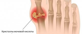
Causes
Salts in a healthy body should be completely excreted in the urine. If one or another failure occurs - for example, associated with metabolic disorders - its crystals begin to stick together, taking on bizarre shapes and sizes. Large ones are considered to be 5mm or more.
As a rule, one does not think about the existing disease, its causes and the need for treatment as long as the crystal remains in a stationary position. When it starts to move, acute pain appears.
The most likely causes of the appearance of renal formations include:
- geographical factor - hot climate, insufficient water consumption or too hard water;
- various pathologies of the genitourinary system, congenital anomalies that cause narrowing of the urinary tract, which makes the outflow of urine difficult;
- vitamin imbalance, lack of ultraviolet radiation, unhealthy diet.
- abnormalities in the functioning of the parathyroid glands, causing disturbances in calcium metabolism;
- various diseases - pyelonephritis, hydronephrosis, kidney prolapse, inflammation of the bladder, prostate adenoma, prostatitis;
- sedentary work.
Often, kidney stones have a different nature, but in more than half of the cases their composition is mixed. They often end up in the bladder or ureter. Much is determined by the nature of nutrition and the age factor.
By what methods are they identified?
The most common method for diagnosing the presence of kidney stones is ultrasound. It clearly shows the size and location of the stone. The only drawback of this method is that it is impossible to determine from ultrasound whether it is a urate stone or some other one. Therefore, a number of additional studies are prescribed :
- General blood analysis. Indicates the possible presence of inflammatory processes (if the level of leukocytes is elevated).
- Analysis of urine. It is used to determine the pH level (acidic, alkaline, neutral). Also, an increased level of white blood cells in the urine may indicate an infection.
- X-ray using contrast agents. A regular x-ray “does not see” urate, and therefore it is necessary to introduce contrast.
- CT (computed tomography). Allows you to determine the density of the formation and, accordingly, its chemical composition.
- Urography – x-ray of the urinary tract.
It is worth noting that a 100% chemical analysis of the stone can only be done after it has been scraped or removed .
Diet food
Diet acts as one of the measures to effectively dissolve urates, and is also used for preventive purposes. It provides for the refusal of:
- fatty meat;
- offal;
- beer and alcohol in general;
- soda containing orthophosphoric acid - Coca-Cola, etc.;
- canned foods.
A significant reduction in diet is necessary:
- legumes;
- sorrel, onion, spinach;
- cocoa products and chocolate;
- seafood and fish;
- table salt;
- strong coffee and tea.
It is necessary to include in food:
- milk and other dairy products;
- fruits and watermelons;
- eggs;
- seeds and nuts;
- wheat and buckwheat cereals.
Portions should be small and taken often - 5-6 times a day.
Menu for the week
Based on the above listed products, we display an approximate menu for the week. Days and meals can be alternated. Here is an example of a weekly diet:
| Monday | Breakfast: carrot and cabbage salad, a slice of bran bread and rosehip decoction. Snack: boiled egg, pumpkin and carrot salad, compote. Lunch: milk soup with noodles, potato-squash pancakes, rowan decoction. Snack: cottage cheese with apple. Dinner: vegetarian stew, milk. |
| Tuesday | Breakfast: cottage cheese, banana, compote. Snack: salad with bell pepper and arugula, juice. Lunch: pearl barley soup with veal, uzvar. Snack: two pears. Dinner: cottage cheese casserole, tea. |
| Wednesday | Breakfast: millet porridge, multivitamin juice. Snack: yogurt with raspberries. Lunch: cabbage soup, vegetable salad, compote. Snack: jelly, biscuits. Dinner: oatmeal, stewed vegetables, tea. |
| Thursday | Breakfast: semolina porridge, two plums, tea. Snack: sweet pepper and cucumber salad, a couple of slices of bread. Lunch: pumpkin and cottage cheese casserole, hawthorn broth. Snack: kefir, baked apple. Dinner: boiled lean meat, baked vegetables (carrots , beets, potatoes). |
| Friday | Breakfast: fruit salad with yogurt. Snack: cottage cheese with sour cream and honey. Lunch: borscht without meat, carrot bits, compote. Snack: kefir, red apple. Dinner: stuffed peppers, pancakes with cottage cheese, milk. |
| Saturday | Breakfast: buckwheat porridge, toast with jam, fruit tea. Snack: tea with marshmallows. Lunch: broccoli soup, zucchini pancakes, compote. Snack: strawberries with cream, mint lemonade. Dinner: cheesecakes with fruit, fruit juice. |
| Sunday | Breakfast: cheesecakes, fruit juice. Snack: blueberries with low-fat cream. Lunch: salmon with baked vegetables, kefir. Snack: tomato juice. Dinner: Greek salad, fermented baked milk. |
Diet also helps a lot: table No. 7 according to Pevzner, if you follow the instructions of this diet, you can easily cure your kidneys and keep your body healthy.
Features of stones
The prevalence of urate stones among patients reaches almost 50% of the total. In appearance, they are rounded formations in a color range from yellow to brown, with a smooth surface.
They form in different organs, and this greatly depends on age: after 45-50 years they are almost always found in the bladder, and in young patients from 20 to 45 years old - in the ureter and kidneys. If this type of formation is not detected in time, serious consequences often arise.
Manifestation of the clinical picture
Unfortunately, the disease usually does not manifest itself for a very long time, and symptoms can only be noticed when the stone reaches a large size and renal colic begins. It manifests itself as follows:
- discomfort that initially begins in the lumbar region and then spreads to the lower abdomen;
- a sharp pain begins in the same area, which does not go away;
- chills;
- nausea and possibly vomiting;
- flatulence;
- pain when urinating, urine may become cloudy, have sediment in the form of sand, blood;
- high body temperature is an alarming sign of the development of inflammation.
Renal colic begins when the formed stone becomes an obstacle to the outflow of urine.
Before such an acute phase, urate stones can manifest themselves in the form of aching pain in the lower back and, in some cases, problems with urination.