The prevalence of neurofibromatosis type 1 (NF1) is approximately 1 case in 3,000 people, but this figure varies markedly depending on the country and in Russia it is 1 case in 7,812 people. The life expectancy of people with neurofibromatosis type 1 is reduced by ~8–21 years, and their cause of death is most often related to malignancy.
NF1 is a genetically determined disease with an autosomal dominant pattern of inheritance, which means that all carriers of germline mutations in the 17q11.2 gene will develop NF1. However, the manifestations of this disease are extremely variable and can differ even among members of the same family who have an identical mutation. By the way, there are quite a lot of different mutations that cause the disease (>3000), which is associated with the large size of the gene (more than 60 exons) and the variety of types of mutations (translocations, deletions, inversions and point mutations) that can occur in this area - this complicates genetic diagnosis. The latter is carried out on DNA material from peripheral blood lymphocytes and/or tumor material, with the exception of segmental neurofibromatosis (sometimes called neurofibromatosis type V), when the lesion affects only one segment of the patient’s body. In the latter case, only tumor material can serve as diagnostic material. .
Figure 1 | Segmental neurofibromatosis. Multiple neurofibromas located within one or more dermatomes.
The 17q11 gene encodes the protein neurofibromin, which is a tumor suppressor. Neurofibromin is produced in nerve cells and specialized neuroglial cells (oligodendrocytes and Schwann cells) and normally suppresses the RAS proto-oncogene, accelerating its transition to an inactive form. The inability to produce functional neurofibromin results in increased cell growth and survival.
Neurofibromas
Benign neoplasms characteristic of NF1. There are cutaneous (subcutaneous), intraneural, plexiform and diffuse neurofibromas. .
Figure 2 | From left to right: cutaneous neurofibromas; intraneural neurofibromas along the peripheral nerve; large intra-abdominal plexiform neurofibroma (asterisk).
Cutaneous neurofibromas originate from dermal progenitor cells, can be painful or painless nodular formations, the “doorbell” symptom is described: when pressed, the neurofibroma completely sinks into the skin.
Intraneural and plexiform neurofibromas are tumor formations from the sheaths of peripheral nerves and nerve plexuses. In mice with biallelic loss of the NF1 gene in Schwann cells, it was possible to model neurofibromas similar to those in people with NF1. Like human plexiform neurofibromas, the mouse tumors were composed of a variety of cell types, including mast cells, macrophages, fibroblasts, neurons, and Schwann cells.
Neurofibromas can develop into malignant nerve sheath tumors, with plexiform neurofibromas, especially those of the brachial and lumbosacral plexuses, presenting the greatest risk. Also, the risk of degeneration is increased in patients with a history of radiation therapy or a family history of malignancy.
A characteristic symptom of neurofibromas is that the formation moves only laterally, but not up and down (since it “sits” on the nerve trunk). Neurofibromas can compress a peripheral nerve, causing loss of function.
Etiology and pathogenesis of neurofibromatosis type 1
The cause of neurofibromatosis type 1 is an autosomal dominant mutation in the NF1 gene. In one half of the cases, it is inherited from the parents, while the full penetrance of the mutation ends after the end of childhood, that is, the probability of carriage is 100 percent. In the other half of cases, a de novo mutation is noted.
A loss-of-function mutation of the NF1 gene on chromosomal segment 17q11.2 causes insufficient synthesis of the tumor suppressor protein neurofibromin. Over 1 thousand different NF1 mutations have been identified. Most mutations result in a severe truncation of the gene product.
Neurofibromin, a member of the GTPase-activating protein family, is responsible for the negative regulation of the activity of the RAS/MAPK signaling pathway by accelerating the hydrolysis of Ras-bound guanosine triphosphate (GTP). Genetic damage disrupts control over cell growth and neuronal development.
Skeletal abnormalities
Normal functioning of bone tissue requires a coordinated interaction between resorbing (osteoclasts) and bone-forming (osteoblasts) cells. The use of a mouse model showed impaired osteoblast function (increased pyrophosphate production, decreased bone morphogenetic protein 2, which promotes osteoblast differentiation) in mice mutant for the NF1 gene. As a result, patients experience impaired bone mineralization, arched deformities of the tibia, kyphotic and scoliotic deformities, chest deformities, pseudarthrosis of large joints, and dysplasia of the sphenoid bones.
The most important
Recklinghausen's disease is a dangerous genetic disease that cannot be completely cured. Its insidiousness is that the symptoms are so versatile that diagnosis is difficult. If you notice more than 6 large pigment spots in yourself or your loved ones in atypical places (armpits, groin, neck), then immediately consult a dermatologist. The specialist will conduct the necessary research and make a diagnosis. It is important to start treatment in the early stages of the development of the pathology in order to avoid dangerous complications that can be life-threatening. With complex therapy and regular visits to the doctor, the patient has every chance of living a long life.
Optic tract glioma
Gliomas are benign tumors of the optic nerve and other parts of the visual analyzer. The location can be any (orbital, intracanal, intracranial - the latter is sometimes divided into pre-, post-chiasmatic and chiasmatic), multiple lesions are possible, symmetrical location of tumors is uncharacteristic. A significant dependence of tumor growth on the tumor environment, which is represented mainly by neuroglial cells, has been shown. The latter influence the proliferation of tumor cells by secreting cytokines, as well as neurotoxins, which damage the axons of the optic nerve, leading to impaired visual acuity. This gives hope that in the future, targeted therapies targeting the tumor environment may be able to slow the growth of optic nerve gliomas. This neoplasm rarely causes death, but significantly affects the quality of life of patients. Girls get sick 5–10 times more often than boys.
In addition, other types of brain tumors (astrocytomas, gliomas) occur more often in patients with NF1 than the average population.
Characteristics of the disease
Recklinghausen's neurofibromatosis is a whole group of hereditary pathologies characterized by the appearance of multiple neurofibromas (tumors of nervous tissue). They are formed due to disruption of the normal development of nerve cells. Neoplasms are localized along the nerve ganglia and their branches. The disease disrupts the functionality of various body systems, which is associated with a gene mutation.
Reference. Recklinghausen's disease affects both sexes in equal proportions. Most often it manifests itself in children from 3 to 16 years old.
This is one of the most common genetic pathologies, it is diagnosed in 1 patient out of 2500 - 7800. If one of the parents suffers from this disease, then with a 50% chance the child will develop it. Situations are also possible when a mutation occurs in the genetic material (egg, sperm) of parents who did not have neurofibromatosis in their family.
Other manifestations
Tumor diseases include pheochromocytoma and iris hamartomas (Lisch nodules).
Mental retardation is common.
The cumulative risk of cancer by age 50 in individuals with neurofibromatosis type 1 is increased by 20–39%, with a lifetime risk of developing cancer of ~60%.
In addition, people with neurofibromatosis type 1 have an exceptionally high risk of developing malignant brain tumors (approximately 40 times the risk of developing high-grade glioma), endocrine cancer (more than 74 times the risk of developing adrenal cancer), peripheral nerve malignancies (>1000 times increased risk of developing) and breast cancer.
Children with neurofibromatosis type 1 have an increased risk of leukemia, acute lymphocytic leukemia, and non-Hodgkin's lymphoma.
In addition, some studies confirm an increased risk of developing multiple sclerosis, epilepsy, macrocephaly, and hydrocephalus. .
Figure 3 |
Lisch nodules.
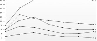
Figure 4 | Manifestations of symptoms of neurofibromatosis type 1 depending on the patient’s age.
The criterion for diagnosing NF type 1 is the presence of two or more signs (according to the National Institutes of Health conference on neurofibromatosis, USA, 1988):
- ≥ 6 café-au-lait spots > 5 mm in diameter prepubertally or 15 mm in diameter postpuberty;
- ≥ 2 neurofibromas of any type or one plexiform neurofibroma;
- Small pigmented spots in the armpits and groin areas, resembling freckles (Crove's symptom);
- Optic nerve glioma;
- ≥ 2 Lisch nodules (iris hamartoma);
- Bone changes: dysplasia of the wing of the sphenoid bone, thinning of the cortical layer of the tubular bones with or without pseudarthrosis;
- Presence of NF type I in first-degree relatives.
List of sources
- Sergeev A. S. Mutation frequency of neurofibromatosis // Abstract of thesis. Ph.D. biol. Sci. M., 1973 (USSR Academy of Medical Sciences, Institute of Medical Genetics).
- Schneider N. A. Neurofibromatosis of the first type: Recklinhausen disease / N. A. Schneider, A. I. Gorelov //Siberian Medical Review. - 2007. - No. 3 (44). — P. 91-95.
- Kozlov A. V. Neurofibromatosis 2 (NF2) // Surgery of skull base tumors / Edited by A. N. Konovalov. - M.: OJSC "Mozhaisk Printing Plant", 2004. - P. 169-170. — 372 s
- Ufimtseva M.A., Bochkarev Yu.M., Galperin A.M., Golovyrina I.L., Gurkovskaya E.P. SKINAL MANIFESTATIONS OF RECKLINGHAUSEN'S DISEASE // Modern problems of science and education. – 2021. – No. 6.
- Popova A. A Clinical and diagnostic aspects of neurofibromatosis // University Medicine of the Urals - 2021 - No. 2.
From the side of the central nervous system
Optic tract glioma can be detected on CT (especially in studies with small slice thickness) as a fusiform or exophytic extension of the optic nerve, and tortuosity of its course may also be observed. However, MRI is a more sensitive technique. Glioma manifests itself with a high T2WI MR signal (a thin hypointense strip along the periphery can be detected - the dura mater pushed aside by the tumor) and an iso- to hypointense MR signal on T1WI; Such tumors often accumulate contrast agent.
Differential diagnosis of optic nerve glioma is primarily carried out with meningioma: the latter is characterized by the presence of calcifications and the “tram rails” symptom, when the edges of the intact optic nerve are visible in the form of parallel stripes against the background of the tumor). Glioma of the chiasmal region must be differentiated from ganglionglioma, meningioma, astrocytoma or hypothalamic glioma, etc.
There is a Dodge classification of the prevalence of optic tract gliomas: stage 1: only the optic nerve is involved in the process. Stage 2: involvement of the chiasm. Stage 3: involvement of the hypothalamus and/or other adjacent structures.
.
Figure 5 |
An axial CT scan reveals an unevenly enlarged right optic nerve (on the scan on the left). .
Figure 6 | Bilateral optic nerve gliomas in a patient with NF1. Bilateral expansion of the optic nerve is determined with an increase in the MR signal from it on T2WI (1) and intense accumulation of the contrast agent (2). In the same patient, areas of increased MR signal were detected in T2WI without clear contours, mass effect or edema of the adjacent brain parenchyma, located in the basal ganglia and thalamus (3).
Folk remedies
Photo: ambafra-pk.org
Traditional medicine will never get rid of neurofibromatosis, but can help with complex treatment prescribed by a doctor. Despite the fact that folk remedies are considered harmless, before using any recipes, it is recommended to consult a doctor for advice, who will tell you all the benefits and possible harms of a particular remedy.
To maintain immunity, do not forget about nutrition. You should increase the content of fermented milk products (milk, kefir, fermented baked milk), seafood (sea fish, seaweed) in your diet; among meat products, preference is given to chicken and beef; green tea is also useful, as it has an antioxidant effect. It is also recommended to get rid of bad habits. This will help strengthen the general condition of the body.
There is a recipe for an infusion of celandine. To prepare it you need to take 1 tbsp. l. herbs and pour three glasses of boiling water. After infusion, you should drink one glass 15 minutes before meals. This plant is considered poisonous in high concentrations, so it is important to maintain proportions when preparing the infusion. It should be noted that the prepared infusion is stored for no more than 48 hours; after this time, consuming the infusion is strictly prohibited.
Burdock infusion is also used. Both stems and roots of the plant are suitable for its preparation. These ingredients are brewed in water at the rate of 30 g per 1 liter of water. After preparation, the infusion is used 3 times a day.
The information is for reference only and is not a guide to action. Do not self-medicate. At the first symptoms of the disease, consult a doctor.
SEARCH FOR TREATMENT AROUND THE WORLD WITH YELLMED
Brain stem gliomas
They make up approximately 8–10% of the total number of CNS neoplasms in NF1. The average age of patients is 8 years. The cerebellum is most often affected, followed by the pons and diencephalon. Concomitant hydrocephalus and brain herniation should be noted. Tumors appear hyperintense on T2WI and usually exhibit contrast enhancement heterogeneously. To avoid missing low-intensity contrast enhancement, subtraction may be used. .
Figure 7 | Brain stem glioma. In the area of the cerebellum, a hyperintense formation is detected on T2WI (a), which on post-contrast T1WI (b) demonstrates weak inhomogeneous accumulation of contrast.
Medicines
Photo: prostatit.guru
Medicines are used in the treatment of neurofibromatosis, but their purpose is only for symptomatic treatment; they do not eliminate the problem itself. The following medications are used:
- ketotifen;
- fenkarol;
- tigazol;
- lidase.
These drugs are prescribed by a doctor strictly individually, since the use of these drugs is not rational in every case.
Tigazol contains vitamin A. There are practically no contraindications to the use of the drug; people with liver or kidney failure, as well as in cases of individual intolerance to the component, should be careful.
Ketotifen is an antiallergic drug and has a membrane-stabilizing effect. Fenkarol also belongs to antiallergic drugs. The drug has a good antipruritic effect, therefore it is prescribed to people who have itching in the clinical picture of the disease. Fenkarol is well tolerated and causes virtually no side effects. Contraindicated for people with gastrointestinal and kidney diseases. It should be remembered that the drug does not have a depressant effect on the central nervous system, but in rare cases a mild sedative effect is observed.
Lidase contains hyaluronidase as an active substance. This is an enzyme whose action is aimed at hyaluronic acid. The drug increases the permeability of tissues, improves their trophism, increases the elasticity of scar areas, promotes the resorption of hematomas and eliminates contractures.
Focal areas of hyperintensity (FASI)
Found in approximately 80% of patients with NF1, they are areas of hyperintense MR signal on T2WI and FLAIR located in the basal ganglia, thalamus, brainstem, cerebellum and subcortical white matter. Morphologically they represent areas of myelinopathy with enlarged vacuoles. The correlation of these changes with the clinic is not fully understood. .
Figure 8 | Asymmetric areas of hyperintense MR signal in T2 are detected in the basal ganglia on both sides. Also noteworthy is the hyperintense formation on the left surface of the neck and head - plexiform neurofibroma.
Treatment
Often, the management of patients with neurofibromatosis is limited to symptomatic treatment. If neurofibromas are located in areas of increased trauma and cause pain, they are removed surgically. In case of multiple formations, chemotherapy is prescribed and radiation is performed. If the musculoskeletal system is damaged, rehabilitation measures are carried out.
The disease has a favorable prognosis. Tumor formations become malignant extremely rarely. When performing special measures, patients maintain a satisfactory quality of life and performance.
Other manifestations from the central nervous system, head and neck area
Figure 9 |
STIR of the cervical spine in sagittal projection. A hyperintense intramedullary formation with indistinct edges is identified—spinal cord astrocytoma. Patients with NF1 have an increased risk of CNS malignancies. .
Figure 10 | Axial T2Fs (a) and coronal T2WI (b) in a patient with NF1. Uneven expansion and hyperintensity of the MR signal from the spinal nerves, caused by intraneural neurofibromas, are determined. .
Figure 11 |
Sagittal T2VI of the lumbar spine. Ectasia (expansion) of the dural sac is determined. .
Figure 12 |
Axial CT scan of the orbits. Dysplasia of the left wing of the sphenoid bone (circle) is determined; The superior orbital fissure is enlarged. Neurofibromas often arise in this area, leading to proptosis on the affected side. .
Figure 13 | 3D reconstruction of CT scan of the skull bones. Dysplasia of the left wing of the sphenoid bone is observed. Also noteworthy is the enlargement of the brain skull.
Forecast
Life expectancy for type 1 Recklinghausen disease is approximately 8 years less than for healthy people. However, this applies to patients with benign tumors who started complex treatment early and are regularly monitored by a doctor. Malignant neoplasms cause early death. This outcome is possible with untimely treatment, then the person dies from multiple complications.
If there are no diseases of the musculoskeletal system, and the patient does not lag behind in development, then his level of performance is normal. The pathology has a chronic course and jerky development. The patient's condition may worsen due to hormonal changes, after serious illness or injury. Neurofibromatosis, which affects peripheral nerves, has the best prognosis.