Pathologies of the respiratory system are among the most common diseases. The harmful effects of bad habits, polluted urban air and a sedentary lifestyle are serious risk factors for the breathing of modern people. The presence of pulmonary symptoms - prolonged cough, sputum discharge in the morning, shortness of breath, often perceived as normal, is often a sign of dangerous diseases. To identify and assess the severity of respiratory system disorders, special studies are carried out, which include spirometry. What kind of analysis this is is of interest to many patients who are prescribed this test. Spirometry allows you to assess lung capacity, inspiratory/expiratory speed, the degree of ventilation failure and obstruction.
Spirometry: indications for performance
Spirometry is used mainly in pulmonology, less often in such areas of medicine as allergology and cardiology.
This study is indicated for patients in the following cases:
- if there are complaints from the respiratory system: chronic cough, shortness of breath, etc.);
- in case of changes in the organs of the respiratory system identified using other diagnostic methods;
- when gas exchange in the body is disrupted (with a decrease in oxygen content in the blood and an increase in carbon dioxide content);
- as a preparatory measure for other studies and surgical interventions (such as bronchoscopy, thoracotomy, etc.).
Spirometry also plays a key role in the diagnosis and differential diagnosis of COPD (chronic obstructive pulmonary disease) and bronchial asthma. The study is used to evaluate the effectiveness of their therapy.
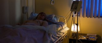
Patients suffering from these pathologies should regularly (at least once a year) undergo spirometry in a medical facility, and at home monitor external respiration indicators with a special device - a peak flow meter.
In addition, spirometry is recommended for people whose work activities are associated with hazardous conditions, heavy smokers and allergy sufferers.
Evaluating the results of the procedure and comparing them with the norm
A table with normal spirography values helps the doctor decipher the results of the procedure.
Carrying out spirography for bronchial asthma, pneumonia, chronic obstructive pulmonary disease and other pathologies will be accompanied by a decrease in these indicators. Depending on the nature of the problem and its severity, the approach to treating the patient differs.
Spirometry: contraindications
Spirometry is contraindicated in patients with the following conditions:
- pneumothorax;
- bleeding from the respiratory tract;
- tuberculosis;
- dissecting aortic aneurysm;
- hypertensive crisis;
- previous myocardial infarction;
- ischemic and hemorrhagic stroke;
- in the postoperative period after ophthalmological or intracavitary surgical interventions (in the first 1.5 months);
- mental disorders (if the patient is unable to follow the recommendations of the diagnostician regarding the quality and speed of breathing during the procedure).
The decision on the need and advisability of conducting the study is made solely by the attending physician. He evaluates the safety of spirometry in each specific case, identifies contraindications to the test, and determines whether they are absolute or relative.
Research methodology
How is spirography performed? After explaining all the nuances of the diagnostic procedure and giving the patient a twenty-minute rest, the doctor begins to perform the technique. The patient is asked to sit up straight. It is important not to tilt your head or bend your torso to prevent distortion of the study results.
Spirography is a diagnostic method that records and evaluates a stream of air released through the mouth. To reliably assess the results, the patient needs to close his nose with a special clip. Once the patient is prepared, the diagnosis can begin.
The person grasps the mouthpiece to direct air into the appropriate tube. Before direct exhalation, the doctor controls the tightness of contact between the mouth and the plastic to prevent loss of part of the gas mixture and distortion of the final result.
The doctor gives instructions to the patient about the nature of breathing.
The most commonly used methods are:
- Normal breathing in a calm mode. Over the course of 6-7 cycles, the computer records the tidal volume (TI) of the patient’s lungs, calculates the number (multiplicity) of chest movements in 1 minute and other parameters;
- Forced exhalation. First, the patient takes as deep a breath as possible. Then, within six seconds, the patient must quickly and under pressure push the air from the lungs into the tube. This sample may be partially modified if necessary. The main thing is to follow the doctor’s instructions;
- Frequent and maximum deep breathing for ten to fifteen seconds. This technique can cause dizziness and even loss of consciousness. The technique is performed with caution in children and elderly people.
Processing of digital data is carried out by a computer with the formation of a corresponding graphic image. After completing the above algorithm of actions and recording the final results of the technique, the doctor makes a written conclusion that helps to establish the final diagnosis and decide on the treatment of a particular patient.
Important! How much does spirography cost? The appropriate diagnosis can be carried out in a public or private clinic. In the first case, if there is a referral from a doctor, the service is provided to the patient free of charge. In private clinics the price ranges from 800 to 1200 rubles. To select a medical institution, you can use reviews with photos of functional diagnostic rooms that are available on the Internet.
Spirometry: preparation for implementation
To obtain the most complete and accurate information, in preparation for spirometry the patient should follow the following recommendations:
- the day before the study, avoid heavy physical activity;
- stop smoking at least four hours before the planned procedure;
- two to three hours before spirometry, refuse to eat;
- several hours before the test (the exact time is determined by the attending physician) do not take medications that dilate the bronchi.
In addition, 24 hours before spirometry, the subject should exclude coffee and other caffeine-containing drinks from the diet. Immediately before the examination, the patient loosens his belt, removes his tie and other items of clothing that restrict breathing; women are advised to remove lipstick from their lips.
Is it possible to do spirography for children?
Spirography can also be done for children from 5 years old. True, it happens that it is difficult to explain to a child how to breathe correctly, in this case it is better to double-check the results.
By the way, our sanatorium even accepts babies under one year old! There are specially designed children's programs. We have already told you why sanatorium treatment is important even for the little ones.
Now we understand more about the prevention of all kinds of diseases, and about spirography and what kind of procedure it is.
Spirometry: how is the study carried out?
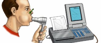
To carry out spirometry, a device of the same name is used - a spirometer, which records the volume and speed of air during inhalation and exhalation. It is equipped with a sensor for sensing air flow, determining the above characteristics, as well as a device for converting their values into digital format and calculating the necessary indicators.
During spirometry, the patient is seated on a chair, a mouthpiece is inserted into his mouth, and a special clip is put on his nose, designed to eliminate distortion of the study data that may occur due to nasal breathing. During spirometry, the patient can breathe only through the nose. The mouthpiece is equipped with a tube that is necessary to allow air to enter the spirometer.
The doctor consults the subject about the essence of the study, after which he turns on the device. The patient must fully comply with the diagnostician's instructions: breathe as the specialist says, which will ensure that a number of tests are performed correctly. To eliminate errors and increase the information content of spirometry, the test is performed several times, and in the conclusion its average value is taken into account.
Most often, spirometry is combined with a test in which a drug is used to dilate the bronchi. This study is carried out to determine the reversibility of the obstructive process, if any. Spirometry is used in the process of differential diagnosis of bronchial asthma with COPD (chronic obstructive pulmonary disease). After classical spirometry, the patient inhales a bronchodilator drug, after which the study is performed again. The diagnostician uses the results obtained to determine the response of the bronchi to a bronchodilator - to identify a decrease in obstruction or the absence of this result.
The results of spirometry are ready almost immediately after the procedure - they are given to the patient five to ten minutes after completion of the study.
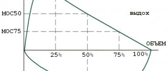
How is a spirogram made?
A spirogram is the result of a study in the form of a diagram with decoding. To receive it, the patient sits next to the device, to which a breathing tube (mouthpiece) is attached. For hygiene purposes, only a replaceable disposable tube is used.
The patient should clasp it tightly with his lips and press a little with his teeth for security. Then he inhales and exhales air several times. In this case, the nasal passages are fixed with clamps so that breathing occurs exclusively through the mouth.
Maneuvers are performed as directed by the doctor - deep or short breathing. A short break of 1-2 minutes is taken between them. Mild dizziness may occur as a side effect. This is a normal phenomenon that goes away on its own within a few minutes.
Spirometry: patient sensations
Most often, patients tolerate spirometry well. They do not have any discomfort or pain. In rare cases, a test using a bronchodilator is accompanied by a feeling of palpitations and slight trembling in the upper and lower extremities. This should not be a cause for concern, since this condition is temporary and not dangerous to the patient's health.
In extremely rare situations, reactions such as a coughing attack or bronchospasm may occur, as a result of which further spirometry must be stopped and medical assistance provided to the patient.
Methodology and principle of research
The procedure is as follows: the patient breathes into a tube connected to a vessel, the bellows are displaced, their movement is recorded, resulting in a curved line called a spirogram. The mixture of gases entering the device during exhalation is purified using filters, and the consumed oxygen is replenished from a reserve tank.
The mechanism of operation of the spirograph and the assessment of vital capacity of the lungs
In modern medicine, the latest developments in spirographs are used, in which the recording of respiratory functionality and its analysis are carried out by a computer program. This greatly increases the accuracy of results and comfort during diagnosis. In order to conduct a comprehensive analysis and exclude pathologies associated with other organs, but having similar or borderline manifestations, the patient is simultaneously given an X-ray of the lungs, an ECG (electrocardiogram) and an echocardiogram.
These diagnostics are considered mutually complementary. With their help, you can detect the presence of pathologies of the respiratory and cardiovascular systems, as well as identify the relationship between them.
Spirometry: where to do the test?
Today there are a huge number of clinics where spirometry is performed. The price in Moscow may be slightly higher than in other regions of Russia and, on average, is about 1,300 rubles.
The highest quality diagnostics of respiratory system diseases is carried out at the Yusupov Hospital. The clinic is equipped with all the necessary modern diagnostic equipment, thanks to which examination errors are minimized. Interpretation of spirometry results with maximum accuracy and in the shortest possible time is performed by highly qualified diagnosticians. Thanks to reliable diagnostics, experienced pulmonologists at the Yusupov Hospital Therapy Clinic - doctors of the highest category - select effective individual therapeutic tactics for each patient.
How to correctly decipher the results?
Interpreting spirography results is a relatively simple process. The doctor uses special tables indicating the norm of a particular indicator. If the values are very different, then a certain severity of the pathological process is recorded.
The main digital indicators used during spirography will be mentioned below.
RR - respiratory rate
Respiratory rate is the number of movements of the chest that are accompanied by gas exchange in the lungs. A healthy person performs 16-20 corresponding cycles per minute. In a young child (up to 3 years old), the figure can reach 30-35 movements.
DO - tidal volume
Tidal volume is the amount of gas mixture (air) that enters and leaves the lungs in 1 quiet cycle. The average figure is 500 ml. Fluctuations from 300 to 900 ml are allowed depending on the individual characteristics of the human body.
MVR - minute breathing volume
In a specific case, we are talking about the amount of gas mixture that circulates through the bronchopulmonary system in 1 minute in a quiet mode. Values – 5-9 l.
Vital capacity - vital capacity of the lungs
The indicator characterizes the largest amount of air available for exhalation at rest after the deepest inhalation. The indicator is individual and depends on the constitution, physical characteristics, etc. The average for men is 4.5-4.9 liters, for women - 3.5-4.0 liters.
FVC - forced vital capacity
A similar indicator to the previous one, which differs in the nature of exhalation. The latter should be forced (as strong as possible). Average values are 3-7 l.
FEV1 - forced expiratory volume in 1 second
FEV1 is an indicator characterizing the amount of air exhaled in the first second. The method of implementation is similar to that for FVC.
IT - Tiffno index
An indicator calculated as a percentage and displaying the ratio of FEV1 to FVC.
MVL - maximum ventilation
The second name is the breathing limit. This indicator reflects the ventilation function of the lungs of a particular patient. The patient is asked to breathe as deeply as possible for a quarter of a minute. The result obtained is multiplied by 4. The norm for healthy people is 70-120 liters per minute. In asthmatics and patients with respiratory failure, the indicator decreases depending on the severity of the pathology.
PSDV - air speed indicator
Another percentage ratio, which is expressed in the formula MVL/VC.