The essence of the electroencephalography method
Back in the mid-19th century, it was discovered that the electrical activity of brain neurons is recorded on the surface of the scalp.
In 1875, independently of each other, the English medical researcher Richard Cato and the Russian physiologist V.Ya. Danilevsky, in experiments on animals, determined the presence of weak brain currents of different frequencies, which are recorded on the surface of the head. Almost half a century later, in 1924, German psychiatrist Hans Berger performed the first electroencephalography (EEG). It took 10 years for the scientific community to realize and acknowledge an amazing fact - the possibility of studying the finest electromagnetic processes in the nervous tissue of the central nervous system without any invasion. Make an appointment/p>
The essence of electroencephalography:
- the functioning of the brain is a huge number of different-frequency electrical charges “running” across the membranes of neurons over time intervals measured in milliseconds;
- since electrical activity is inextricably linked with the propagation of electromagnetic waves, such activity can be monitored directly on the surface of the scalp (for this, electrodes are used, connected to certain areas of the head);
- Over decades of practical research, certain typical electrical rhythms of the human brain have been identified, which are designated by Greek letters: alpha, beta, gamma, etc.
- EEG has one enormous advantage over all other methods (MRI, positron emission tomography or PET) - this method allows scientific and medical observations of the fastest electrical impulses in the brain directly in real time, that is, the time resolution here is measured in milliseconds, whereas MRI signals are time-measured in seconds, tenths of a second at best.
Due to the last feature of the EEG:
- only an EEG can show how the brain reacts to a particular stimulus in the very first moments;
- only EEG can show what the pattern of electrical activity of the central nervous system is in very short periods of time;
- Only with the help of EEG can primary microchanges in the frequency of electrical impulses of neurons be detected in neurological and mental pathologies.
What it is
Let's take a closer look at the procedure itself.
During an EEG, the doctor places small sensors on the patient's head that are connected to an encephalograph. The equipment records the received information and translates it into graphs that reflect EEG rhythms. The specialist evaluates the result obtained and forms his conclusion.
In some cases, the doctor asks the patient to perform certain actions, such as opening and closing his eyes. This is required in order to analyze indicators determined by current activity.
No special preparation is required before an EEG, which distinguishes it from many other studies.
It is only recommended to abstain from tea, coffee, energy drinks and other drinks that contain caffeine. They may influence diagnostic results.
Try to avoid stress and emotional stress on the eve of the study. These processes can affect brain activity.
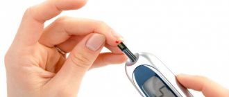
During the examination, the patient must remove metal objects from the body, as they may affect the results.
An EEG can detect not only the presence of epilepsy, but also other serious illnesses, for example:
- consequences of injuries and surgical interventions;
- neoplasms;
- vascular pathologies;
- some other diseases of the brain and central nervous system.
All these diseases are very dangerous for the patient himself. That is why, if they are detected, you must immediately contact a doctor.
Areas of study within EEG
What exactly can be studied using electroencephalography? Examples:
- Various aspects of perception. Let's say a person recognizes the characteristics of certain objects incredibly quickly. For example, the brain detects the difference between the image of an animal and a person within 150-200 milliseconds after the image appears before the eyes.
- Memory. Electroencephalography shows the characteristics and level of quality of both short-term and long-term memory. But especially short-term, fast memory.
- You can get a deep characterization of cognitive functions and learning abilities based on the level of activity of theta and gamma rhythms. This dependence is interesting: beta rhythms work to strengthen new neural connections, while theta rhythms, on the contrary, weaken them. Let’s say that if the problem was solved correctly, then the brain remembers the algorithms used, and increased activity of beta rhythms is observed. If it is incorrect, theta rhythms intensify. These are just isolated examples. The general processes of memory and learning are immeasurably more complex, and they involve dozens, if not hundreds of rhythms, many of which have not yet been discovered.
- The EEG demonstrates an individual's ability to make various adaptations. For example, to increased background noise.
- EEG opens up great prospects for the study of human circadian rhythms, reflexes and the ability to instantly respond to a changing situation.
Among the disadvantages of EEG, two points can be identified:
- mainly impulses passing in the cerebral cortex are observed, but the activity of the deep structures of the central nervous system on the EEG is much less visible;
- strong dependence on the patient’s psycho-emotional state - stress, lack of sleep, nervous overexcitation and even simple anxiety can distort the results of the study.
The legislative framework
The procedure for undergoing medical examinations is constantly changing.
So, the last of them happened in 2021. The new procedure was introduced by order of the Ministry of Health No. 344. This document extends its force to the territory of the entire Russian Federation.
The new rules came into force in March 2021. The previous procedure for undergoing medical examinations was canceled by this order.
Read the explanation of the categories of the new driver's license here.
It will apply to both driver candidates and those who are re-obtaining their licenses. The examination itself is carried out to identify medical contraindications for driving the vehicle.
The presence of a contraindication is established in accordance with Government Decree No. 1604.
Diseases detected by EEG
But why is EEG included in the list of mandatory medical examinations when receiving a medical certificate? As already mentioned, EEG can indicate numerous pathologies. First of all, of a neurological nature. And the main place here is occupied by epilepsy. Since epilepsy is a common medical reason for many types of work: with large construction or industrial equipment, with high-tech equipment, with potentially dangerous technologies (for example, working as a nuclear reactor operator). The potential danger is that a person may have an epileptic attack while working, and this will lead to an emergency situation.
Most people have undergone an EEG at least once in their lives. And everyone remembers how you had to look at the rapidly blinking light bulb. This is a kind of test for epilepsy. In epileptics, exposure to flickering light can lead, for example, to generalized bilaterally synchronous arrhythmic peak-wave complexes. On the graph of electrical rhythms, this is clearly visible even to a non-specialist: a relatively uniform curve turns into a spasmodic one. True, there are types of epilepsy that are not associated with a reaction to flickering light. However, EEG still turns out to be a fairly highly accurate method for identifying this pathology.
In addition to epilepsy, EEG can reveal:
- various sleep pathologies (numerous types of insomnia - chronic and situational);
- inflammation of brain tissue (meningitis and encephalitis);
- chronic mental disorders - depression, neuroses, OCD, panic attacks, etc.
In this regard, an EEG can also become part of a psychiatric examination of a patient.
For what purpose is it being checked?
Passing a medical examination before issuing a driver's license is a familiar procedure for everyone.
Doctors check whether a particular person has contraindications for driving a vehicle. If none have been identified, then he receives a driver’s license.
The condition of a particular person is assessed by various doctors. The need to undergo an EEG in 2014 came as unexpected news for drivers.
This study is quite specific and is used to determine a limited range of diseases.
Not all hospitals have special equipment for performing EEG. Passing it is an additional cost for drivers.
It should be noted that this rule did not last long. In 2021, the obligation to undergo an EEG remains only for drivers of categories C and D.
But keep in mind that this examination can be prescribed to any patient if the neurologist decides so.
Why undergo an EEG? Such a study allows us to establish the presence of serious pathologies. All of them pose a great danger when driving a vehicle.
Identifying these ailments makes it possible to protect both the driver and other road users.
How long does the electroencephalogram procedure take?
A regular encephalogram (routine EEG or diagnosis of a paroxysmal state) takes from 20 to 30 minutes.
During the examination, a number of tests are carried out:
- rhythmic photostimulation;
- hyperventilation;
- load in the form of slow blinking.
If it is necessary to evaluate certain brain functions, the specialist adds additional tests, which he informs the patient about in advance. Such tests include:
- clenching your fingers into a fist;
- being in the dark;
- sleep deprivation for a certain period;
- night sleep monitoring.
Decoding values in different age intervals
An EEG recording of a premature baby at 25–28 gestational weeks looks like a curve in the form of slow flashes of delta and theta rhythms, periodically combined with sharp wave peaks 3–15 seconds long with a decrease in amplitude to 25 μV. In full-term infants, these values are clearly divided into three types of indicators. During wakefulness (with a periodic frequency of 5 Hz and an amplitude of 55–60 Hz), the active phase of sleep (with a stable frequency of 5–7 Hz and a fast low amplitude) and quiet sleep with flashes of delta oscillations at a high amplitude.
Over the course of 3-6 months of a child’s life, the number of theta oscillations is constantly growing, while the delta rhythm, on the contrary, is characterized by a decline. Further, from 7 months to a year, the child develops alpha waves, and delta and theta gradually fade away. Over the next 8 years, the EEG shows a gradual replacement of slow waves with fast ones - alpha and beta oscillations.
Until the age of 15, alpha waves predominate, and by the age of 18, the BEA transformation is complete. Over the period from 21 to 50 years, stable indicators remain almost unchanged. And from 50, the next phase of rhythmicity restructuring begins, which is characterized by a decrease in the amplitude of alpha oscillations and an increase in beta and delta.
After 60 years, the frequency also begins to gradually fade, and in a healthy person, manifestations of delta and theta oscillations are noticed on the EEG. According to statistics, age indicators from 1 to 21 years, considered “healthy,” are determined in subjects 1–15 years old, reaching 70%, and in the range of 16–21 – about 80%.
Indications for an encephalogram
The doctor will definitely prescribe an electroencephalography if the patient has one or more alarming symptoms:
- frequent fainting;
- headache;
- disturbances in sleep phases, falling asleep, insomnia;
- head injuries;
- seizures;
- epileptic seizures;
- neuroses;
- acute cerebrovascular accident;
- degenerative diseases of the brain;
- neuroses;
- nausea and vomiting for no apparent reason;
- hypertension;
- suspicion of stroke, microstroke, cerebral infarction;
- suspected development of a brain tumor.
An encephalogram of the brain is an informative method for diagnosing infectious diseases of the brain and its membranes: encephalitis, meningitis. Electroencephalography may be prescribed during the treatment of these diseases in order to monitor its effectiveness.
An electroencephalogram is necessarily performed as part of preparing the patient for brain surgery.
Electroencephalography is widely used in pediatric practice. The diagnostic method is used for such conditions in children as:
- delayed mental, emotional and speech development;
- somnambulism;
- nervous tic;
- stuttering;
- head injuries;
- convulsive syndrome.
An encephalogram of a child’s brain is an important study in the diagnosis and treatment of children with diseases such as autism, Down syndrome, and cerebral palsy. With the help of an encephalogram, the diagnosis and its severity are clarified, how quickly the pathology progresses, and the effectiveness of treatment.
Due to the fact that the encephalograph does not emit impulses, but only records them, the procedure has no contraindications. The exception is when the patient has wounds on the head that prevent the installation of the electrodes of the device to obtain an encephalogram.
Night video monitoring of EEG
Video EEG monitoring is the only objective method for diagnosing many diseases of the central nervous system. With the help of research, neurologists determine specific disturbances in the initial phase of EEG recording during an attack. The system allows for a long-term examination. The following information is synchronously recorded on the hard drive:
- EEG recording on a digital electroencephalograph;
- physiological signals of the body through polygraphic channels;
- audio information from two microphones;
- video information from two video cameras;
- markers reflecting certain events that the doctor notes during the recording.
EEG night monitoring, the cost of which in Moscow starts from 1200 rubles, is carried out in the following cases:
- if necessary, confirm the diagnosis of epilepsy;
- to clarify the form of epilepsy and localization of epileptic foci;
- In order to confirm drug remission of epileptic disease and the adequacy of the antiepileptic treatment;
- to resolve the issue of discontinuing antiepileptic therapy;
- for paroxysmal disorders of consciousness and paroxysmal movement disorders;
- to clarify the cause of a single epileptic seizure;
- in order to clarify the cause of rare attacks (incorrect diagnosis, incomplete compensation);
- with relapse of attacks due to constant use of medications.
Night EEG video monitoring is performed for speech delay of unknown origin, progressive decline in cognitive functions, and minimal brain dysfunction. The study should be done for patients suffering from affective-respiratory paroxysms, febrile convulsions, daytime and nighttime sleep disorders. Indications for night video EEG monitoring are:
- enuresis, encopresis (bedwetting of urine and feces);
- bettolepsy (transient disturbances of consciousness that occur at the peak of a coughing attack);
- nonepileptic paroxysms (night terrors, narcolepsy, sleepwalking);
- paroxysms of dizziness of unknown origin that occur without objective symptoms.
Video EEG monitoring (price depends on the duration of the procedure) is performed by neurophysiologists for myoclonus and similar conditions: sleep myoclonus, autonomic myoclonus, opsoclonus (dancing eyes syndrome), benign infantile myoclonus.
How to prepare for research
The option of night EEG monitoring is completely safe for the patient. But it will have to be refused if there are open wounds in the head area. The period of exacerbation of mental illness is considered as a relative contraindication. When the patient becomes unstable, the doctor may use sedatives.
If the subject is taking tranquilizers or anticonvulsants, they will be temporarily withdrawn after prior consultation with the treating doctor.
Preparation for EEG monitoring includes the following points:
- 24 hours before the study, it is necessary to avoid the consumption of alcohol, tea and coffee;
- before the procedure - approximately 2 hours before the start - you need to eat a hearty meal;
- Smoking is prohibited on the day of the EEG;
- You must wash your hair and do not use any hair cosmetics;
- It is necessary to remove all jewelry - rings, earrings, chains, bracelets, piercings.
If necessary - depending on the chosen EEG format - the patient will have to remain awake for 24 hours. Before the procedure begins, the patient will be offered a mild sedative, which will allow him to relax and fall asleep for the entire period of the study.
Stages of implementation
The procedure for conducting an EEG is not particularly complicated and requires preliminary preparation. The patient has several requirements:
• the scalp should be clean. This will ensure better contact between the parting sensors and the patient's skin;
• for several days before the study, you should stop drinking alcohol or taking medications.
It is best to conduct research in the first half of the day. Small sensors fixed to an elastic mesh are attached to the scalp. Reference electrodes are fixed on the ears. Pulses are recorded for about 15 minutes.
After the EEG recording is processed and deciphered by the doctor who conducted the research, the result is given to the patient or transferred to his attending physician.
Decoding of electroencephalogram indicators
In order to decipher the EEG and not miss any of the smallest manifestations in the recording, the specialist needs to take into account all the important points that may affect the indicators being studied. These include age, the presence of certain diseases, possible contraindications and other factors.
Upon completion of the collection of all research data and their processing, the analysis is completed and then a final conclusion is formed, which will be provided for making a further decision on the choice of therapy method. Any disturbance in activity may be a symptom of diseases caused by certain factors.
Alpha rhythm
The normal frequency is determined in the range of 8–13 Hz, and its amplitude does not go beyond 100 μV. Such characteristics indicate a healthy state of a person and the absence of any pathologies. The following are considered violations:
- constant fixation of the alpha rhythm in the frontal lobe;
- exceeding the difference between the hemispheres by up to 35%;
- constant violation of wave sinusoidality;
- presence of frequency dispersion;
- amplitude below 25 μV and above 95 μV.
The presence of disturbances in this indicator indicates a possible asymmetry of the hemispheres, which may be the result of oncological tumors or pathologies of cerebral circulation, for example, stroke or hemorrhage. A high frequency indicates brain damage or TBI (traumatic brain injury).
A complete absence of the alpha rhythm is often observed in dementia, and in children, deviations from the norm are directly related to mental retardation (MDD). Such a delay in children is evidenced by: disorganization of alpha waves, shift of focus from the occipital region, increased synchrony, short activation reaction, overreaction to intense breathing.
These manifestations can be caused by inhibitory psychopathy, epileptic seizures, and a short reaction is considered one of the primary signs of neurotic disorders.
Beta rhythm
In the accepted norm, these waves are clearly detected in the frontal lobes of the brain with a symmetrical amplitude in the range of 3–5 μV, recorded in both hemispheres. A high amplitude leads doctors to think about the presence of a concussion, and when short spindles appear, to the occurrence of encephalitis. An increase in the frequency and duration of spindles indicates the development of inflammation.
In children, the pathological manifestations of beta oscillations are considered to be a frequency of 15-16 Hz and a high amplitude present - 40-50 µV, and if its localization is the central or anterior part of the brain, then this should alert the doctor. Such characteristics indicate a high probability of delayed development of the baby.
Delta and theta rhythms
An increase in the amplitude of these indicators above 45 μV on a constant basis is characteristic of functional brain disorders. If the indicators are increased in all brain regions, then this may indicate severe dysfunction of the central nervous system.
If a high amplitude of the delta rhythm is detected, a tumor is suspected. Inflated values of the theta and delta rhythm recorded in the occipital region indicate a child’s lethargy and a delay in his development, as well as impaired circulatory function.
Rhythms of a waking person
Brain activity recorded on the EEG in an adult has several types of rhythms, characterized by certain indicators and states of the body.
- Alpha rhythm. Its frequency remains in the range of 8–14 Hz and is present in most healthy individuals – more than 90%. The highest amplitude values are observed when the subject is at rest, in a dark room with his eyes closed. It is best identified in the occipital region. It is fragmentarily blocked or completely subsides during mental activity or visual attention.
- Beta rhythm. Its wave frequency fluctuates in the range of 13–30 Hz, and the main changes are observed when the subject is active. Pronounced fluctuations can be diagnosed in the frontal lobes under the obligatory condition of active activity, for example, mental or emotional arousal and others. The amplitude of beta oscillations is much less than alpha.
- Lambda rhythm. It has a small range - 4–5 Hz, and is triggered in the occipital region when it is necessary to make visual decisions, for example, when searching for something with open eyes. The vibrations disappear completely after concentrating your gaze on one point.
- Mu rhythm. Defined by the interval 8–13 Hz. It starts in the back of the head, and is best observed in a calm state. Suppressed when starting any activity, not excluding mental activity.
EEG classification according to Luders
The EEG classification according to Lüders1 is very detailed, it includes both artifacts and rare types of EEG.
- Slow activity Slow down background activity
- Transient slowdown
- Continued deceleration
- Spikes
- Excessive fast activity
- Alpha Coma
- Temporal deceleration in the elderly
Rhythms in sleep
A separate category of types of rhythms that manifest themselves either in sleep conditions or in pathological conditions includes three varieties of this indicator.
- Delta rhythm. Characteristic of the deep sleep phase and for comatose patients. It is also recorded when recording signals from areas of the cerebral cortex located on the border with areas affected by oncological processes. Sometimes it can be recorded in children 4–6 years old.
- Theta rhythm. The frequency interval is within 4–8 Hz. These waves are triggered by the hippocampus (information filter) and appear during sleep. Responsible for high-quality assimilation of information and forms the basis of self-learning.
Based on the results obtained during EEG recording, an indicator is determined that characterizes a complete all-encompassing assessment of the waves - bioelectrical activity of the brain (BEA). The diagnostician checks the EEG parameters - frequency, rhythm and the presence of sharp flashes that provoke characteristic manifestations, and on these grounds makes a final conclusion.
EEG classification according to Jung
Jung (1953) distinguishes four variants of an unchanged EEG:
- The alpha electroencephalogram contains a well-defined alpha rhythm, the frequency of which fluctuates by no more than 1–1.5 waves per second. Low amplitude beta waves occur in short bursts in the precentral region. Theta waves are barely visible.
- The beta electroencephalogram is dominated by waves with a frequency of 16 to 25 per second with an amplitude of 20–30 μV, recorded partly continuously in the recording, partly in the form of separate groups or series.
- A flat EEG contains a very rare low-amplitude alpha rhythm, a beta rhythm that is small and difficult to distinguish, and flat theta waves are also found. Similar EEGs occur in more than 10% of healthy people. Flat EEGs with an accelerated main type of activity can be detected during mental stress. Transient flattening of the EEG is also detected when alpha waves are blocked during the stage of falling asleep (referred to by some authors as stage zero, for example, Roth, 1962).
- An irregular EEG contains an alpha rhythm, the average frequency of which fluctuates ± 1.5 waves per second. The maximum amplitude of the alpha rhythm in the occipital leads is not clearly expressed. Theta waves, superimposed on alpha waves, are more pronounced in the anterior and temporal regions than in the parietal regions, where they are sometimes visible as components of the lower harmonics of the alpha rhythm.
Such irregular EEGs occur in healthy people as often as flat EEGs. They are more common in youth and in older age and present difficulties in distinguishing them from EEGs with pronounced frequency instability, which represent a transition to dysrhythmia. Transient EEG irregularity occurs during hyperventilation and during the stage of falling asleep.
How is the examination carried out?
The brain examination procedure is absolutely painless and does not require special preparation. During the procedure, the patient must keep his head level while lying or sitting so that the doctor receives the most accurate data. In order to reduce possible interference, the patient's skin is lubricated with a special gel that conducts ultrasound well. The examination usually lasts no more than a quarter of an hour. Doctors perform an encephalogram in two modes - transmission (two sensors are used) and emission (one sensor is used).
After the procedure is completed, the doctor receives data that he needs to decipher. Experienced specialists of the SANMEDEXPERT clinic are able to give an accurate interpretation, which will help make the correct diagnosis and prescribe appropriate treatment.