Human immunity is directly related to the intestinal microflora, respiratory system and genital area. Tests for dysbacteriosis are a reliable and affordable way to clarify the origin of ailments and erect a reliable barrier to pathogenic strains. An imbalance, the disappearance of beneficial microorganisms, is often the root cause of many chronic diseases. Leads to the accumulation of toxins, atrophy of the gastrointestinal mucosa, poor absorption of vitamins and microelements, and activates the action of viruses.
When do you need to take a microbiological diagnosis of intestinal dysbiosis test?
- Diarrhea.
- Atopic dermatitis, chronic diarrhea, intestinal damage due to the action of ionizing radiation or chemicals.
- Long recovery period after intestinal infections.
- Long-term intestinal disorders in which pathogenic microorganisms cannot be isolated.
- Intestinal dysfunction in patients exposed to prolonged exposure to radiation, chemicals, etc., intensive antibiotic and/or immunosuppressive, chemotherapy and hormonal therapy.
- The presence of bacteremia, purulent-inflammatory foci that are difficult to treat (pyelitis, cholecystitis, ulcerative colitis, enterocolitis, sluggish pneumonia).
- Surgical treatment performed in the presence of risk factors for the development of intestinal dysbiosis.
Other examination methods
There is another, less common, but more accurate and faster method of microbiological examination - chromatography-mass spectrometry of microbial markers (CMS, or as it is also called MSMM). It was developed by Doctor of Biological Sciences G.A. Osipov and is used in some laboratories.
This method allows you to study the microbiological composition of various biological materials - blood, urine, saliva, vaginal discharge, etc. The analysis is carried out within 3 hours and allows you to determine the presence of 50 microorganisms in the test material without inoculation, using the so-called signal molecules (microbial markers).
Detailed description of the study
Examination of stool for dysbacteriosis is carried out using a microbiological method. Used to assess the state of intestinal microflora and identify the cause of diarrhea.
Examination of stool for intestinal dysbiosis allows us to assess the composition of the microflora and identify deviations in it, as well as develop an individual treatment plan.
Dysbacteriosis is a change in the qualitative and quantitative composition of the intestinal microbiocenosis. Various reasons can lead to this condition:
- change in diet;
- inflammatory processes;
- long-term antibiotic therapy;
- surgery;
- prolonged stress;
- immunodeficiency;
- living in an uncharacteristic climatic zone (high mountains, Arctic).
Normal gastrointestinal bacteria:
- bifidobacteria;
- lactobacilli;
- typical forms of E. coli;
- enterococci;
- bacteroides (anaerobes).
In addition to “useful” bacteria, there is opportunistic flora in the intestines. These microorganisms can normally be present in the biotope, but an increase in their number entails a change in the ratio of microorganisms in the biocenosis of the rectum and can lead to clinical manifestations of dysbiosis. Such microorganisms include:
- non-fermenting bacteria;
- fungi;
- enterobacteria;
- clostridia;
- staphylococci.
Normally they are not detected, but pathogenic bacteria are found in diseases. These include:
- pathogenic Escherichia;
- shigella;
- salmonella.
The study allows us to determine the nature of changes in the intestinal microflora and identify the pathology that led to its development.
So, there is no such thing as “dysbacteriosis”?
Of course have. For example, pseudomembranous colitis - severe inflammation of the colon after an antibiotic - is a real dysbiosis: competitors have died, and therefore Clostridium difficile multiplies. Just to treat this, there is absolutely no need to state the obvious - the composition of bacteria in the intestine has changed. It is enough to confirm the infection (identify C. difficile toxins) and prescribe treatment.
Intestinal microflora, without a doubt, affects all processes in our body. By transplanting stool from an obese mouse to a mouse of normal weight, we induce obesity in the latter. The composition of gut bacteria is fundamentally different between people with anxiety and depression. Well, adding the probiotic Bacteroides fragilis to mice that have been artificially induced to have autism improves their social skills. Read the popular book “Look What's Inside You” by the famous microbiologist Rob Knight: our knowledge about microflora is colossal, but we are just beginning to apply it in practice (that is, to treat diseases).
The composition of bacteria can and should be studied. This is the subject of an ambitious international study, the Human Microbiome Project, with a budget of $115 million. Naturally, no “stool cultures” are used. Metagenomics methods are used to analyze the microbial jungle of the gut. They make it possible to describe how many unique DNA sequences are present in a particular person, which groups of bacteria predominate and which are absent. By the way, when such technologies (for example, 16S rRNA sequencing) appeared, it turned out that 75% of the species detected during genetic analysis of the same feces are not known to science at all.
What else is prescribed with this study?
Adenovirus (diarrhea syndrome), antigen test
157.0. Feces 2 days
1,000 ₽ Add to cart
Determination of pathogen sensitivity to antibacterial drugs (DDM)
01. 1 day
490 ₽ Add to cart
Determination of pathogen sensitivity to bacteriophages
03. 1 day
220 ₽ Add to cart
Determination of pathogen sensitivity to an expanded range of antibacterial drugs
02. 2 days
600 ₽ Add to cart
Determination of pathogen sensitivity to an expanded range of antibacterial drugs, with determination of the minimum inhibitory concentration (MIC, MIC)
13. 2 days
960 ₽ Add to cart
RULES FOR FECAL COLLECTION DURING STUDIES
INTESTINAL MICROFLORA (DYSBACTERIOSIS)
- All examined patients, 1-3 days before taking the sample, should be on a diet that excludes the intake of foods that enhance fermentation processes in the intestines and lactic acid products, as well as alcohol, antibiotics and bacterial preparations (containing bifidobacteria, lactobacilli, E. coli, etc.) .d.).
- The material is feces after natural defecation, which is collected in a clean disposable container with a screw cap and spoon.
- It is not recommended to collect feces from the toilet.
- Collect feces on a clean surface, which can be a clean new sheet (bag) of polyethylene or paper (this method is preferred).
- When using a vessel, it is first washed well with soap and a sponge, rinsed repeatedly with tap water, and then doused with boiling water and cooled.
- Feces are taken mainly from the middle portion with a special spoon mounted in the lid of a sterile container, in an amount of no more than 1/3 of the container’s volume. Do not fill the container to the top. Close the lid carefully.
- The material must be delivered to the laboratory no later than 2 hours from the moment of collection. If necessary, delivery time can be increased to 3 hours, storage conditions for the material are 4 degrees C.
- To correctly assess the results, it is recommended to conduct 2-3 studies with an interval of 1-2 days.
RULES FOR URINE COLLECTION FOR WOMEN
- Before collecting urine, prepare 6-10 clean cotton balls, a vessel with warm soapy water (use regular toilet soap), a vessel with warm boiled water and a container for collecting urine (open the lid of the container so that it can be removed with one hand).
- Wash your hands with soap.
- Sit comfortably on the toilet and spread your knees as wide as possible.
- Wash the external genital area by successively replacing 4 cotton balls soaked in soapy water. Each ball must be passed from the pubis to the anus only once, trying to penetrate all folds.
- Rinse the soapy area using two or more cotton balls soaked in warm boiled water. Each ball must be passed from the pubis to the anus only once, trying to penetrate all folds.
- To prevent vaginal discharge from getting into the urine, it is recommended that sexually active women insert a tampon into the vagina when collecting urine.
- Remove the lid from the container and hold it in your hand, being careful not to touch the edges. Prepare to collect urine.
- Keeping your labia apart, release a little urine into the toilet, stop urinating, and then, placing the container under the stream of urine, fill it to half capacity. Try not to touch the container to your body.
- Close the container carefully with the lid.
- The urine should be delivered to the laboratory within 2 hours. If immediate sowing is not possible, it should be stored in the refrigerator for up to 18 hours.
RULES FOR URINE COLLECTION FOR MEN
1. Wash your hands with soap.
2. Pull back the foreskin (if it is not circumcised), wash the head of the penis with soap and warm boiled water, dry with a clean napkin.
3. Prepare the container by opening the container lid so that it can be removed with one hand. Do not touch the inside of the container or lid with your hands.
Release a small amount of urine into the toilet. Stop urinating.
4. Holding the foreskin in a retracted position, direct a stream of urine into the container and fill it halfway, being careful not to touch the edges of the container.
5. Carefully close the container with the lid.
6. Deliver the urine to the laboratory within 2 hours. If immediate sowing is not possible, it should be stored in the refrigerator for up to 18 hours.
RECOMMENDATIONS FOR PREPARING FOR AN ULTRASOUND
ABDOMINAL ORGANS
1. The study is carried out on an empty stomach (refusal to eat 6-8 hours before the study). It is not recommended to drink or smoke before the test.
2. 2-3 days before the study, it is advisable to limit the intake of foods that cause flatulence (whole milk, brown bread, cabbage, other fresh vegetables and fruits, juices, sweets).
3. To reduce flatulence, it is recommended to take espumizan on the day before the study, 2 capsules 3 times a day and 2 capsules in the morning on the day of the study, or activated charcoal 2 tablets 3 times a day.
RECOMMENDATIONS FOR PREPARING FOR AN ULTRASOUND
PROSTATE, FEMALE GENITAL ORGANS TRANSABDOMINAL SENSOR
1. The study is carried out with a full bladder.
2. To fill the bladder an hour before the test, you need to drink 1.5 liters. liquids (mineral or drinking water without gas).
References
- Methodological recommendations “Bacteriological diagnosis of intestinal dysbiosis”, approved. Ministry of Health of the RSFSR - 1977.
- Industry standard “Protocol for the management of patients with intestinal dysbiosis.” OST 91500.11.0004-2003.
- Determination of dysbiotic changes in the gastrointestinal tract using markers of intestinal contents. Federal clinical guidelines - 2005.
- Clinical recommendations of the national association of specialists in the control of infections associated with the provision of medical care and the all-Russian public non-profit organization “Association of Coloproctologists of Russia” for the diagnosis, treatment and prevention of Clostridium difficile-associated diarrhea (CDI) - 2021.
- Chan BK, Abedon ST, Loc-Carrillo C. Phage cocktails and the future of phage therapy. Future Microbiol. 2013 Jun;8(6):769-83.
- Robles Alonso V, Guarner F. Linking the gut microbiota to human health. Br J Nutr. 2013 Jan;109 Suppl 2:S21-6. Review.
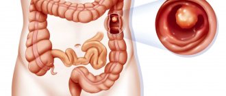
Analysis of stool for helminth eggs and protozoan cysts
Helminths (worms) are parasitic worms that live in the body of humans and animals and cause diseases called helminthiasis (helminthic infestations). The disease in humans is most often caused by roundworms (nematodes) and flatworms (platytodes), which are divided into tapeworms - cestodes and flukes - trematodes. Helminths change their host in the process of life development, and the range of hosts, routes and mechanisms of transmission are individual.
Clinical manifestations are largely determined by the localization of the parasite, its number and feeding habits. Helminths, parasitizing in the human body, can have a mechanical effect, damaging the tissues and organs of the host, toxic and allergic phenomena, metabolic disorders, closing the lumen of the intestines, excretory ducts of the liver, gall bladder, pancreas, disrupting intestinal motility, irritating the receptors of the intestinal wall.
Protozoa are single-celled organisms. Like helminths, during the development process protozoa go through several stages and change hosts. Many protozoa have two forms of existence - vegetative and cystic. The cyst has a durable shell and ensures the survival of the protozoan in unfavorable, and even extreme, conditions. It is cysts that usually infect humans.
A stool test for helminth eggs and protozoan cysts must be taken if a parasitic infection is suspected, to monitor the effectiveness of antiparasitic treatment, before hospitalization, when preparing a medical record, or certificates. For example, for children before visiting the pool.
In the CMD laboratory, stool analysis for helminth eggs and protozoan cysts is carried out using two different methods:
- Feces for helminth eggs and protozoan cysts: microscopic method; "thick smear" method and Kato staining
- Modified sedimentation method using PARASEP concentrators (enrichment method). Analysis of feces for helminth eggs and protozoan cysts using a Parasep concentrator: the use of PARASEP concentrators makes it possible to identify helminths and protozoa even in small quantities.
Stool analysis for carbohydrates
Stool analysis for carbohydrates is used to diagnose congenital or acquired deficiency of the lactase enzyme in children of the first year of life.
Lactase is an enzyme that is produced by intestinal epithelial cells (enterocytes) and participates in the biochemical reaction of the breakdown of lactose, otherwise known as milk sugar. When consuming milk and dairy products, if there is not enough lactase, digestion is disrupted and unpleasant symptoms appear: diarrhea, flatulence, cramps, etc.
Causes of lactase deficiency:
- decrease in enzyme production by intact enterocytes due to genetic defects or physiological immaturity of enzyme systems in children of the first year of life - primary, or congenital, lactase deficiency;
- a decrease in enzyme production due to damage to enterocytes during an inflammatory or other (food allergy, tumor) process - secondary, or acquired, lactase deficiency.
Indications for taking a stool test for carbohydrates:
- the presence in children of symptoms such as flatulence, frequent regurgitation, diarrhea, cramps, abdominal pain;
- control of correct diet selection.