September 9, 2021 Rhinopharyngitis or nasopharyngitis is an inflammation of the mucous membrane of the nasal cavity, pharynx and their lymphoid formations.
Our upper respiratory tract has no clear boundaries. All boundaries between the pharynx, larynx, nose and other parts of the respiratory system are very conditional.
In this regard, inflammation of the pharynx almost always, regardless of the cause, spreads to neighboring areas of the respiratory tract.
The nasal cavity serves as the main entry point for infection. From the nasal cavity, viruses, bacteria, fungi and other pathogens very quietly and quickly penetrate the pharynx and spread through the respiratory tract.3
Causes of nasopharyngitis
Rhinopharyngitis is predominantly an infectious disease. In more than 80% of cases, especially in the autumn-winter period, it is caused by viruses - rhinovirus, adenoviruses, influenza viruses, etc. Bacterial nasopharyngitis occurs slightly less frequently. In this case, the causative agents of the disease are chlamydia, mycoplasma, streptococcus and staphylococcus.
Separately, we can distinguish allergic rhinopharyngitis, which develops under the influence of allergens that penetrate the upper respiratory tract - plant pollen, animal hair, tobacco smoke, household chemicals, etc.
Risk factors for developing a runny nose: hypothermia, weak immunity, the presence of chronic ENT diseases or pathologies of internal organs.
Diagnostics
The primary diagnosis and treatment according to the general scheme are determined by the patient at the first visit to the doctor. A preliminary diagnosis is made based on a visual examination of the pharynx and anamnesis. Next, more serious diagnostics are required to determine the causative agent of the disease and assess the general condition of the patient.
1. General blood test. Needed to determine the degree of inflammation and general condition.
2. General urine analysis. Necessary for assessing the functioning of the kidneys and a number of metabolic processes in the body.
3. A swab from the throat with further inoculation on a nutrient medium. This study allows you to identify the causative agent of the disease and determine which antibiotic will be most effective against it.
If necessary, the doctor can refer the patient for consultation to other specialists if complications of the disease are determined or that it is a consequence of another pathology. Treatment for bacterial pharyngitis can sometimes be quite broad.
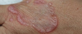
Symptoms of nasopharyngitis
The disease develops acutely:
- the nasal mucosa swells;
- copious watery or mucous discharge appears;
- nasal breathing becomes difficult;
- there is a burning and itching sensation in the nose;
- your throat starts to hurt;
- body temperature rises.
Swelling of the mucous membrane contributes to nasal congestion, which leads to a decrease or loss of smell, a change in the timbre of the voice (a specific nasal sound appears). The patient often sneezes and his head begins to hurt. The mucus formed in the nose flows down the back wall of the throat, causing a reflex cough. Lymph nodes in the neck may become enlarged.
Rhinopharyngitis in children has a similar clinical picture. In addition, children are often capricious and refuse to eat.
If left untreated, chronic nasopharyngitis develops. Nasal congestion occurs periodically, most often it occurs on the half of the nose on the side of which the person is lying (left - on the left side and vice versa). When lying on your back, nasal breathing becomes completely difficult. Other symptoms characteristic of the acute form of the disease are usually absent. If atrophic rhinitis develops, patients complain of dryness and itching in the nasal cavity, and crusts with an unpleasant odor form in the nose. In the case of hypertrophic chronic rhinitis, the nose is constantly blocked.
Acute Nasopharyngitis
22.04.2019
| 1C Strong recommendation based on low-quality evidence | The benefits are likely to outweigh the possible risks and costs, or vice versa | Evidence based on observational studies, anecdotal clinical experience, results of RCP performed with significant shortcomings. Any estimate of effect is considered uncertain. | Relatively strong recommendation, subject to change as higher quality evidence becomes available |
| 2A Weak recommendation based on high-quality evidence | The benefits are comparable to the possible risks and costs | Reliable evidence based on well-executed RCPs or supported by other compelling data. Further research is unlikely to change our confidence in the benefit-risk assessment. | Weak recommendation. The choice of the best tactics will depend on the clinical situation(s), patient or social preferences. |
| 2B Weak recommendation based on moderate quality evidence | The benefits are comparable to the risks and complications, but there is uncertainty in this assessment. | Evidence based on results of RCT performed with significant limitations (inconsistent results, methodological flaws, indirect or incidental), or strong evidence presented in some other form. Further studies (if conducted) are likely to influence and may change our confidence in the benefit-risk estimate. | Weak recommendation An alternative strategy may be a better choice for some patients in certain situations. |
| 2C Weak recommendation based on low quality evidence | Ambiguity in assessing the balance of benefits, risks and complications; the benefits may be weighed against the possible risks and complications. | Evidence based on observational studies, anecdotal clinical experience, or RCP with significant limitations. Any estimate of effect is considered uncertain. | Very weak recommendation; alternative approaches may be used equally. |
*In the table, the numerical value corresponds to the strength of recommendations, the letter value corresponds to the level of evidence
DEFINITION
Acute respiratory viral infection (ARVI) is an acute, in most cases, self-limiting infection of the respiratory tract, causing catarrh syndrome of the upper respiratory tract (URI - upper respiratory infection) in English literature), occurring with fever, runny nose, sneezing, cough, pain in the throat, a violation of the general condition of varying severity.
As a diagnosis, the term “ARVI” should be avoided, using the term “acute nasopharyngitis” (in the English-language literature the term “common cold” is used - a cold), since the pathogens of ARVI also cause laryngitis (croup), tonsillitis, bronchitis, bronchiolitis, which follows indicate in the diagnosis. These syndromes are discussed in detail separately (see FKR on the management of children with acute tonsillitis and stenosing laryngotracheitis (croup).
Acute nasopharyngitis is diagnosed when there is an acute runny nose and/or cough, while influenza and lesions of other localizations are excluded:
- acute otitis media (corresponding complaints, otoscopy);
- acute tonsillitis (predominant involvement of the tonsils, plaque);
- bacterial sinusitis (swelling, hyperemia of the soft tissues of the face, orbit, and other symptoms);
- damage to the lower respiratory tract (increased or difficult breathing, obstruction, retractions of the pliable parts of the chest, shortening of the percussion sound, wheezing in the lungs);
In the absence of these signs, a viral infection of only the upper respiratory tract is likely (ARVI - rhinitis, nasopharyngitis, pharyngitis), often accompanied by conjunctivitis. The “red eye” sign is simple to evaluate and, at the same time, very specific for excluding a bacterial infection, not inferior in diagnostic value to laboratory markers of inflammation.
ICD-10 CODE J00 - acute nasopharyngitis (runny nose).
EPIDEMIOLOGY
ARVI is the most common human infection: children aged 0-5 years suffer, on average, 6-8 episodes of ARVI per year [1], in kindergartens the incidence is especially high in the 1st-2nd year of visit - at 10-15 % higher than that of disorganized children, but at school the latter get sick more often [2]. The incidence is highest in the period from September to April and is (registered) 87-91 thousand per 100 thousand population. Among frequently ill children, many have an allergic predisposition and/or bronchial hyperreactivity, which causes a more pronounced manifestation of even a mild respiratory infection.
ETIOLOGY
ARVI is caused by about 200 viruses, most often rhinviruses, which have more than 100 serotypes, as well as PC virus, parainfluenza viruses, adenoviruses, bocavirus, metapneumovirus, and coronaviruses. Some non-polio enteroviruses can cause similar manifestations. Rhino-, adeno- and enteroviruses cause stable immunity, which does not exclude infection by other serotypes; RS, corona and parainfluenza viruses do not leave lasting immunity.
The spread of viruses most often occurs through self-inoculation onto the nasal mucosa or conjunctiva from hands contaminated by contact with a patient (handshake!) or with surfaces contaminated with the virus (the rhinovirus remains on them for up to a day).
Another way is airborne droplets - when you inhale particles of an aerosol containing the virus, or when larger droplets get on the mucous membranes during close contact with a patient.
The incubation period for most viruses is 24-72 hours. The release of viruses by patients is maximum on the 3rd day after infection, sharply decreases by the 5th day; Low-intensity virus shedding can persist for up to 2 weeks.
PATHOGENESIS
Symptoms of nasopharyngitis are the result not so much of the damaging effects of the virus as of the reaction of the innate immune system. Affected epithelial cells release cytokines, incl. interleukin 8 (IL 8), the amount of which correlates both with the degree of attraction of polynuclear cells into the submucosal layer and epithelium, and with the severity of symptoms. An increase in nasal secretion is associated with an increase in vascular permeability; the number of leukocytes in it can increase 100-fold, changing its color from transparent to white-yellow (accumulation of leukocytes) or greenish (peroxidase) - there is no reason to consider a change in the color of the secretion a sign of a bacterial infection. Coronaviruses leave nasal epithelial cells intact; the cytopathic effect is inherent in adenoviruses and influenza viruses.
The assumption that with any viral infection the bacterial flora is activated (“viral-bacterial etiology of acute respiratory infections” based, for example, on the presence of leukocytosis in the patient) is not confirmed by practice: in most patients, acute respiratory viral infections proceed smoothly without the use of antibiotics. Bacterial complications of ARVI occur rarely (1-5% of cases). As a rule, they are already present on the 1st-2nd days of illness; in later periods they occur most often as a result of superinfection. One should keep in mind streptococcal pharyngitis, which may not be accompanied by the classic “sore throat”; the bright, “scarlet” color of the palatine arches and especially the back wall of the pharynx may indicate a streptococcal infection. In such cases, a diagnostic ex-press test can help. It is also necessary to remember about “silent” pneumonia, which is difficult to identify clinically (especially if the patient is not percussed).
CLASSIFICATION
The division of nasopharyngitis according to severity is possible depending on the level of temperature and the severity of general nonspecific symptoms.
CLINICAL PICTURE
It varies widely, the manifestations of viral infections of various etiologies overlap each other. Infants usually have fever, discharge from the nasal passages, and sometimes there is restlessness, difficulty feeding and falling asleep. In older children, typical manifestations are: runny nose, difficulty in nasal breathing (peak on the 3rd day, duration up to 6-7 days), in 1/3-1/2 patients - sneezing and/or cough (peak on the 1st day , average duration - 6-8 days), less often - headache (20% on the 1st and 15% until the 4th day) [4, 5]. In a number of children, after suffering from acute respiratory viral infection, some symptoms, such as cough, may persist until the 10th day or even longer:
The vast majority of patients have a normal or subfebrile temperature, and among those hospitalized, febrile fever is more often detected, which in 82% of patients decreases on the 2-3rd day of illness; Febrility lasts longer (up to 5-7 days) with influenza and adenovirus infection [6]. Maintaining such a temperature for more than 3 days (in the absence of signs of influenza or adenovirus infection) should alert you to a bacterial infection. A repeated rise in temperature after a short-term improvement may indicate the same thing, although more often it is a sign of superinfection.
COMPLICATIONS
Complications of nasopharyngitis are observed infrequently, are associated with the addition of a bacterial infection and are manifested by the following symptoms:
- persistence of nasal congestion for longer than 10-14 days, deterioration of the condition after improvement, the appearance of pain in the face may indicate the development of bacterial sinusitis;
- painful “clicks” in younger patients, a feeling of “stuffiness” in the ear in older children is a consequence of dysfunction of the cochlear tube due to a viral infection, caused by changes in pressure in the middle ear cavity, which can lead to the development of acute otitis media.
ARVI and, especially, influenza predispose (the more often the younger the child) to infection of the lungs, primarily with pneumococcus with the development of pneumonia. In addition, respiratory infection is a trigger for exacerbation of chronic diseases - most often bronchial asthma and urinary tract infections.
DIAGNOSTIC EXAMINATION
Examination of a patient with nasopharyngitis is aimed at identifying bacterial foci that are not detected by clinical methods. Urinalysis (including using test strips on an outpatient basis) is mandatory for all febrile children, because 5-10% of infants and young children with urinary tract infection also have viral co-infection with clinical signs of ARVI.
A blood test is warranted for more severe general symptoms. Leukopenia, characteristic of influenza and enteroviral infections, is usually absent in other acute respiratory viral infections, in which in 1/3 of cases leukocytosis reaches a level of 10-15 • 109/l and even higher. Such figures in themselves cannot justify the prescription of antibiotics, but may be a reason to search for a bacterial focus, first of all, “silent” pneumonia, for which the predictive value (PPR) of leukocytosis >15-109/l reaches 88% , and SRV >30 mg/l - almost 100%. But in children in the first 2-3 months of life and with acute respiratory viral infections, leukocytosis can reach 20 T09/l or more.
Indications for chest x-ray are:
- maintaining febrile temperature for more than 3 days,
- detection of the above high levels of inflammatory markers,
- the appearance of physical symptoms of pneumonia (see FKR on the management of pneumonia in children).
It should be remembered that the identification of increased bronchovascular pattern and shadow of the roots of the lungs and increased airiness in the images are not an indication for antibacterial therapy.
Otoscopy is a routine method and is indicated for all patients with symptoms of nasopharyngitis.
X-ray of the paranasal sinuses is not indicated for patients with ARVI in the acute period (the first 10-12 days) - it often reveals virus-induced inflammation of the sinuses, which spontaneously resolves within 2 weeks [6,7].
Routine virological and/or bacteriological examination of all patients does not make sense, because does not affect the choice of treatment, with the exception of a rapid test for influenza in highly febrile children and a rapid test for streptococcus for tonsillitis.
EXAMPLES OF DIAGNOSIS
- Acute nasopharyngitis, conjunctivitis, severe course.
- Acute nasopharyngitis, mild course.
In the presence of nasopharyngitis, as well as other manifestations of a viral infection (laryngitis, bronchitis, etc.), bacterial complications, ARVI should not be included in the diagnosis. The diagnosis is formulated in accordance with the nosological form.
TREATMENT
ARVI is the most common reason for the use of various medications and procedures, most often unnecessary, with unproven effects, often causing side effects. Therefore, it is very important to explain to parents the benign nature of the disease and the expected duration of symptoms, as well as to reassure them that minimal interventions are sufficient.
Antiviral therapy, which is absolutely justified for influenza, is less effective for ARVI and in most cases is not required. It is possible to prescribe interferon-alpha (ATX code: L03AB05) no later than 1-2 days of illness, however, there is no reliable evidence of its effectiveness. It may be justified to administer it in the form of drops into the nose - 1-2 drops 3-4 times a day; rectal suppositories (interferon alpha-2b) are also used for 2-5 days:
- neonates: gestational age <34 weeks 150,000 IU three times daily, >34 weeks up to 150,000 IU twice daily;
- children aged 1 month to 7 years - 150,000 IU twice a day;
- children over 7 years old - 500,000 IU twice a day.
Interferonogens are sometimes recommended for ARVI:
umifenovir (ATX code: J05AX13): children 2-6 years old 0.05, 6-12 years old - 0.1, >12 years old - 0.2 g 4 times a day, tilorone (ATX code: J05AX): 60 mg/ days on the 1st, 2nd, 4th and 6th days of illness.
However, in children over 7 years of age, when interferonogens are used, the febrile period is reduced by only 1 day, i.e. their use in most acute respiratory viral infections with a short febrile period is not justified (2A) [8].
For influenza A and B: in the first 24-48 hours of illness, neuraminidase inhibitors are effective (1A):
- Oseltamivir (ATX code: J05AH02) children from 1 year 4 mg/kg/day for 5 days or
- zanamivir (ATX code: J05AH01) children >5 years old, 2 inhalations (total 10 mg) 2 times a day for 5 days.
These drugs have no effect on other viruses that do not secrete neuraminidases.
In extremely severe cases of influenza, the administration of intravenous immunoglobulin (ATX code: J06BA02), which contains antibodies to influenza viruses, is justified.
Antibiotics are not used for the treatment of uncomplicated ARVI and influenza (1A), incl. if the disease is accompanied in the first 10-14 days of illness by rhinitis, conjunctivitis, darkening of the sinuses on radiography, laryngitis, croup, bronchitis, broncho-obstructive syndrome [2, 9, 23]. Antibacterial therapy in the case of an uncomplicated viral infection not only does not prevent bacterial superinfection, but contributes to its development due to the suppression of normal pneumotropic flora, which “restrains the aggression” of staphylococci and intestinal flora.
Antibiotics may be indicated for children with chronic lung pathology, immunodeficiency, who are at risk of exacerbation of the bacterial process; their choice of antibiotic is usually predetermined in advance by the nature of the flora.
The results of studies on the effectiveness of the use of immunomodulators for respiratory infections, as a rule, show an unreliable effect. Drugs recommended for the treatment of more severe infections, such as viral hepatitis, are inappropriate for ARVI with a short acute period. For the treatment of ARVI, so-called “over-the-counter drugs” are not recommended for both children under 6 years of age (1A) and children 6-12 years of age (2A) [17].
Symptomatic (supportive) therapy is the basis of treatment of ARVI. Adequate hydration helps liquefy secretions and facilitate their passage (2C). Elimination therapy is effective and safe. Injecting saline into the nose 2-3 times a day removes mucus and restores the functioning of the ciliated epithelium (2C) [2, 10]. It is better to administer saline solution while lying on your back with your head thrown back to irrigate the nasopharynx and adenoids. In young children with copious discharge, aspiration of mucus from the nose using special manual suction followed by the administration of saline is effective. The position in the crib with the head end raised helps the mucus to drain from the nose. In older children, sprays with isotonic saline solution are justified.
Vasoconstrictor nasal drops (decongestants in a short course of up to 2-3 days) do not shorten the duration of a runny nose, but can make it easier to combat nasal congestion (2C), as well as restore the function of the auditory tube. In children 0-6 years old, phenylephrine (ATX code: R01AB01) 0.125%, xylometazoline (ATX code: R01AB06) 0.5%, oxymetazoline (ATX code: R01AB07) 0.01-0.025% are used, in older children - more concentrated solutions. The use of systemic drugs containing decongestants (for example, pseudoephedrine) is highly discouraged; drugs in this group are allowed only from the age of 12 years.
Reducing the temperature: a feverish child should be uncovered, wiped with water (T° 25-30°C), (for chills and trembling, give paracetamol). Antipyretic drugs in healthy children >3 months are justified at temperatures above 39.5 ° C. These drugs are administered at lower temperatures (38-38.5 ° C) in children under 3 months, in children with chronic pathology, as well as temperature-related discomfort. Regular (course) intake of antipyretics is undesirable; a repeat dose is administered only after a new increase in temperature. Prescribing antipyretics at T° >38.0°C for more than 3 days may complicate the diagnosis of a bacterial infection. Antipyretics together with antibiotics are fraught with masking the ineffectiveness of the latter and, accordingly, delaying their replacement. In order to reduce fever in children, it is permissible to use only 2 drugs - paracetamol (ATX code: N02BE01) up to 60 mg / day or ibuprofen (ATX code: M01AE01) 25-30 mg / day.
In children, metamizole acetylsalicylic acid and nimesulide are not used for antipyretic purposes. Cough relief: since cough in nasopharyngitis is most often caused by irritation of the larynx with flowing secretions, nasal toilet is the most effective method of its relief. A cough associated with a “sore throat” due to inflammation of the mucous membrane of the pharynx or its drying out when breathing through the nose, is eliminated by warm, sweet drinks (2C) [13] or, after 6 years, by using lozenges or lozenges containing antiseptics (2C).
Antitussives, expectorants, mucolytics, including numerous patented drugs with various herbal remedies, are not indicated for “colds” due to ineffectiveness (2C), which has been proven in randomized studies [13, 14, 15].' Steam and aerosol inhalations showed no effect in randomized studies [11, 12] and are not recommended by the World Health Organization (WHO) for the treatment of “colds” (2B) [13]. Antihistamines with atropine-like effects in randomized trials did not show effectiveness in reducing runny nose and nasal congestion (2C) [14]. Taking vitamin C (200 mg/day) from the onset of ARVI does not affect the course (2B) [18].
MANAGEMENT OF CHILDREN
Semi-bed rest with a quick transition to general after the temperature drops. A repeat examination is necessary if the temperature persists for more than 3 days or the condition worsens. Hospitalization is required for severe cases and the development of complications.
PREVENTION
Controlling transmission - Thorough hand washing after contact with someone who is sick is of utmost importance. It is also important to wear masks, wash surfaces around the patient, in the remote control room - quickly isolate sick children, adhere to the ventilation regime and the duration of walks. Hardening protects against infection with a small dose of infection and, probably, contributes to a milder course of ARVI.
Vaccination. Despite the fact that there are no vaccines against respiratory viruses yet, annual vaccination against influenza from the age of 6 months. reduces the incidence of ARVI [19]. In children of the first year of life from risk groups (prematurity, bronchopulmonary dysplasia (FCR for the provision of medical care to children with BPD), congenital heart defects (CHD), neuromuscular disorders) palivizumab is used to prevent PC viral infection in the autumn-non-winter season - intramuscularly, at a dose of 15 mg/kg monthly - from 3 to 5 injections hBacterial lysates (code ATX J07AX; code ATX L03A; code ATX J07AX; code ATX A01AB11; code ATXR07AX; code ATXL03AX) in children who frequently suffer from ARVI, especially acute respiratory viral infections -organized into children's groups can reduce the incidence of illness [2].
There is no reliable evidence of a decrease in respiratory morbidity under the influence of immunomodulators (tactivin, inosine pranobex, etc. [20]), herbal preparations [21, 22] or vitamin C [17].
OUTCOMES AND PROGNOSIS
As stated above, ARVI, in the absence of bacterial complications, is fleeting, although it can leave symptoms such as discharge from the nasal passages and cough for 1-2 weeks. The opinion that ARVI, especially frequent ones, lead to the development of “secondary immunodeficiency” is unfounded.
Return to list
How to treat nasopharyngitis?
Considering the main symptom of nasopharyngitis, treatment is almost always accompanied by the use of vasoconstrictor drops or sprays that relieve swelling and normalize nasal breathing. These drugs contain mainly oxymetazoline, phenylephrine, naphazoline or xylometazoline as active substances - Galazolin, Nazik, Snoop, Xilen, Sanorin, Tizin, Nazivin, DLYANOS, Babyfrin, etc. Depending on the dosage, the drugs can be used for the treatment of acute nasopharyngitis in children and adults.
If the disease is caused by a viral infection, it is recommended to use interferon-based nasal agents - Nazoferon, Grippferon, Genferon light. The drug based on the natural components of ARVI Active has an immunostimulating and indirect local antiviral effect.
If the disease is bacterial in nature, the doctor prescribes antibacterial drugs for nasopharyngitis, taking into account the type of pathogen and its sensitivity to the antibiotic.
If rhinorrhea is caused by allergies, antihistamines such as loratadine, suprastin, fexofenadine, levocetirizine or cetirizine are required.
To eliminate pain and sore throat, antiseptic medications for nasopharyngitis are used - Ingalipt, Grammidin, Agisept, Strepsils.
Main pathogens
Doctors name the following as the main pathogenic bacteria that can cause pharyngitis:
- streptococci;
- staphylococci;
- Neisseria;
- pneumococci.
Normally, they are completely suppressed by the human immune system and cannot actively develop and cause disease. Only if there is no sufficient protective response do they begin to actively invade mucosal tissue. In the chronic form of the disease, they are in a depressed state until their immunity decreases, even short-term.
Rarer pathogens that can also cause the problem include: meningococci, Pseudomonas aeruginosa, gonococci and chlamydia. Tests that can be prescribed by a doctor can accurately determine the causative agent of the pathology.
Complications
If acute nasopharyngitis is not treated, it can become chronic.
There is also a risk of developing the following complications:
- addition of a bacterial infection;
- laryngitis;
- sinusitis;
- sinusitis;
- tracheitis;
- inflammation of the middle ear (which can lead to hearing loss);
- bronchitis;
- pneumonia.
Respiratory infections can cause exacerbation of existing chronic diseases, such as bronchial asthma.
During pregnancy
A pregnant woman experiences a natural decrease in immune activity so that the body does not reject the child as a foreign object. Because of this, inflammation of the pharynx due to bacterial infection is quite common. Lizobact is not used for treatment of pharyngitis in pregnant women.
The particular danger of bacterial damage is due to the fact that it can lead to severe developmental disorders of the fetus if therapy is carried out with errors. Also, due to tissue swelling, the volume of oxygen in the blood decreases. Severe intoxication of the body worsens the condition and increases the likelihood of premature birth or miscarriage.
Self-medication for this disease in pregnant women is impossible. To eliminate the problem, you will need to use approved antibiotics, as well as antiseptic and anti-inflammatory drugs, which only a doctor can correctly prescribe. For mild forms of the disease, therapy is carried out on an outpatient basis, under medical supervision. If there is a threat of miscarriage or the woman’s condition is assessed as moderate or severe, then mandatory hospitalization is indicated.
Types of tracheitis
There are several classifications of this disease:
- according to duration, tracheitis is divided into acute and chronic, the latter most often occurs with periods of exacerbation and remission;
- according to its etiology (reason), it can be infectious or non-infectious;
- Based on the nature of changes in the mucous membrane, they distinguish between catarrhal (when the surface layer of the mucous membrane becomes inflamed), hyperplastic (when the mucous membrane becomes inflamed and thickens) and atrophic tracheitis (when the mucous membrane becomes thinner and loses a significant number of cells capable of performing its functions).
The approach to its treatment depends on the type of disease.
Predisposing factors
The development of tracheitis is promoted by:
- general hypothermia;
- decreased immunity;
- lack of vitamins;
- rickets in a child;
- impaired nasal breathing (with chronic runny nose, severe curvature of the nasal septum, adenoids);
- tendency to allergic reactions;
- long-term use of inhalers with glucocorticosteroids (hormonal anti-inflammatory drugs), which is sometimes necessary in the treatment of bronchial asthma;
- living in an area with unfavorable environmental conditions;
- presence of a smoker in the family.
All this does not cause clinically significant inflammation of the tracheal wall, but disrupts its barrier function and facilitates the penetration of pathogens.
Prevention
By following simple recommendations from doctors, you can avoid nasopharyngitis and its many complications.
Advice from an otolaryngologist:
- timely treatment of viral and infectious diseases;
- rational nutrition and drinking regime;
- preventive intake of vitamin-mineral complexes in the spring-autumn period;
- flu vaccination;
- use of oxolinic ointment in winter;
- compliance with personal hygiene rules;
- strengthening the immune system.
Related services: Pediatric otolaryngologist ENT combine