Indications for the procedure
A referral for a barium X-ray of the intestines is issued by an oncologist or gastroenterologist; in some cases, a general practitioner can refer you for the procedure. An examination is prescribed in cases where the doctor suspects a disease that poses a threat. Therefore, a referral for an X-ray of the intestines with barium should be taken with particular seriousness.
There is a whole range of signs of dangerous diseases of the digestive tract, in which a specialist prescribes an examination of the intestine with a barium-based contrast agent using radiography.
Serious weight loss for no significant reason
Severe weight loss without lifestyle or diet changes clearly indicates pathology. The patient often experiences lethargy and apathy. The source of the symptom may be a disruption of the intestines. To find out the cause, your doctor may order a barium X-ray of your small or large intestine.
The occurrence of loose stools for no reason
Loose stools for no apparent reason indicate a disruption in the digestive process. With long-term problems, it can cause intestinal damage. Therefore, the symptom requires prompt diagnosis and proper treatment.
Change in stool color
Loose, black stools are also called “tarry” stools. The discharge resembles a black paste with a pungent, unpleasant odor. The symptom indicates bleeding in the initial part of the duodenum or in the small intestine. If the bleeding is heavy, the patient may show signs of blood loss.
These conditions are extremely dangerous. Therefore, at the first appearance of tarry stools, it is better to immediately contact a specialist and undergo an intestinal examination with barium as prescribed; X-rays will show the source of bleeding and help the doctor make a diagnosis.
The appearance of severe pain in the abdominal area
Pain in the abdominal area can be caused by intestinal deformation or dysfunction. Often pain in the abdominal area indicates an ulcer. To determine the source of pain, various procedures are prescribed to examine the intestines, including the use of barium during x-rays.
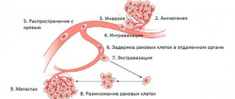
Serious pathologies (emerging tumor processes)
Various neoplasms can interfere with normal intestinal motility, leading to pain, impaired defecation, loss of appetite, etc. To localize the neoplasm and establish its nature, the patient is prescribed an intestinal X-ray; the use of barium in an X-ray helps to obtain a contrast and informative image.
Conditions for performing fluoroscopy of the gastrointestinal tract
X-rays are taken at intervals of twenty minutes to an hour. The process may take several hours. The subject periodically needs to change position on the table (lying, standing, on his side) and wait from half an hour to an hour for the contrast to move to the next section of the digestive canal.
Depending on the areas studied, there are several types of fluoroscopy of the stomach and intestines :
- irrigoscopy - x-ray of the colon (the suspension is administered by enema);
- fluoroscopy – images of the upper gastrointestinal tract (pharynx, esophagus, stomach, first part of the small intestine);
- esophagography – pictures of the esophagus, pharynx.
During intestinal fluoroscopy, general photographs and targeted ones are taken from different angles for a better examination of pathologies or disorders of the examined area. A suspension of pure barium sulfate is used as a contrast.
Contrast in intestinal fluoroscopy
X-ray diagnostics can be conventional (contrast), with double and air contrast.
- The patient takes part of a pre-prepared barium suspension orally. To better distribute the solution throughout the gastric mucosa, the patient’s abdomen is thoroughly massaged.
- Double contrast - taking the solution through a perforated tube. X-ray of the intestines and stomach with 2nd contrast allows you to study in detail the disturbances in the relief of the organ lining.
- Air contrast - taking a solution of crystalline soda instead of barium.
What does an x-ray show?
Radiography, which uses a contrast agent, is considered one of the most informative methods for studying internal organs.
Intestinal shape
Using radiography, a specialist visualizes the shape of the intestine, its appearance and motor functions. Also, barium X-rays of the small intestine and colon are often used because they show areas of narrowing and obstruction.
Location and size of the lumen
Based on how barium fills the lumen of the colon during an examination of the intestines and how it looks on an x-ray, a specialist can diagnose the pathology and prescribe treatment. For example, if the contrast is deposited in flakes, the absorption function of the intestine is impaired; if barium is unevenly distributed on the x-ray, this is a clear sign of a neoplasm.
Elasticity degree
When filling the intestines with a barium-based contrast agent, specialists can use x-rays to evaluate the elasticity of the tissues and their ability to stretch.
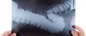
X-ray examination of the intestine (irrigoscopy)
Irrigoscopy
is an x-ray method of examining the large intestine using a contrast agent, usually barium sulfate, administered through the rectum.
Irrigoscopy determines:
1. The shape, location and diameter of the lumen of the colon, 2. The extensibility and elasticity of the intestinal wall, the function of the bauginian valve (this is an intestinal fold located at the junction of the ileum and the colon. Normally, it allows intestinal contents to pass in only one direction - from the small intestine into the thick one, and if its function is impaired, there is a reflux in the opposite direction. During irrigoscopy, this is clearly visible from the movement of the contrast). 3. Functional state of different parts of the intestine 4. Relief of the mucous membrane. This indicator is crucial in the diagnosis of ulcerative lesions, diverticulosis, fistulas, tumors, as well as congenital malformations and cicatricial narrowings of the large intestine.
Irrigoscopy is a painless and non-traumatic procedure. Therefore, if there is a need for an X-ray examination of the large intestine, this technique is used.
Indications for irrigoscopy
Examination of the large intestine using irrigoscopy is used to clarify the diagnosis in the presence of the following complaints:
- Bleeding from the rectum;
- Copious mucous or purulent discharge from the intestines;
- Pain in the anus and along the colon;
- Chronic constipation or diarrhea;
- This method is also used if for some reason it is impossible to perform a colonoscopy or if questionable results are obtained during it. And another indication is a suspicion of an intestinal tumor in a patient with a family history or previously treated for this disease.
Contraindications to irrigoscopy
Irrigoscopy - the contraindications to which are not so numerous - is not performed in the following cases:
- In severe general condition of the patient, for example, with severe heart failure or tachycardia;
- If perforation of the intestinal wall is suspected;
- During pregnancy;
- Caution – in case of acute inflammatory bowel diseases (diverticulitis or ulcerative colitis).
Preparation for irrigoscopy
Like any other intestinal examination, the irrigoscopy procedure requires certain preparation. The colon must be free of feces so that filling with contrast is optimal, and the examination itself is informative: 2–3 days before the procedure, waste products are excluded from the diet, that is, those that cause bloating and heavy stool. It is forbidden to eat some porridges (barley, millet and oatmeal), greens and fresh vegetables (beets, carrots, cabbage, legumes), fruits (apricots, peaches, bananas, apples, oranges), and bread made from dark flour. Meat broths should not be rich, and it is recommended to steam or boil all dishes. The day before the examination, lunch should be light, it is not recommended to have dinner at all, and not to have breakfast on the day of the procedure. In addition to dietary restrictions, it is necessary to cleanse the intestines using enemas or special laxatives. A cleansing enema is done the day before the examination and in the morning on the day of the procedure. At one time, at least a liter of water is injected into the intestines and enemas are repeated until fecal impurities disappear from the rinsing waters. You can also cleanse the intestines with the help of special laxatives. Drugs such as Fortrans, Duphalac, Flit will help the patient prepare for the examination very comfortably and ensure high-quality irrigoscopy. They begin to be taken according to a certain schedule on the eve of the procedure and are completed in the morning on the day of the examination.
Contraindications
Methods for examining the intestines using barium, including x-rays, have a number of contraindications. One part of them is dictated by the impact of ionizing radiation, the other – by the contrast agent.
Pregnancy
Ionizing radiation and barium can have a negative effect on the development of the fetus. Therefore, during the period of bearing a child, it is impossible to conduct an examination of the intestines using barium, especially using X-rays.
Intestinal perforation
Perforation (through damage) of the intestine makes it impossible to use barium, as it can enter the abdominal cavity. If there is a suspicion of such damage to the large or small intestine, contrast is not used during the examination.
The patient is in a serious or even unconscious state
When the patient is in serious condition, especially with acute inflammatory processes, the contrast can be harmful to his health. Therefore, examination of the intestines using barium using radiography in a pre-fainting state or in acute inflammatory processes is not carried out.
Ulcerative colitis
With ulcerative colitis, inflamed areas appear on the intestinal mucosa, which makes the use of barium in X-rays impossible. In this case, conventional radiography is prescribed.
Tachycardia
If barium is used to examine the intestines, various complications are possible that will lead to stress on the heart, so the use of contrast when X-raying a patient with tachycardia is not recommended.
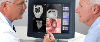
Features of colonoscopy
The procedure is carried out using an endoscopic device - a colonoscope. The main feature is the need for careful preparation, including a special diet and taking medications that help cleanse the intestinal mucosa. Preparation lasts at least 2 days.
The study reveals:
- Oncological diseases.
- Sources of intestinal bleeding.
- Assess intestinal motility.
- Diagnose inflammatory processes in the intestine.
Advantages of carrying out X-rays of the chest cavity at the CELT clinic
We have our own diagnostic center with a full range of equipment necessary to conduct research in a wide variety of areas. Modern X-ray equipment not only makes the procedure safe, but also takes it to a new quality level. Our patients can count on:
- affordable prices;
- accurate diagnosis;
- the procedure is performed by radiologists with over 15 years of experience;
- use of effective safe means;
- use of modern gentle technologies.
You can find out our prices and make an appointment by calling in advance.
Conditions for obtaining reliable diagnostic results
To avoid distorted results of hardware diagnostics of the stomach and intestines, patients are advised to follow a simple diet the day before. Alcohol should be discontinued one day before the test, tobacco and medications that can affect intestinal motility – 12 hours before.
to undergo fluoroscopy of the intestines on an empty stomach, after a three-day slag-free diet. There should be no metal objects in the clothes or on the patient.
The results can be distorted by:
- increased gas exchange;
- full stomach (metabolic disorders, in this case an enema is given a few hours before fluoroscopy of the stomach and intestines);
- metal on the patient (jewelry, crowns, prosthetic plates).
At the end of the study, barium suspension will be released for another 2-3 days. It can cause constipation or mechanical obstruction of the canal, which will aggravate the disease, if any. Therefore, if you experience serious discomfort (bloating, pain, nausea) during this period and (especially) after 72 hours, you should contact your doctor.