Polymerase chain reaction ( PCR
) is one of the most striking achievements in the field of molecular biology. The method has become widespread in various fields of science. Due to its very high specificity and sensitivity, the PCR method is used in medicine, biology, veterinary medicine, forensics, sanitary service and other fields of human activity. For PCR analysis, you can use any biological materials that contain nucleic acids (DNA or RNA molecule).
Principle of PCR research
Every living creature, at least on our planet, has a unique “imprint” - DNA (deoxyribonucleic acid), which is responsible for the transmission of hereditary factors from ancestors to descendants. Structurally, this molecule consists of two strands of nitrogen base molecules, held next to each other by chemical bonds and twisted into a spiral (it is believed that for compactness). From your biology course, you may remember names such as adenine (A), guanine (G), thymidine (T), and cytosine (c). These are 4 nucleotides that create the DNA sequence. Viruses store their genetic information in another nucleic acid, RNA. Information about already studied DNA and RNA is stored in scientific databases of laboratories. After the PCR method
, for many pathogens of various diseases (bacteria, fungi and viruses) their own specific genetic detectors (primers) have been created - unique nucleotide sequences characteristic only of a particular pathogen. And if you place them in a test tube with the test material, if it contains DNA or RNA of “living” pathogens, the primers trigger a replication reaction - the creation of a huge number of copies, which can be identified visually. Those. they start copying their DNA/RNA dozens of times. And when calculating the results, laboratory staff can understand whether the desired bacteria and viruses are in the test sample or not, which is why PCR results are most often qualitative, i.e. "detected" or "not detected".
Features of the PCR test for the new coronavirus
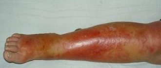
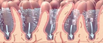
The SARS-CoV-2 virus, like all respiratory agents, enters through the mucous membranes of the nose, eyes or mouth. It is fixed in cells, clinging with specific spikes to receptors on their membranes. The tissues most vulnerable to damage are the respiratory system, urination, digestion, central nervous system, and heart. Their surface contains ACE2 and ACE2 receptors, to which coronavirus proteins are tropic.
In the first days after infection, SARS-CoV-2 actively multiplies in the epithelium of the nasopharynx. The infection is contained in the mucous secretion and saliva. Carriers, without knowing it, spread it by sneezing, coughing, or deep breathing. Even without symptoms, such people are dangerous to others. Expressed COVID in most cases does not differ from ordinary acute respiratory viral infections; it has no specific manifestations. Only PCR can determine whether a person is infected with a new pathogen.
Coronaviruses do not circulate in the blood and lymph. Samples for analysis are taken from the mucous membranes of the nasopharynx. The experience of doctors has shown that in people with asymptomatic and manifest disease, the accuracy of the study is about 80%; the probability of false negative results is quite high. The pathogen does not stay on the epithelium for long, being subject to immune attacks. For this reason, it is rational to repeat the examination several times. And if the virus is not detected for a long time, people with suspected COVID should donate blood for antibodies to the infection using the ELISA method.
To whom do we owe the emergence of the PCR method?
According to American biochemist Kary Mullis, the idea of identifying living organisms by a short section of their genetic code (DNA) came to his mind in 1983, on his way home from work. And this idea was based on the work of another American biochemist, Arthur Kornberg, which at one time did not find a response from the scientific community.
Kerry allowed the possibility of taking a DNA molecule of any organism, using high temperature to “unravel” its helix into two strands, using specific markers-primers to mark sections of DNA unique to this microorganism, and then, using the enzyme DNA polymerase, to create from two strands of two new DNA molecules. But they already contain labeled primers. And then all that remains is to simply look for these areas in the diagnostic material.
As a result, the CETUS corporation, where Mulis worked, allocated him a team of scientists. And in 1985, in the publication of the American Society of Human Genetics, a publication appeared with a theoretical justification for PCR as a method for identifying the genetic material of living organisms.
Who needs diagnostics
Since PCR allows us to detect even hereditary diseases that migrate from generation to generation in their latent phase, conducting such a study will probably be useful for everyone. Of course, such knowledge does not seem urgent and necessary to everyone here and now. But it seems that as civilization develops, the successes of medicine and genetics will also increase. And new discoveries will allow us to make great progress in achieving longevity and health. By that time, a test for heredity may well become a routine and mandatory analysis for everyone.
Until this happens, the test is most often necessary in the following cases:
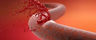
- If there is a suspicion of contracting the disease through sexual contact, or through an injection, contact with contaminated blood on mucous membranes or open wounds (a small cut is enough), an attack by wild animals, accidental injury to surrounding objects, for example, stepping on an old nail in a field.
- To clarify a diagnosis made by other means, especially in cases where treatment gives a non-specific reaction of the body.
- To establish family ties to claim inheritance rights, or in alimony litigation.
- To identify a person's identity.
Other tests are more related to areas of individual interest. Who is interested in knowing what the transcript of all the nationalities in his family shows? Someone is trying to plan for a future child and is being tested for compatibility. Someone decided to replace the DNA test with a nutritionist and receive individual instructions and advice on proper nutrition.
How it all happens in the laboratory
DNA extraction
First, a sample of biological material is prepared: centrifuged, sedimented, etc. Then laboratory technicians need to extract DNA from the resulting biological concentrate. Amplification (increasing the number of DNA copies) The most important stage of the study. It is carried out in a thermal cycler and it is here that all processes that fall under the definition of a polymerase chain reaction take place: denaturation, annealing, elongation. Denaturation The very first stage is to unfold (denature) nucleic acids to make them available for further work. It is carried out by heating the reaction mixture to 80-90 °C. Annealing Denatured (dissolved) DNA/RNA is treated with primers—short chains of nucleic acids made in the laboratory. Thanks to the programmed region, primers attach only to those nucleic acids for which they were created. For example, a primer for herpes simplex virus type 1 will never bind to the DNA of another virus, microorganism or cell. It is the primers that determine the extremely high specificity of PCR
– the ability to react only to nucleic molecules of specific types, types of classes and even strains of microorganisms. Or certain types of cells of living organisms.
Elongation
Or synthesis. After completion of the annealing process, conditions for polymerase activity are created in the reaction mixture. The enzyme, focusing on primer molecules (and not the original nucleic acids), begins the synthesis of new DNA/RNA strands. Which become copies of the original, sought-after nucleic acid molecules. This temperature cycle is carried out 30 or more times. As a result, even with an initially small amount of the desired genetic material, a significant number of nucleic acids “labeled” with primers accumulate in the reaction mixture (it grows exponentially, doubling with each cycle). It is much easier to detect large amounts of DNA/RNA, due to which another The advantage of PCR is its highest sensitivity.
Detection
PCR results are assessed in several ways
:
- Electrophoresis in a viscous medium. The idea is that DNA/RNA is electrically charged and moves towards one of the electrodes. A DNA dye (for example, ethidium bromide) is added to the medium (agar or polyacride gel). During the electrophoresis session, nucleic acid molecules move and form clusters, tinted with ethidium. Under ultraviolet light, it appears in the form of stripes of varying thickness and brightness.
- Hybridization method. Primers that are pre-labeled with a phosphor (fluorophore) are used. After the required number of temperature cycles, a special device is used - a fluorescence detector. Due to the fact that fluorophores for different targets can be added to a sample (they will glow under ultraviolet light in different colors), the hybridization method is suitable for diagnosing several targets in one sample.
- PCR diagnostics in real time (real-time PCR). It differs in that detection is carried out directly during the amplification process. To do this, you need phosphor probes (from the previous paragraph) and special DNA amplifier devices. These devices evaluate the increase in phosphor brightness after each temperature cycle and subsequently calculate the initial number of desired nucleic acids in the sample.
Electrophoresis and hybridization are suitable only for qualitative assessment, that is, they answer the question of whether the sample contains the desired material. Real-time PCR is the only quantification method available. If there are no targets for primers in the sample, then the temperature cycles will go idle and a negative result will be obtained during detection.
Antibody test for COVID-19
When a pathogenic agent invades the body, the immune system begins to produce specific immunoglobulin proteins (Ig) that can remember and neutralize this microorganism. Thus, a repeated encounter with this pathogen will not be dangerous. The body quickly inactivates it.
When diagnosing COVID-19, two types of antibodies are important: IgM and IgG. IgM is detected in the analysis on days 7-10 after infection (from the 5th day of the onset of clinical signs of the disease) and remains in the body for up to two months. Since immunoglobulins do not begin to be produced from the first day of infection, the test for antibodies may initially be negative, because the study can be carried out at a time when the body has not yet had time to form an immune response. It is logical to detect IgM at the height of the disease and immediately after regression of symptoms.
IgG is produced later, from about 21-28 days after infection, and remains in the body for a longer time, for several months or years. Consequently, they can be detected only at the stage of recovery or at a later date. By the presence and level of IgG in the blood, one can judge both the fact of a past infection and determine the presence of specific immunity to a given pathogen.
Which diagnostic method is better?
Considering the difference between PCR and antibodies for COVID, the difference in these studies, it is impossible to unambiguously determine which diagnostic method is preferable. If there is contact with a sick or infected person, as well as if there are symptoms that fit the picture of coronavirus, PCR diagnostics is recommended. It takes more time, but will provide fairly reliable data on the presence of an infectious pathogen in the body.
If you need to clarify whether you have already had COVID-19, ELISA diagnostics are recommended to determine the level of antibodies. The analysis will be informative if the disease occurred even in an asymptomatic form.
Advantages of the PCR technique
In total, more than 10 different amplification methods have been developed, used by laboratories depending on the initial conditions and goals. What they have in common is high sensitivity (for a positive result, 40 (!) or less of the desired DNA copies in 1 ml of sample are enough, that is, the probability of a false negative response is negligible. And very high specificity: the probability of a false positive response is less than 1%. But the accuracy of the results is very depends on the quality of collection of diagnostic material, careful compliance with all technical requirements for each stage and the quality of equipment, consumables (buffer, primers, washing solution, etc.).
Progress of the reaction
One reaction cycle occurs in three stages:
- Heating stage. The DNA helix in the sample is heated for a period of half to two minutes at 94-96 degrees. When exposed to high temperatures, DNA chains break down into separate fragments
- At the second stage, the necessary fragments are caught using primers and fused. This stage takes place at a temperature slightly lower, 4-5 degrees, and is called “annealing.” The duration of the stage also varies from half to two minutes.
- A special enzyme is added to start the self-reproduction (synthesis) of the “caught” gene region - DNA polymerase. And this enzyme starts the fragment doubling reaction, using the starting fragment as a template for synthesizing a copy. The reaction takes place at 72 degrees for 10 to 12 minutes.
Typically, one study consists of 20 to 35 similar cycles.